CH12 - Central Nervous System
1/57
There's no tags or description
Looks like no tags are added yet.
Name | Mastery | Learn | Test | Matching | Spaced | Call with Kai |
|---|
No analytics yet
Send a link to your students to track their progress
58 Terms
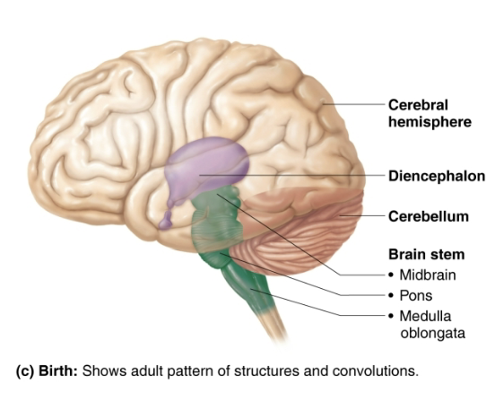
List the four regions of an Adult Brain
Cerebral hemispheres (Cerebrum)
Diencephalon
Brain stem
Midbrain
Pons
Medulla
Cerebellum
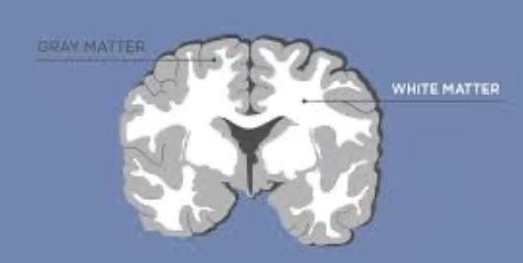
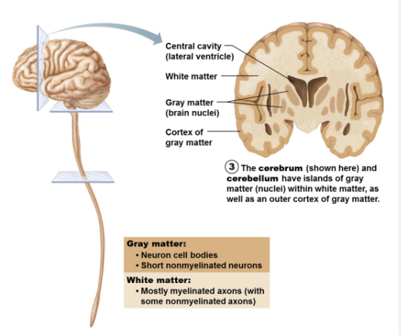
Gray Matter vs White matter
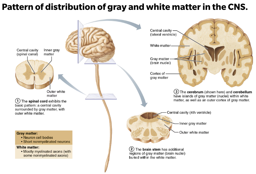
Gray matter
Short, non-myelinated neurons and cell bodies
White matter
Myelinated and non-myelinated axons
Describe the basic pattern found in CNS in regards to brain regions and organization
CNS (Brain & Spinal cord)
Central cavity surrounded by gray matter, white white matter external to gray matter
Spinal cord
Exhibits this basic pattern → however pattern changes with ascent to brainstem
T/F: Most neuron cell bodies are located in PNS
→ False
Most neuron cell bodies are located in CNS
Nuclei: clusters of neuron cell bodies in CNS
Ganglia: clusters of neuron cell bodies in PNS
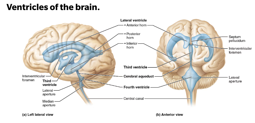
Structural and Function of Ventricles
STRUCTURE
Fluid-filled chambers that are continuous to one another and to central canal of spinal cord
Filled with cerebrospinal fluid (CSF)
Lined by ependymal cells (neuroglial cells)
Line the central cavities of the brain and spinal column
Produce cerebrospinal fluid (CSF)
Paired lateral ventricles (first and second) are large, C-shaped chambers located deep in each hemisphere
Pair is separated by membranous septum pellucidum
Third ventricle is in diencephalon
Fourth ventricle is between brain stem and connects with central canal of spinal cord
FUNCTION
Produce and secrete cerebrospinal fluid to protect and maintain your central nervous system
CSF is constantly bathing the brain and spinal column, clearing out toxins and waste products released by nerve cells
EXAMPLE
The waste product - Amyloid A-b peptide → increases the risk of Alzheimer’s disease if too much accumulates in the brain
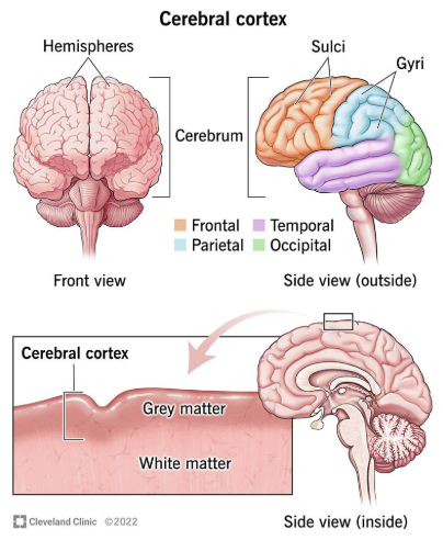
Structural and Function of Cerebral Hemispheres (Cerebrum)
Regions of an Adult Brain (1/4)
STRUCTURE
Left and right hemisphere
Accounts for 83% of brain mass
Separated by a groove, the longitudinal fissure
Each of these hemispheres has an outer layer of grey matter, the cerebral cortex
The cerebral cortex is supported by inner layer of white natter
The hemispheres are linked by the corpus callosum, a very large bundles of nerve fibers
FUNCTION
Form superior part of brain
List the major lobes of the Cerebral Hemispheres (Cerebrum)
Frontal lobe
Parietal lobe
Temporal lobe
Occipital lobe
Insular lobe
Buried under portion of temporal, parietal and frontal lobes
Functions of Frontal Lobe
Executive functions
Planning, organizing, problem-solving, decision-making, attention, and self-control
Motor control
Initiating and coordinating voluntary movements
Langauge
Producing and understanding speech
Emotional regulation
Controlling emotions, such as anger, fear, and sadness
Personality
Expressing personality traits and social behavior
Working memory
Holding and manipulating information in the short term
Creativity and innovation
Generating new ideas and solutions
Functions of Parietal Lobe
Somatosensory perception
Processing sensations such as touch, pain, temperature, and proprioception (body awareness)
Spatial orientation
Understanding and interpreting the position and movement of objects in space
Attention and focus
Directing attention to specific stimuli and maintaining focus
Movement planning
Initiating and coordination voluntary movements
Language processing
Contributing to the understanding and production of language
Number and calculation
Processing numerical information and performing calculations
Functions of Temporal Lobe
Auditory Processing
The primary auditory cortex, which receives and processes auditory information
Language
Understanding and producing spoken language
Memory
Hippocampus, plays a vital role in forming and storing memories
Emotion
The amygdala processes and regulates emotions, including fear, pleasure, and anger
Visual Processing
Contributes to visual processing by aiding in object recognition, face recognition, and the integration of visual information with other sensory inputs
Functions of Occipital Lobe
Receiving visual information
Receives visual information from the retina via the thalamus
Mapping visual input
It helps with spatial reasoning and visual memory
Color determination
Helps determine the color of the items you see
Depth perception
It is involved in assessing distance, size, and depth
Object and face recognition
It plays a crucial role in identifying familiar faces and objects
Motion detection
The occipital lobe is also involved in processing motion
Functions of Insular Lobe
Interception
Integrates information about the internal state of the body, including pain, temperature, and visceral sensations
Taste
It’s involved in the perception and processing of taste sensations
Emotional Awareness
Experience and recognition of emotions, including happiness, sadness, fear, disgust, and empathy
Social Cognition
The insula contributes to social decision-making, trust, and other aspects of social behavior
Self-Awareness
The insula plays a role in our sense of our own body in space and our relationship to it, and awareness of self
Addiction
Implicated in the processes of addiction, including and reward-seeking behavior
Clinical - Explain Dementia
Refers to a decline in cognitive function, including memory, thinking, and reasoning, to the point where it interferes with a person’s ability to perform daily activities
Dementia is not one specific disease, but rather a group of symptoms cause by various brain diseases
While many diseases can cause dementia, Alzheimer’s disease is the most common
Other causes include vascular dementia, Lewly body dementia, frontotemporal dementia, and Parkinson’s disease dementia
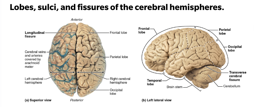
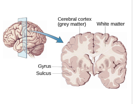
List and describe the Surface Markings on the Cerebral Hemispheres (Cerebrum)
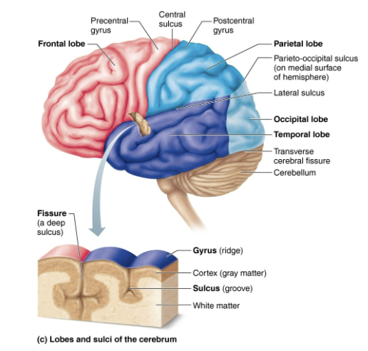
Gyri
Ridges
Sulci
Shallow grooves
Fissures
Deep grooves
List the major Fissures of the Cerebral Hemispheres (Cereburm)
Fissure → deep grooves
Longitudinal fissure
Separates two hemispheres
Transverse cerebral fissure
Separates cerebrum and cerebellum
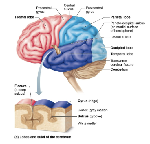
List the major Sucli that divide lobes
Central sulcus
Separates pre-central gyrus of frontal lobe and post-central gyrus of parietal lobe
Parieto-occipital sulcus
Separates occipital and parietal lobes
Lateral sulcus
Outlines temporal lobes
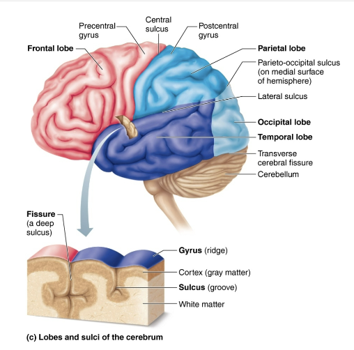
List the three basic regions of the Cerebral Hemisphere (Cerebrum)
Cerebral cortex of gray matter superficially
White matter internally
Basal nuclei deep within white matter
Structural and Function of Cerebral Cortex
BASIC REGIONS of the Cerebral Hemisphere (Cerebrum) (1/3)
STRUCTURE
Thin (2-4 mm) superficial layer of gray matter
Composed of neuron cell bodies, dendrites, glial cells, and blood vessels
40% of mass of brain
FUNCTION
Site of conscious mind
Awareness
Sensory perception
Voluntary motor initiation
Communication
Memory storage
Understanding
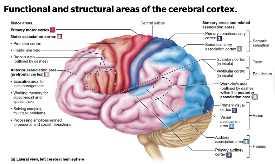
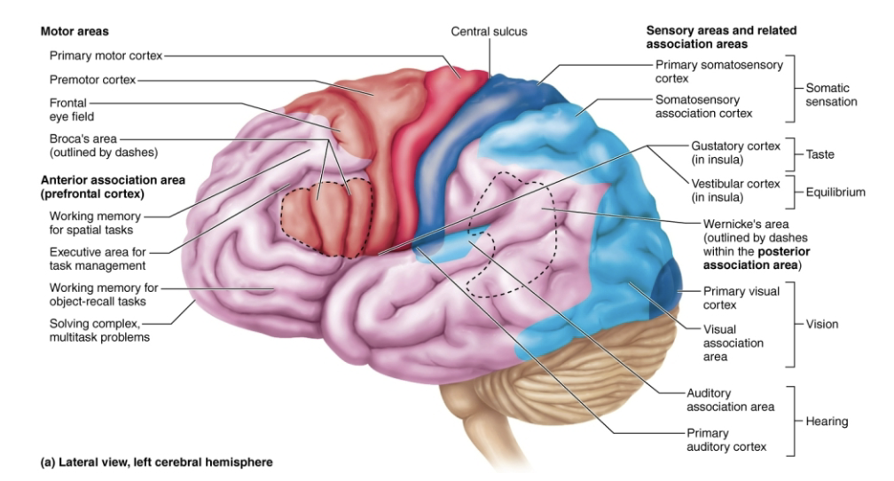
List the functional areas of the cerebral cortex
BASIC REGIONS of the Cerebral Hemisphere (Cerebrum) (1/3)
No functional area of the cortex acts alone, and conscious bheavior involves the entire cortex in one way or another
Motor areas
Control voluntary movement
Sensory areas
Conscious awareness of sensation
Association areas
Integrate diverse information
Visual areas
Auditory areas
Responsible for complex processing that goes on between the arrival of sensory input cortices and the generation of behavior
T/F: All neurons in the cortex are interneurons
→ True
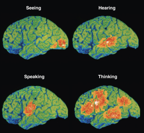
Explain Cerebral Cortex Domains
Specific motor and sensory functions located in discrete cortical areas
Higher functions are spread over many areas
Shown on Functional imaging (PET and MRI)
Explain Contralateral vs Lateralization of cortical function
EACH hemisphere is concerned with contralateral (opposite) side of body
Lateralization (specialization) of cortical function can occur in only ONE hemisphere
Conscious behavior involves entire cortex
Describe the location, function, structure of Motor Areas in Cerebral Cortex
Functional areas of the cerebral cortex (1/3)
LOCATION
In frontal lobe
FUNCTION
Motor areas act to control voluntary movement
STUCTURE
Primary motor cortex
Premotor cortex
Broca’s area
Frontal eye field
List the components of Motor Area
Functional areas of the cerebral cortex (1/3)
Motor areas in Cerebral Cortex
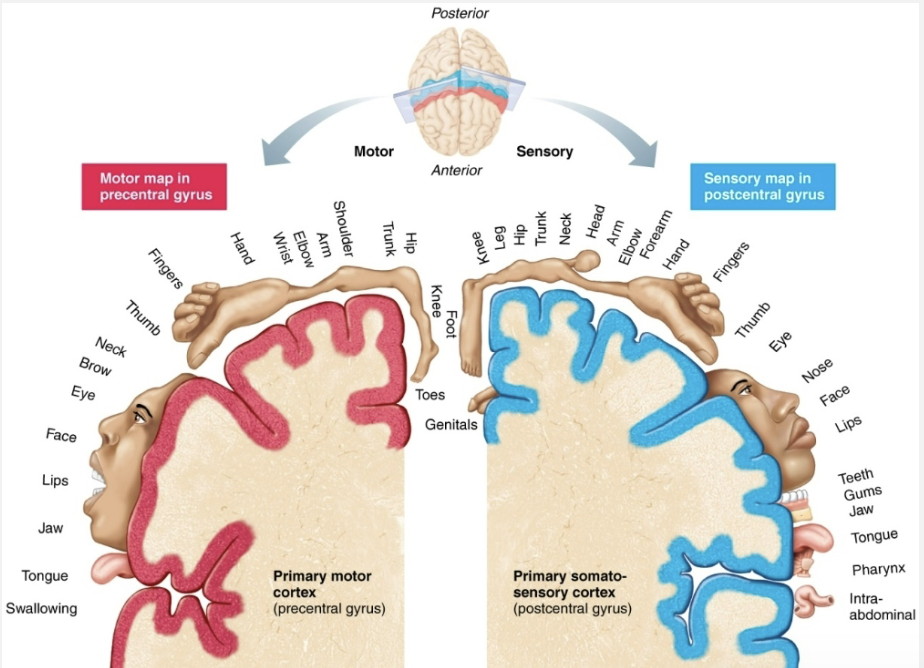
Primary (somatic)Motor Cortex
LOCATION
In pre central gyrus of each frontal lobe
FUNCTION
Initiating voluntary movements
Somatotopy → All muscles of body can be mapped to area on primary motor crotex
Motor homunculi → Upside-down caricatures represent contralateral motor innervation of body regions
Premotor cortex
LOCATION
Anterior to the pre-central gyrus of each frontal lobe
FUNCTION
Helps to plan and coordinate complex movements
Broca’s area
LOCATION
Anterior to inferior premotor area
Present in one hemisphere (usually left)
FUNCTION
Motor speech area that directs muscles of speech production
Active in planning speech and voluntary motor activities
Clinical - Examples to damages to area Primary Motor Cortex
Damage to areas of primary motor cortex, as seen in a stroke, paralyzes muscles controlled by those areas
Paralysis occurs on opposite side of body from damage
Only voluntary control is lost, however, as the muscles can still contract reflexively
Clinical - Examples to damages to area Premotor Area
Muscle strength or ability to PERFORM discrete individual movements is not impaired; ONLY CONTROL over movements is lost
EX: Damage to premotor area controlling movement of fingers would still alllow fingers to move, but voluntary control needed to type would be lost
Other premotor neurons can be reprogrammed to rake over skill of damaged neurons
Would require practice, just as the initial learning process did
Describe the location, function, structure of Sensory areas in Cerebral Cortex
Functional areas of the cerebral cortex (2/3)
LOCATION
Occur in parietal, insular, temporal, and occipital lones
FUNCTION
Areas of cortex concerned with conscious awareness of sensation
STUCTURE
Primary somatosensory cortex
Location and function of Primary somatosensory cortex
Functional areas of the cerebral cortex (2/3)
Sensory areas in Cerebral Cortex
LOCATION
In postcentral gyri of parietal lobe
FUNCTION
Receives sensory information from skin and proprioceptors of skeletal muscle, joints, and tendons
Capable of spatial discrimination → identification of body region being stimulated
Somatosensory homunculus → upside-down caricatures represent contralateral sensory input from body regions
Describe the location, function, structure of Assoication areas in Cerebral Cortex
Functional areas of the cerebral cortex (3/3)
Visual association areas in Cerebral Cortex
LOCATION
Surronds primary visual cortex
FUNCTION
Uses past-visual experiences to interpret visual stimuli (color, form, or movement)
EX ability to recognize faces
Complex processing involves entire posterior half of cerebral hemispheres
STRUCTURE
Primary visual (striate)
Cortex located on extreme posterior tip of occipital lobe
Auditory association areas in Cerebral Cortex
LOCATION
Located posterior to primary auditory cortex
FUNCTION
Stores memories of sounds and permits perception of sound stimulus
STRUCTURE
Primary auditory cortex
Superior margin of temporal lobes
Interprets information from inner ear as pitch, loudness, and location
Clinical - Damage to the Primary Visual Cortex
Primary visual cortex → results in blindness
Visual association area → can see, but they do not comprehend what they are looking at
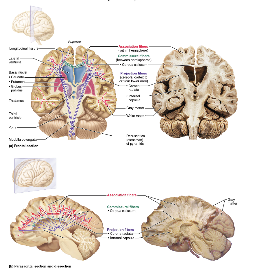
Describe the general structure function of the Cerebral White Matter
BASIC REGIONS of the Cerebral Hemisphere (Cerebrum) (2/3)
STURCURE
Second of three basic regions of cerebral hemispheres
Consists of myelinated fibers bundled into large tracts
FUNCTION
Responsible for communication between cerebral areas and between cortex and lower CNS
Classified according to direction they run”
Association
Commissural
Projection fibers
Describe the general structure function of the basal nuclei (basal ganglia)
BASIC REGIONS of the Cerebral Hemisphere (Cerebrum) (3/3)
STRUCTURE
Third of the three basic regions of cerebrum
FUNCTION
Influence muscle movements
Play role in cognition and emotion
Filter out incorrect/inappropriate responses
Clinical - Disorders of the basal nuclei
Parkinson’s disease
Brain disorder that causes unintended or uncontrollable movements, such as shaking, stiffness, and difficulty with balance and coordination
Huntington’s disease
An inherited disorder that causes nerve cells (neurons) in parts of the brain to gradually break down and die
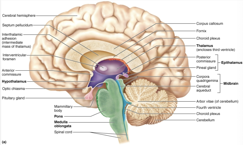
Describe the location of the diencephalon and the name its subdivisions
Consists of three paired gray matter structures:
Thalamus
Hypothalamus
Epithalamus
LOCATION
All three enclose third ventricle
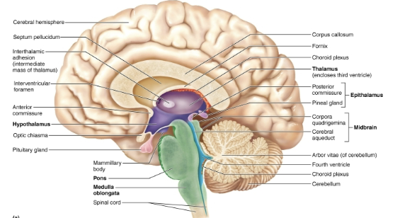
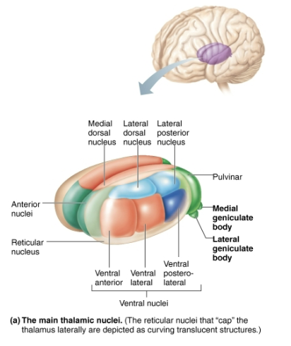
Describe the structure and function of the Thalamus
Diencephalon Subdivision (1/3)
STRUCTURE
Bilateral egg-shaped nuclei that form superolateral walls of third ventricle
Makes up 80% of diencephalon
FUNCTION
Act as relay station for information coming into cortex
Sorts, edits, and relays ascending input such as:
Impulses from hypothalamus for regulating emotion and visceral function
Impulses for memory or sensory integration
Overall it acts to mediate sensation, motor activities, cortical arousal, learning, and memory
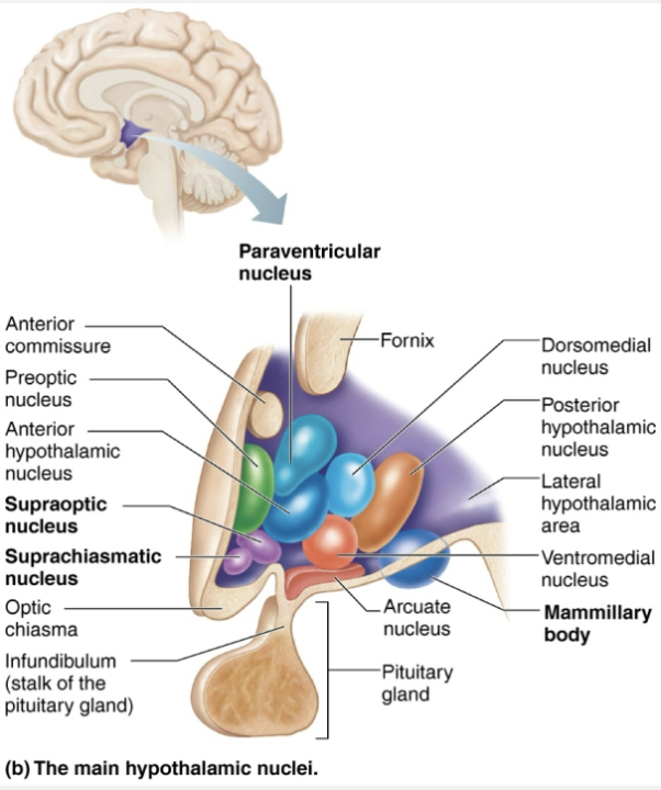
Describe the structure and function of the Hypothalamus
Diencephalon Subdivision (2/3)
STRUCTURE
Located below thalamus
Forms cap over brain stem and forms inferolateral walls of third ventricle
Contains many important nuclei such as:
Mammillary bodies → paired anterior nuclei that act as olfactory relay stations
Infundibulum → stalk that connects to pituitary gland
FUNCTION
Main visceral control and regulating center that is vital to homeostasis
Chief homeostasis controls:
Controls autonomic nervous systems
EX: BP, rate and force of heartbeat, digestive tract motility, pupil size
Initiates physical responses to emotions
Part of limbic system → perceives pleasure, fear, rage, biological rhythms, and sex drive
Regulates body temp
Sweating or shivering
Regulates hunger and satiety in response to nutrient blood levels or hormones
Regulates sleep-wake cycle s
Suprachiasmatic nucleus of thalamus sets out biological clock
Controls endocrine system function
Secretions of anterior pituitary gland
Production of posterior pituitary hormones
Describe the structure and function of the Epithalamus
Diencephalon Subdivision (3/3)
STRUCTURE
Most dorsal portion of dicencephalon
Forms roof of third ventricle
Contains pineal gland (body)
Extends from posterior border
FUNCTION
Pineal gland → secretes melatonin that helps regulate sleep-wake cycle
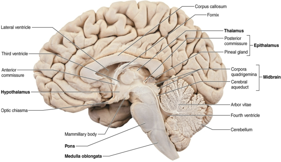
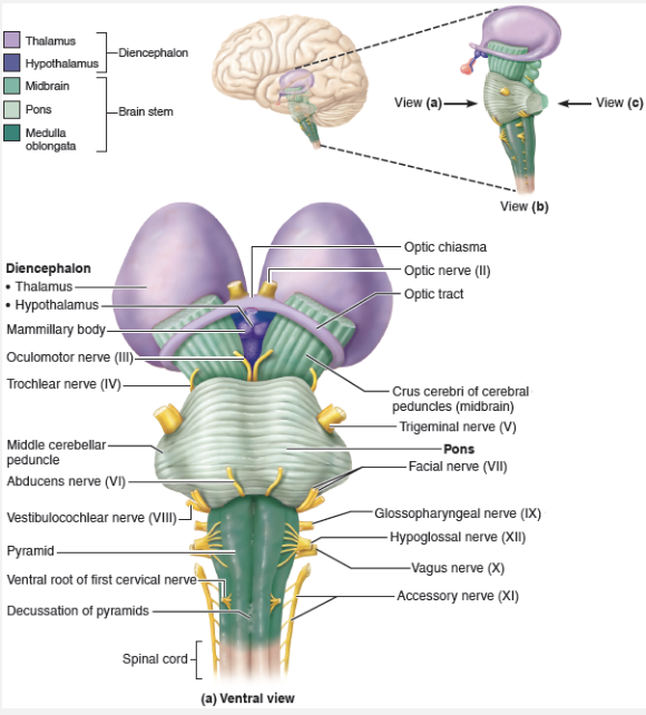
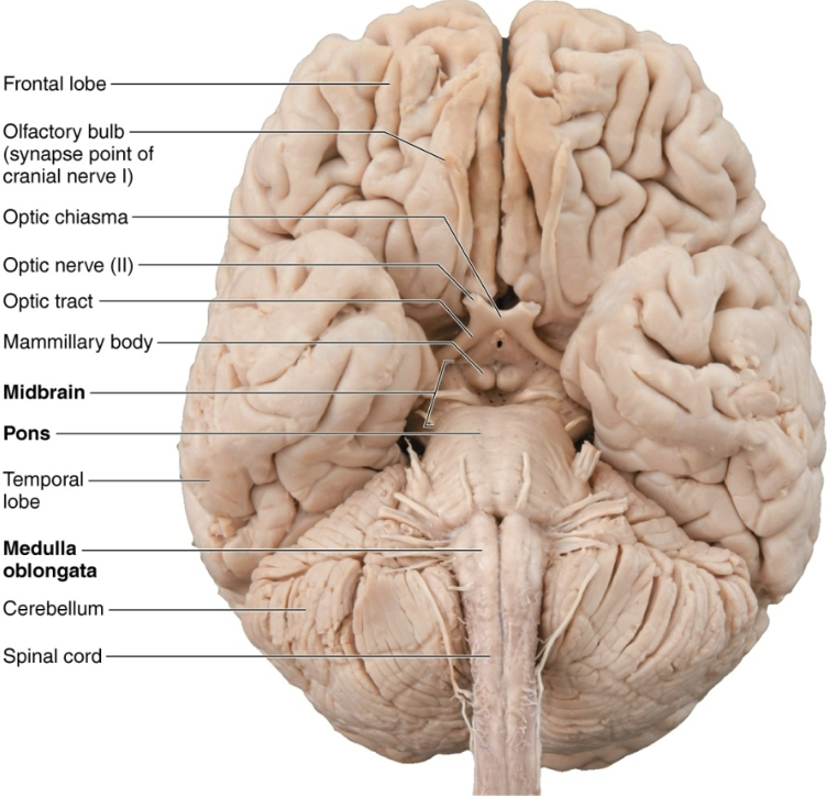
List the three regions of the Brain Stem
Midbrain
Pons
Medulla oblongata
Describe the strucutre of the Brain stem
Similar in structure to spinal cord BUT contains nuclei embedded in white matter
Contains fiber tracts connecting higher and lower neural centers
Nuclei are associated with 10 of the 12 pairs of cranial nerves
Describe the structure and function of the Midbrain
Brain Stem (1/3)
STRUCTURE
Midbrain nuclei scattered throughout white matter include:
Substantia nigra → functionally linked to basal nuclei
Parkinson’s disease is degeneration of this of this area
Describe the structure and function of the Medulla Oblongata
Brain Stem (2/3)
STRUCTURE
Blends into spinal cord at foramen magnum
Contains fourth ventricle
Continuation of central canal of spinal cord
Medulla and pons from ventral wall
Contains choroid plexus → capillary-rich membrane that forms cerebral spinal fluid
FUNCTION
Medulla is an autonomic reflex center
Many functions overlap with hypothalamus → hypothalamus relays instructions via medulla
List the functional groups of Medulla Oblongata
Cardiovascular center:
Force and rate of heart contraction
Vasomotor center adjusts blood vessel diameter for BP regulation
Respiratory centers
Generate respiratory rhythm
Control rate of depth of breathing
Various other centers regulate:
Vomiting
Hiccuping
Swallowing
Coughing
Sneezing
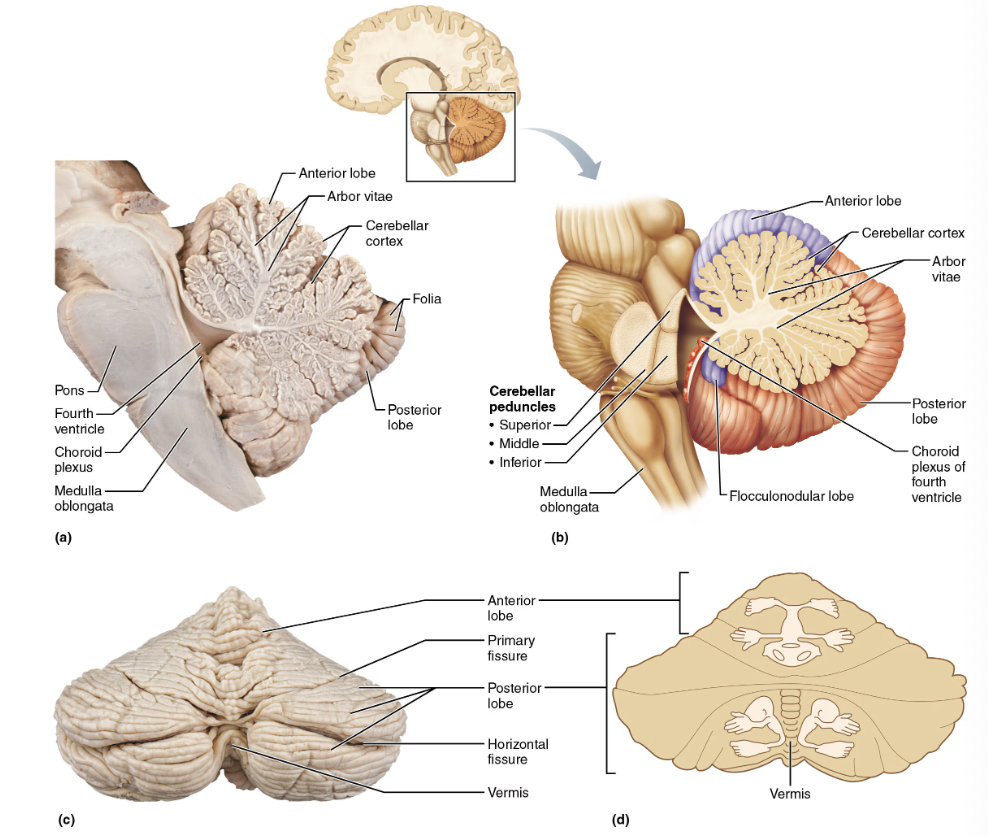
Describe the structure and function of the cerebellum
STRUCTURE
11% of brain mass
Located dorsal to pons and medulla
FUNCTION
Processes input from cortex, brain stem, and sensory receptors to provide precise, coordinated movements of skeletal muscles
Plays a major role in balance
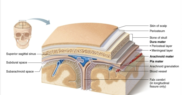
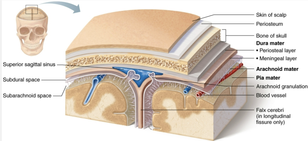
List structures that protect the brain
Meninges
Dura mater
Arachnoid mater
Pia mater
Cerebrospinal fluid (CSF)
Blood barrier
List the layers of Meninges from external to internal
Dura mater
Arachnoid mater
Pia mater
Describe how the cerebrospinal fluid, meninges protect the brain
Dura mater
STRUCTURE
Made up of two layers of fibrous connective tissue → two layers are mostly fused, but separate in certain areas to form dural venous sinuses
FUNCTON
Strongest meninx
Sinuses collect venous blood from brain, empty into jugular veins of neck
Extends inward in several areas to form flat partitions that divide cranial cavity
Partitions referred to as dural septa
Act to limit excessive movement of brain
Arachnoid mater
STRUCTURE
Middle layer with spiderweb-like extensions
Separated from dura mater by subdural space
Subarachnoid space contains CSF and largest blood vessels of brain
Arachnoid granulation protrude through dura mater into superior sagittal sinus
FUNCTION
Arachnoid granulation → permit reabsorption pf CSF back into venous blood
Pia mater
STRUCTURE
Delicate connective tissue that clings tightly to brain, following every convolution
FUNCTION
Contain many tiny blood vessels that feed brain
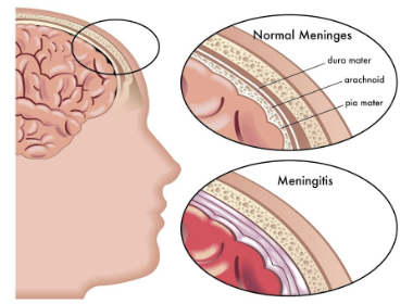
Clinical - Meningitis & Encephalitis
Meningitis: Inflammation of the meninges
May spread to CNS → which would lead to inflammation of the brain → encephalitis
Meningitis is usually diagnosed by observing microbes in sample of CSF obtained via lumbar puncture
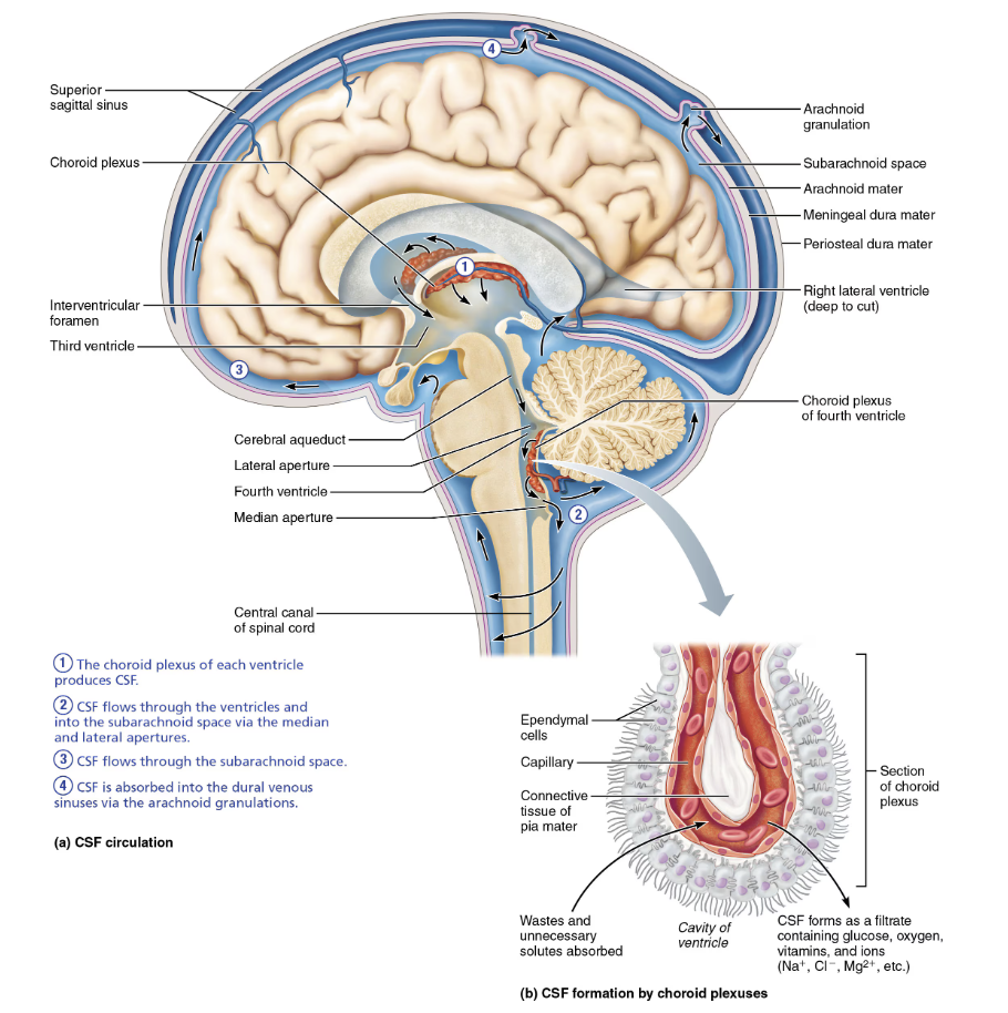
Explain how cerebrospinal fluid (CSF) is formed and describe its circulatory pathway
The choroid plexus of each ventricle produces CSF
CSF flows through ventricles and into the subarachnoid space via the median and lateral apertures
Arachnoid mater
CSF flows through subarachnoid space
Arachnoid mater
CSF is absorbed into the dural venous sinuses via the arachnoid granulations
Dura mater
Describe how the blood-brain barrier protect the brain
STRUCTURE
Substances from blood must first pass through continuous endothelium of capillary walls before gaining entry into neurons
Tight junctions ensure substances pass through, not around endothelial cells
Feet of astrocytes and smooth muscle-like pericytes surround endothelial cells → Help to promote thigh junction formation in endothelial cells
FUNCTION
Helps maintain stable environment for brain
Chemical variations could lead to uncontrollable neuron firings
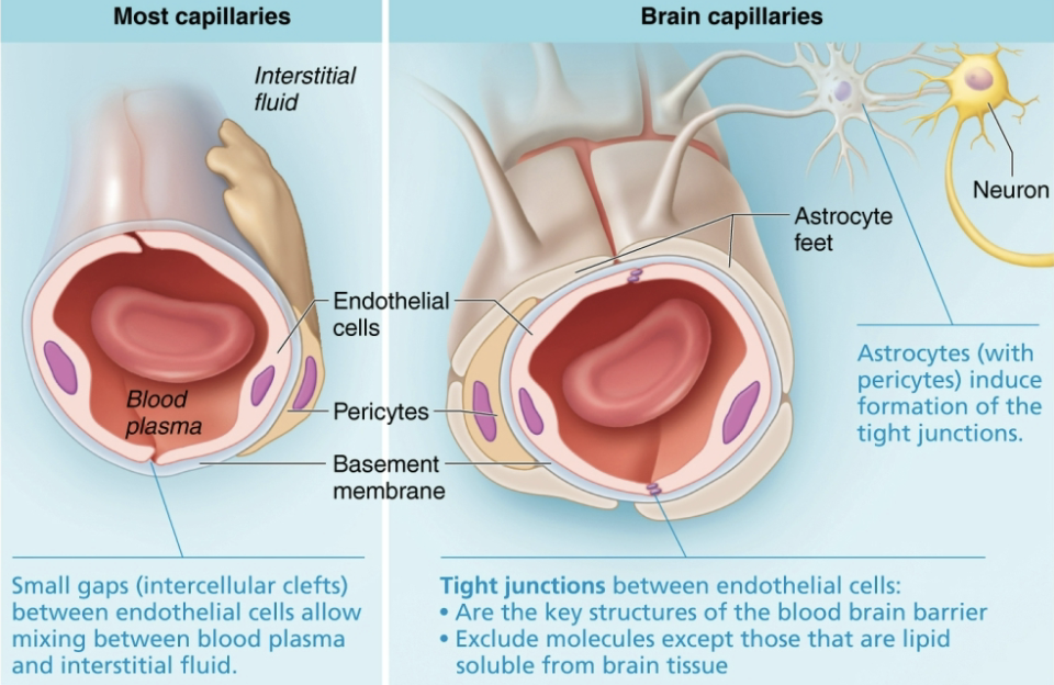
List how substances move through endothelial cells
Simple diffusion
Allows lipid-soluble substances, as well as blood gases to pass freely through cell membrane
Specific transport mechanism
Facilitated diffusion moves substances important to the brain such as glucose, amino acids and specific ion
Transcytosis moves larger substances into and out of brain
What is the last part of Blood-Brain Barrier substances must pass through?
Thick basement membrane surrounding capillaries is last part of barrier substances must pass through
Contains enzymes that destroy certain chemicals that would activate brain neurons
Absent in some areas, such as vomiting center and hypothalamus
Necessary to monitor chemical composition and temperature of blood
List and describe the types of Brain Injuries
Concussion
Alteration in brain function, usually temporary, following a blow to the head
Contusion
More serious concussion can bruise brain
Cerebrovascular Accidents (CVAs)
Also referred to as “strokes”; tissue deprived of blood supply, leading to death of brain tissue (ischemia)
Can be caused by blockage of cerebral artery by blood clot
Describe the cause (if known) and symptoms of Concussion vs Contusions, and cerebrovascular accidents (strokes)
Concussion
Widespread damage to the brain
CAUSE
Only caused by head trauma
SYMPTOMS
Some cause bleeding in the brain but not alll
Often experience cognitive and psychological effects such as memory loss and irritability
Contusion
Localized injury that damages a limited area of the brain
Typically more severe
CAUSE
Mostly caused by head trauma but can also be the result of having high BP as a older person, bleeding problems, taking blood thinning medicines or some illegal drugs
SYMPTOMS
Causes bleeding, clotting and pooling of blood int he brain
Cerebrovascular Accidents (CVAs)
Strokes
CAUSE
Hemiplegia (paralysis on one side) or sensory and speech deficits
SYMPTOMS
Loss of sensation
Seizures
Speech and Associated problems
Describe the structure and function Spinal cord
STRUCTURE
Spinal cord is enclosed in vertebral column
Begins at the foramen magnum
Ends at L1 or L2 vertebra
FUNCTION
Provides two-way communication to and from brain and body
Major reflex center → reflexes are initiated and completed at spinal cord
How is Spinal Cord protected?
Protected by bone, meninges, and CSF
Spinal dura mater is one layer thick
Does not attach to vertebrae
Describe the gross structure of the spinal cord
Epidural space
Conus medullaris
Filum terminale
Spinal nerves
Cauda equina
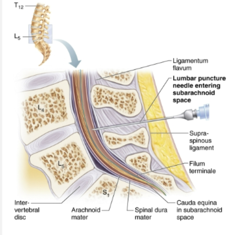
Structure and function of Epidural space
Gross Anatomy of the Spinal Cord
STRUCTURE
CSF fills subarachnoid space between arachnoid and pia maters
Dural and arachnoid membranes extend to sacrum, beyond end of cord at L1 or L2
→ Site of lumbar puncture or tap
FUNCTION
Cushion of fat and network of veins in space between vertebrae and spinal dura matter
Describe the gross and microscopic structure of the spinal cord