Hip and Lumbar Special Tests
1/11
There's no tags or description
Looks like no tags are added yet.
Name | Mastery | Learn | Test | Matching | Spaced |
|---|
No study sessions yet.
12 Terms
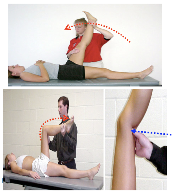
Thomas Test
Pt: Supine + knees bent at end of table
Clinician: standing at side (non-testing limb)
hand1: under lordotic curve
hand2: behind opposite knee
then passive hip flx
(+) test: observe the opposite side
knee Ext = rectus femoris tightness
thigh lift off = hip flexor tightness
Ely’s Test
Pt: prone
Clinician: standing at side (testing limb)
hand1: anterior distal tibia
then passive knee flx
(+) test: ipsilateral hip flx
Implication: rectus femoris tightness
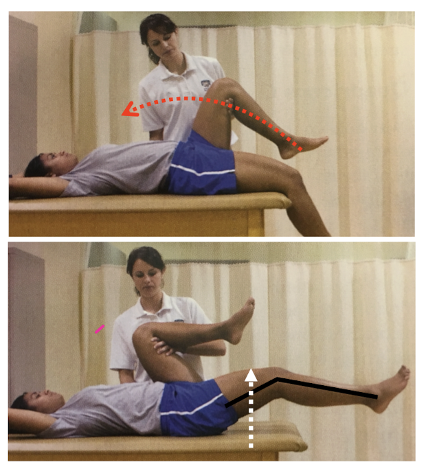
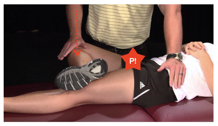
90-90 straight leg raise test
Pt: supine w/ knee & hips flexed at 90
grasps behind the knees to stabilize hip
Clinician: standing at side
Then active knee extension
(+) test: p! w/ knee extension <20 degrees
Implication: hamstring pathology
Piriformis Test
Pt: side-lying on non-testing + hip 90 & knee 90 flex
Clinician: standing in front
hand1: stabilizing hip
hand2: lateral aspect of knee
then downward pressure on knee
(+) test: p! in lower butt & hamstring &/or sciatica
Implication: piriformis syndrome
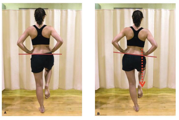
Trendelenburg’s Test
Pt: standing
Clinician: standing behind
Then, single-leg stance (opposite side)
(+) test: hip drops of the non-weight bearing
Implication: gluteus medius weakness
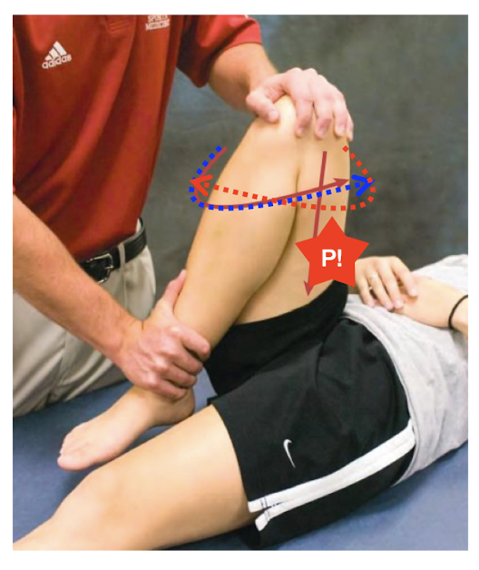
Patrick’s (FABER) Test
“Flx Abd ER”
Pt: supine + figure 4 of the testing foot
Clinician: standing beside
hand1: on opposite ASIS
hand2: on medial aspect of flexed knee
then downward overpressure on knee
(+) test: P! in SI joint or hip
Implication: labral tear or SI joint sprain
Hip Scouring Test
Pt: supine
Clinician: standing beside + passive hip & knee full flx
hand1: on knee
hand2: on anterior aspect of distal tibia
then downward + hip IR & ER in multiple ankles of flx
(+) test: P! in reproduced symptoms
Implication: labral tear or osteochondral defects, OA
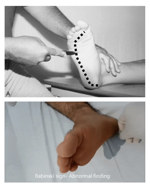
Babinski Test
Procedure: scrape a firm pointed object along lateral inferior border of the foot
(+) test: great toe extension and splaying other toes
Indication: upper motor neuron pathology
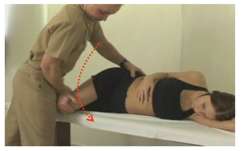
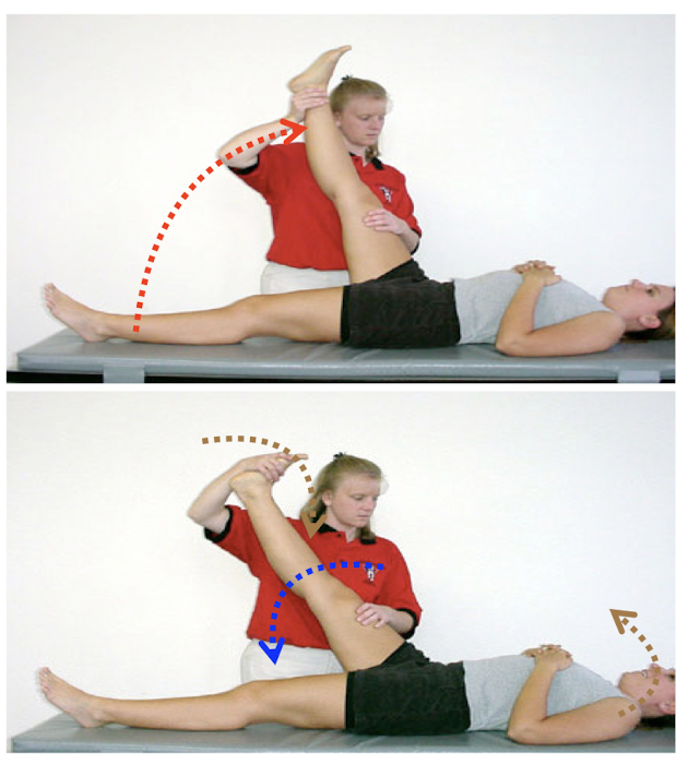
Straight Leg Raise Test
Pt: Supine
Clinician: standing at side
hand1: resting on distal thigh
hand2: support posterior ankle
then passive hip flx to point of tightness or pain
slowly return to point of comfort (hip ext)
lastly, forced ankle DF/neck flx
(+) test: reproduction of Sx before 70 degrees on either side
Implication:
same side: sciatic nerve irritation/ disc herniation
opposite side: disc herniation
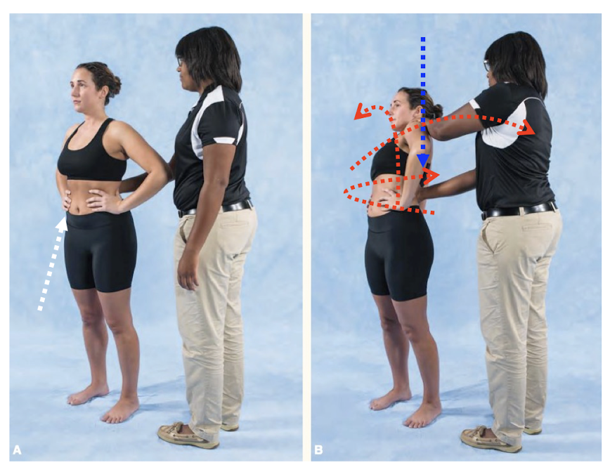
Quadrant Test
Pt: standing w/ feet shoulder width apart
Clinician: standing behind & grasping shoulders
Then, active spinal Ext + Lat Flx + Rot to affected side
examiner overpressure through shoulder
(+) test: reproduced symptoms
Implication:
radiating p! = NRI
local p! = facet jt pathology
p! at PSIS = SI dysfunction
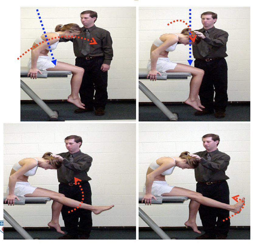
Slump Test
Pt: seated w/ lower leg off the table
Clinician: standing lateral
Then, slump + overpressure w/ neck flx + overpressure w/ knee ext + ankle DF
(+) test: reproduced symptoms at any point in test
Implication: disc herniation, NRI
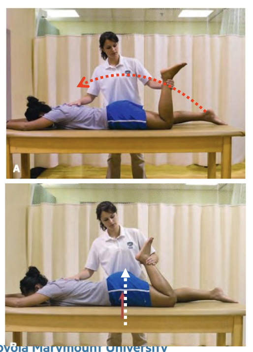
Bowstring Test
Pt: Supine
Clinician: standing lateral
hand1: supporting heel
hand2: anterior distal thigh
Then, SLR to point of tightness or pain
passive knee flx to point of comfort
lastly, pressure popliteal fossa
(+) test: reproduced symptoms at any point in test
Implication: sciatic nerve irritation