Lecture Exam 1 Study BSCI202
 Call Kai
 Learn
Learn Practice Test
 Spaced Repetition
 Match
Match Flashcards
 Knowt Play
1/132

Earn XP
Description and Tags
sTOPPED ON 14 OF IMMUNE
Name | Mastery | Learn | Test | Matching | Spaced | Call with Kai | Chat |
|---|
No analytics yet
Send a link to your students to track their progress
133 Terms
What type of tissue is blood?
Connective
ORIGIN OF BLOOD TISSUE
Blood is a fluid, specialized connective tissue, containing formed elements, proteins that can form fibers, and fluid ground substance.
BODY’S FLUID COMPARTMENTS
In humans (age, sex differences), approx 55-80% is water… Not all organs are equal!
ICF - 67%
ISF - 25%
IVF - 8%
Intra Cellular Fluid ICF = 2/3
ECF = 1/3
(Inter Stitial Fluid ISF = ¾ and Intra Vascular Fluid IVF = 1/4)
OVERVIEW OF BLOOD
8% total body weight
Males 5-6L; Females 4-5L
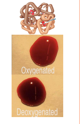
Color varies with O2 content
High O2 - scarlet; Low O2 - dark red
Normal Blood pH: 7.4 (7.35–7.45)

CELLULAR BODY COMPOSITION
84% of our cells are RBCs Importance of transport of O2 for survival → cellular respiration → energy producing mechanism
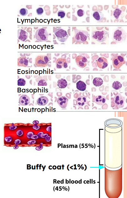
Plasma - 55% , Red Blood Cells - 45%
FUNCTION OF BLOOD
Distribution & elimination (gases, hormones, nutrients, etc) (waste elimination)
Regulation (pH, Temperature, electrolytes)
Protection (Immune cells, immunoglobulins, complement system, etc)
Blood Composition
Plasma – non-living fluid matrix
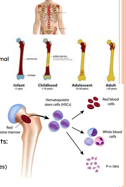
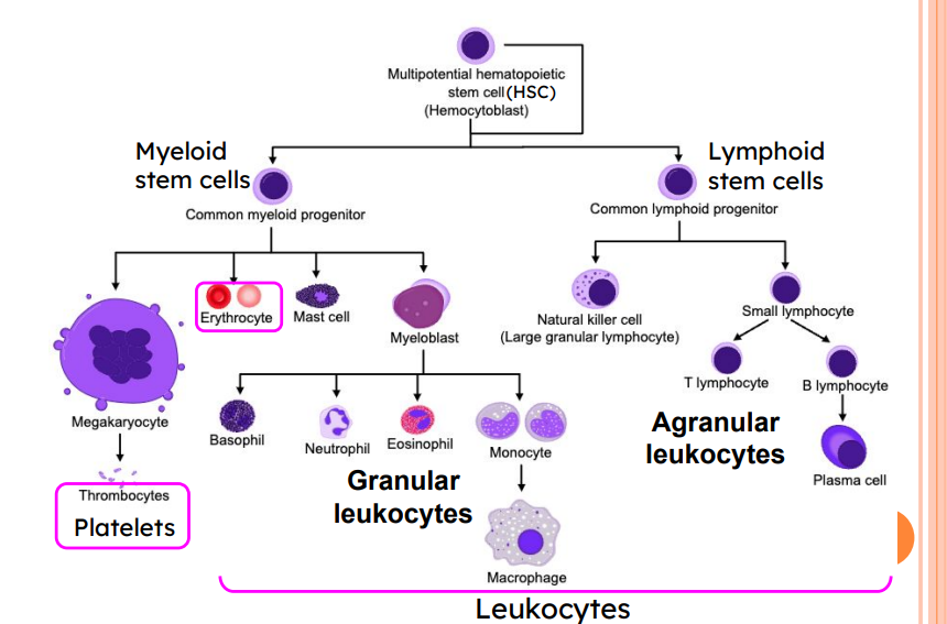
Formed elements – living blood "cells", all derive from Hematopoietic Stem Cells (HSC), formed in the red bone marrow:
⚫ Erythrocytes (red blood “cells”, or RBCs)
⚫ Leukocytes (white blood cells, or WBCs)
⚫ Platelets (“thrombocytes”, cell fragments)

HEMATOPOIESIS
Blood cell formation in red bone marrow
In adults,
axial skeleton, girdles, and proximal epiphysis of humerus and femur
Hematopoietic stem cells (Hemocytoblasts)
Give rise to all formed elements:
RBC
Platelets
WBC (granulocytes, agranulocytes)
FORMED ELEMENTS – HEMATOPOIESIS
Plasma
55% total blood volume
>90% water
>100 dissolved solutes
Nutrients, gases, hormones, wastes, proteins, inorganic ions
Plasma proteins: most abundant solutes (7-9%)
albumin → osmotic balance and buffers pH
globulins → immune response & lipid transport
fibrinogen → blood clotting
Serum is plasma without clotting factors

FORMED ELEMENTS – 1. ERYTHROCYTES
Simple Biconcave discs contribute to gas transport
Biconcave: high surface to Vol ratio → ↗ gas exchange
Filled with hemoglobin (Hb) (>97% content) → carries O2
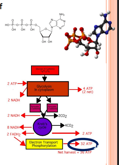
High flexibility (spectrin) for passing through thin capillaries No mitochondria; ATP production by anaerobic fermentation; do not consume O2 they transport
No protein synthesis, growth, or division Erythropoietin regulates erythropoiesis 4.2-6.1 million cells / μL of blood Life span: 100–120 days
Complementarity of structure and function
Erythropoietin regulates erythropoiesis
4.2-6.1 million cells / μL of blood
4.2-6.1 million cells / μL of blood
ERYTHROCYTES: GAS TRANSPORT
Hemoglobin: protein binds reversibly to oxygen:
reversible, cooperative
4 hemes / hemoglobin (Hb); 1 iron ion / heme
1 O2 mlc / iron ion → 4 O2 mlc per hemoglobin complex

STRUCTURE OF HEMOGLOBIN
Hemoglobin consists of 4 globins (two alpha and two beta polypeptide chains) and four heme groups.
The heme group is an Iron-containing pigment that can bind 1 O2 molecule.

HEMOGLOBIN (HB)
O2 loading in lungs
Produces oxyhemoglobin (ruby red)
O2 unloading in tissues
Produces deoxyhemoglobin or reduced hemoglobin (dark red)
CO2 loading in tissues
20% of CO2 in blood binds to Hb → carbaminohemoglobin
Carb/amino/hemoglobin
FORMED ELEMENTS – 2. PLATELETS
Cytoplasmic fragments of megakaryocytes
Granules: serotonin, Ca2+, enzymes, ADP, platelet-derived growth factor (PDGF), and more
Act in clotting process
aka thrombocytes
Normal = 150,000 – 400,000 platelets /μl of blood
Age quickly; degenerate in about 10 days
Thrombopoiesis: Formation of platelets regulated by thrombopoietin
“Thrombo” = relating to the clotting of blood “poiesis” = poi → to make , esis → process “in” = substance (protein)
FORMED ELEMENTS – 3. LEUKOCYTES
Complete cells
Make up <1% of total blood volume
“Buffy coat”
4,800 – 10,800 WBCs/μl blood (1/1,000,000th of a liter)
Leukocytosis: WBC count >11,000/μl
Immune System: defense against disease
⚫ Diapedesis (aka extravasation)
Can live hours, days, or decades
CONCEPT REVIEW
Blood is formed of:
Plasma: 55% of blood volume (BV): fluid carrying dissolved solutes, including large proteins important for oncotic (colloid osmotic) pressure
Formed elements:
Leukocytes (aka, White blood cells) cells of the immune system (part of buffy coat, <1% BV)
Platelets (aka thrombocytes) fragments of cells important in coagulation (hemostasis) (part of buffy coat, <1% BV)
Red blood cells (aka erythrocytes) cells that lost their nucleus, and transport O2 . 45% of BV
Blood is required to transport O2 and CO2 , distribute nutrients, eliminate waste, distribute/regulate heat, regulate pH, transport immune cells, hormones, etc
All cells in the blood develop from Hematopoietic Stem Cells (HSC), in the red bone marrow
CONCEPT REVIEW: ERYTHROCYTES
Erythrocytes are the most numerous cells in the body.
RBC have a biconcave shape that is adapted to transport and exchange gases across their membrane.
Spectrin is a protein that provides flexibility to the RBC such that it can travel through very thin capillaries and regain its shape.
RBCs contain hemoglobin (Hb) which transports O2 bound to the iron in its heme.
They do not carry mitochondria, hence can only produce ATP by anaerobic fermentation.
Hb transports 20% of the blood’s CO2 bound to its amino acid for release via the lungs
REGULATION OF ERYTHROPOIESIS
> 2 million RBCs made per second
Too few → tissue hypoxia
Too many → ↑ blood viscosity
HypoOxia = Low Oxygen
Balance of production and destruction
Hormonal controls
Nutrient access (limiting factors)
iron (Fe2+) → Heme
amino acids → globin, and
B vitamins: B12 & Folic Acid necessary for DNA replication
Absorption of B12 requires intrinsic factor expressed by the stomach’s parietal cells
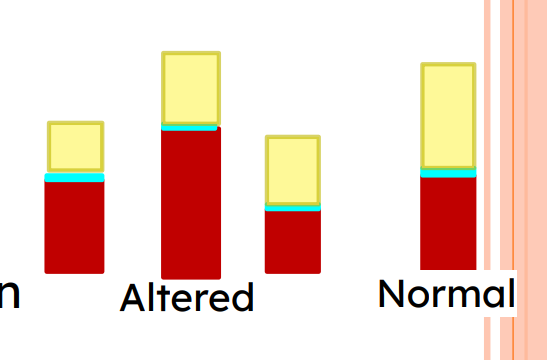
HEMATOCRIT (PACKED CELL VOLUME)
Depends on rates of production and destruction of RBC Percent of blood volume that is RBCs
45% of blood volume on average:
47% ± 5% for males;
42% ± 5% for females
Why is hematocrit taken? What can it tell us?

T/F You can have a normal hematocrit (45%), but still be in hypoxia*.
True
Why / Why not?
Volume RBC? Volume total blood * 100 = Hematocrit
Hematocrit is a relative value, not an absolute value.
Hematocrit does not always correlate with the distribution of O2 to the tissues *Hypoxia: Not enough O2 in the tissues
WHAT CAN A LOW HEMATOCRIT MEAN?
Low hematocrit
Not enough healthy red blood cells (anemia)
Volume RBC/Volume total blood times 100 = Hematocrit
High number of white blood cells (long term illness)
Volume RBC/Volume total blood times 100 = Hematocrit

WHAT CAN A HIGH HEMATOCRIT MEAN?
High hematocrit
Dehydration
Volume RBC/ Volume total blood times 100 = Hematocrit
Disorder/disease
Polycythemia vera
bone marrow disease creates too many red blood cells
Lung/heart
low oxygen in blood triggers red blood cell production
Volume RBC/ Volume total blood X 100 = Hematocrit

HORMONAL CONTROL OF ERYTHROPOIESIS
Hormone Erythropoietin (EPO)
Direct stimulus for erythropoiesis
Released by kidneys (some from liver)
When kidneys sense low O2 in renal tissue
Increases circulating reticulocyte (premature RBC that finish maturation in the blood) in 1-2 days
Testosterone enhances EPO production
→ higher RBC counts in males
males ≅ 47% ; females ≅ 42%
ERYTHROPOIETIN: MECHANISM FOR REGULATING ERYTHROPOIESIS
1) Stimulus: Hypoxia (inadequate O2 delivery)
2) Kidney (and liver to a smaller extent) releases erythropoietin
3) Erythropoietin stimulates red bone marrow
4) Enhanced erythropoiesis increases RBC count
5) O2 carrying ability of blood rises
QUICK FIRE QUESTION
Kidneys filter blood and make urine (more on this later in the semester).
When kidneys fail, patients are put on dialysis to artificially filter waste from the blood. What other impacts might kidney failure have on blood?
Which is likely the case for patients on dialysis…
Low RBC
Kidneys → EPO → Erythropoiesis → RBC
HYPOXIA: LOW O2 IN TISSUES
What causes hypoxia?
hemorrhage (RBC loss)
increased destruction of RBC
decreased production of RBC
Insufficient hemoglobin per RBC (e.g., iron deficiency)
Reduced availability of O2
Why are you at increased risk of stroke (blocked artery) while at high altitudes?
Low environmental oxygen levels
→ tissue hypoxia
→ production more red blood cells
→ increased hematocrit
→ thicker blood
→ increased viscosity
→ blood flows slow (clot!)
→ higher block risk
FATE AND DESTRUCTION OF ERYTHROCYTES
Produced in red bone marrow, under EPO regulation
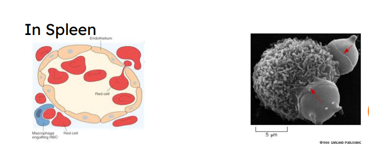
Old RBCs become fragile; hemoglobin (Hb) begins to degenerate
Get trapped in smaller circulatory vessels (capillaries) especially in spleen
Macrophages engulf dying RBCs in spleen
LIFE CYCLE OF RED BLOOD CELLS
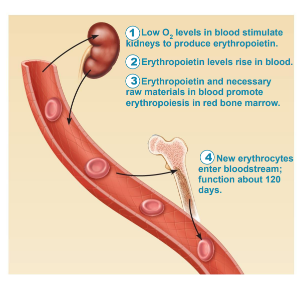
1) Low O2 level in blood stimulate kidneys to produce erythropoietin
2) Erythropoietin levels rise in blood
3) Erythropoietin and necessary raw materials in blood promote erythropoiesis in red bone marrow
4) New erythrocytes enter blood stream function about 120 days
RBC DESTRUCTION & RECYCLING
RBC Destruction and Recycling
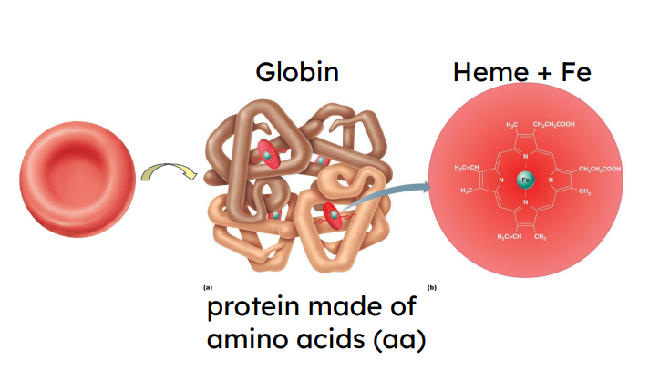
Heme and globin are separated
Heme: Degraded to yellow pigment bilirubin → liver
Liver secretes bilirubin
Serves in production of bile → helps in lipid digestion in small intestine
Degraded to pigment
Pigment leaves body in feces
Brown feces
Iron salvaged for reuse
Globin metabolized into aa Released into circulation Re-uptake for production of proteins
Released into circulation
Re-uptake for production of proteins

ERYTHROCYTE DISORDERS
Anemia
Blood has abnormally low O2 - carrying capacity
Four broad causes
Blood loss (hemorrhage)
Low RBC production
High RBC destruction
Hemoglobin abnormalities

CAUSES OF ANEMIA: LOW RBC PRODUCTION
Iron-deficiency anemia
Renal anemia
Lack of EPO production
Often accompanies renal disease
Treated with synthetic EPO
Pernicious anemia
Autoimmune disease against stomach parietal cells that produce intrinsic factor
Lack of Vit B12, prevents maintaining replication pace of committed cells
CAUSES OF ANEMIA: LOW RBC PRODUCTION
Aplastic anemia
Destruction or inhibition of red bone marrow
Treatment
Hypoplastic Anemia (decline)

CAUSES OF ANEMIA: HIGH RBC DESTRUCTION (1)
Hemolytic anemias
Premature RBC lysis
Caused by
Hb abnormalities
Incompatible blood transfusions
Infections
CAUSES OF ANEMIA: HIGH RBC DESTRUCTION (2)
Sickle-cell anemia
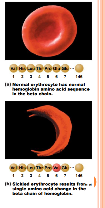
One amino acid mutation in a globin beta chain of hemoglobin (HbS)
RBCs with crescent shape and block small vessels
RBCs rupture easily and reduced O2 carrying-capacity

SICKLE-CELL ANEMIA
More prevalent in people from African malarial belt and descendants
MALARIA
300–500 million cases each year
Kills ~2 million each year
Plasmodium parasite
If 2 alleles of β chain Hb mutated → lesser survival rate
If 1 allele mutated → greater survival rate than WT with malaria
ERYTHROCYTE DISORDERS: HIGH RBC NUMBERS
Polycythemia vera (permanent)
Bone marrow cancer → excess RBCs
Severely increased blood viscosity
Secondary polycythemia (temporary)
Less O2 available (high altitude) or EPO production increases → higher RBC count
Lower plasma volume
Blood doping
⇒ High RBC numbers increase blood’s viscosity ⇒ increased chances of blood clotting, embolism.
YOU PLAY 3: CRS
What is EPO?
1- a biogenic amine hormone produced by the liver if high O2
in blood
2- a steroid hormone produced in males by the gonads if low O2
in blood
3 - a peptide hormone produced primarily by the
kidney if low O2 in blood
4- a steroid hormone produced by the liver when no access to O2
3 - a peptide hormone produced primarily by the kidney if low O2 in blood
METABOLISM: CONVERT INGESTED FOOD INTO USEABLE ENERGY
Metabolism = transformation of energy into biological useable forms
Adenosine-5'-triphosphate (ATP)
Energy is maximized through aerobic respiration (w/↑ O2 )
No negative byproducts as in anaerobic fermentation
CONCEPT REVIEW
Erythropoiesis is the process of RBC formation, it is highly dependent on Erythropoietin (EPO), and will determine hematocrit (% vol of RBC in blood)
Imbalances (high or low) in hematocrit have damaging consequences:
low hematocrit may be caused by an increase in RBC destruction or decrease in RBC production
High hematocrit may be caused by an increase in RBC production or a decrease in RBC destruction. '
They can be temporary or permanent:
Low hematocrit: Anemias, low EPO
High hematocrit: Polycythemia vera, secondary polycythemia

Recall: testosterone increases EPO
HEMOSTASIS ≠ HOMEOSTASIS
HEMOSTASIS:
Series of reactions to stop bleeding
Requires clotting factors & substances released by platelets and injured tissues
Three steps
1. Vasoconstriction by vascular spasm
2. Platelet plug formation
3. Coagulation (blood clotting)
HEMOSTASIS 1. VASOCONSTRICTION
Constriction of vessels by contraction of smooth muscle surrounding arterioles, even muscular arteries if needed.
→ Limit the flow… to limit the loss
HEMOSTASIS 2: PLUG FORMATION
Activated platelets → temporary platelet plug
Stick to collagen fibers
von Willebrand factor (plasma protein)
Swell, become spiked and sticky,
release chemical messengers
ADP
Serotonin
thromboxane A2
Ca2+
Positive feedback loop: activated platelets further activate platelets
INTACT BLOOD VESSEL
Connective tissue
Smooth Tissue
Connective
HEMOSTASIS 3: COAGULATION
Intrinsic Pathway = triggered by factors from within blood
Extrinsic Pathway = triggered by factors from outside
Fibrin & clotting factors → Blood clot (aka, Thrombus)
COAGULATION PATHWAYS
Contact activation or Intrinsic pathway
Internal trauma
Triggered by negatively charged surfaces
Contact with collagen
Activated platelets
Tissue Factor or Extrinsic pathway
Triggered by exposure to tissue factor (TF) (aka, coagulation factor III) released by lysed tissue cells
Faster (few reactions
Ca 2+ is an essential coagulation factor (cf IV)
COAGULATION: COMMON PATHWAY
Common Pathway activated by Intrinsic and Extrinsic pathways
Reinforces platelet plug with fibrin meshwork
Blood transformed from liquid to gel
Three phases of coagulation
1. Prothrombinase formation
2. Prothrombin converted to enzyme thrombin
3. Thrombin catalyzes fibrinogen → fibrin → fibrin mesh
EVENTS OF HEMOSTASIS
Step 1 Vascular spasm
• Smooth muscle contracts, causing vasoconstriction
Step 2 Platelet plug formation
Injury to lining of vessel exposes collagen fibers; platelets adhere.
Platelets release chemicals that make nearby platelets sticky; platelet plug forms
Step 3 Coagulation
• Fibrin forms a mesh that traps red blood cells and platelets, forming the clot.

CLOT RETRACTION & VESSEL REPAIR
Actin and myosin in platelets contract
Contraction pulls on fibrin strands, squeezing serum from
clot
Clot retraction stabilizes clot, by excluding fluid
Draws ruptured blood vessel edges together
Platelet-derived growth factor (PDGF)
→ proliferation of smooth muscle and fibroblasts (CT)
→ rebuild blood vessel outer wall
Vascular endothelial growth factor (VEGF)
→ restores endothelial lining
FIBRINOLYSIS
Removes unneeded clots after healing
Begins within two days; continues for several days
Plasmin is a fibrin-digesting enzyme
Produced by activating Plasminogen
Trapped during clot formation → Negative feedback loop
⚫ Plasmin deficiency leads to thrombosis, due to inadequate
degradation of clots.
DISORDERS: THROMBOEMBOLIC CONDITIONS
Thrombus = blood clot Embolus = unattached mass that travels in the bloodstream

DISORDERS: BLEEDING DISORDERS
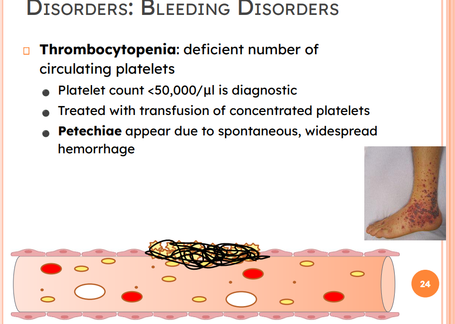
Thrombocytopenia: deficient number of circulating platelets
Platelet count <50,000/μl is diagnostic
Treated with transfusion of concentrated platelets
Petechiae appear due to spontaneous, widespread
hemorrhage
Bleeding Disorders
Inability to synthesize procoagulants
due to impaired liver function
Causes: vitamin K deficiency*, hepatitis, and cirrhosis (most clotting factors are produced in the liver)
BLEEDING DISORDERS
Hemophilia includes several similar hereditary bleeding
disorders
Hemophilia A: factor VIII deficiency
77% of all cases
Hemophilia B: factor IX deficiency
Hemophilia C: mild type; factor XI deficiency
DISSEMINATED INTRAVASCULAR COAGULATION (DIC)
Widespread clotting in intact vessels
And
Severe bleeding
by reduced availability of clotting factors and platelets
residual blood unable to clot
Causes: Usually pregnancy complication; incompatible blood transfusions = transfusion reaction
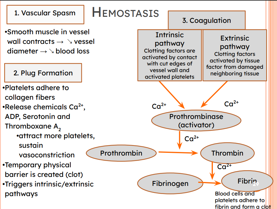
HEMOSTASIS
1. Vascular Spasm
Smooth muscle in vessel wall contracts → ↘ vessel diameter → ↘ blood loss
2. Plug Formation
Platelets adhere to collagen fibers
•Release chemicals Ca2+ , ADP, Serotonin and Thromboxane A2
•attract more platelets, sustain vasoconstriction
•Temporary physical barrier is created (clot)
Triggers intrinsic/extrinsic pathways
3. Coagulation
Intrinsic pathway
Clotting factors are activated by contact with cut edges of vessel wall and activated platelets
Extrinsic pathway
Clotting factors activated by tissue factor from damaged neighboring tissue
(Ca2+) + (Ca2+)
Prothrombinase (activator)
Prothrombinase (activator) → Thrombin →Fibrinogen →Fibrin
Blood cells and platelets adhere to fibrin and form a clo
WHY IS ASPIRIN PROHIBITED BEFORE MOST SURGERIES?
Aspirin = Acetylsalicylic Acid (ASA)
→ antithrombotic drug (blood thinner)
→ Prevents synthesis of
Thromboxane and Prostaglandin
1) platelet aggregation
2) pain information temperature regulation inflammation
BLOOD TRANSFUSIONS
Multiple types of transfusion
Whole blood transfusion
plasma, RBC, platelets, WBCs
Packed red blood cells transfused to restore oxygen-carrying capacity (plasma, WBCs, platelets removed)
Transfusion of incompatible blood can be fatal
RBC PRESENT ANTIGENS ON THEIR MEMBRANE
Blood groups are determined by the type of Antigen (Ag) at the surface of RBC.
Antigens are glycoproteins that can generate an immune response by triggering the generation of antibodies
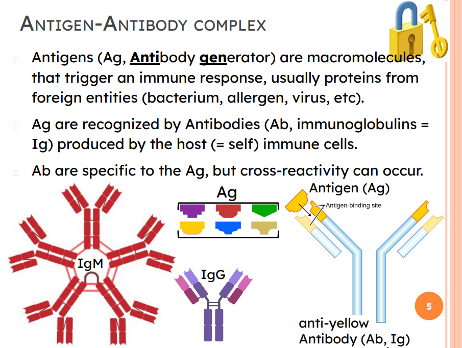
ANTIGEN-ANTIBODY COMPLEX
Antigens (Ag, Antibody generator) are macromolecules, that trigger an immune response, usually proteins from foreign entities (bacterium, allergen, virus, etc).
Ag are recognized by Antibodies (Ab, immunoglobulins = Ig) produced by the host (= self) immune cells.
Ab are specific to the Ag, but cross-reactivity can occur.
HUMAN BLOOD GROUPS: ABO
RBC membranes bear glycoprotein antigens (A, B)
if Ag present → Promote agglutination; called agglutinogens
co-dominance of IA & IB alleles vs i (recessive)
Do not express MHC surface proteins → won’t induce T-cell mediated immunity
Presence or absence of each agglutinogen is used to classify blood cells into different groups: A, B, AB, O
ABO BLOOD GROUP & AB EXPRESSION
Person 1: Type A Blood: A agglutinogens (Ag) + anti-B antibodies expressed at birth (agglutinins; IgM)
ANTI B= anti-B antibodies (agglutinins, ⍺B IgM)
Person 1: Type A Blood: A agglutinogens (Ag) + anti-B antibodies (agglutinins; IgM)
Receives Type B blood from donor.
A = “A” Antigen (agglutinogens, A Ag)

T/F A person who carries RBC with B Ag … will carry anti-B Ab
False
T/F2. A person who is AB type can donate plasma that will have no anti-A nor anti-B Ab.
True
T/F3. A person who is type O, can receive packed RBC from type A or type B, but cannot receive whole blood transfusions from these donors.
False
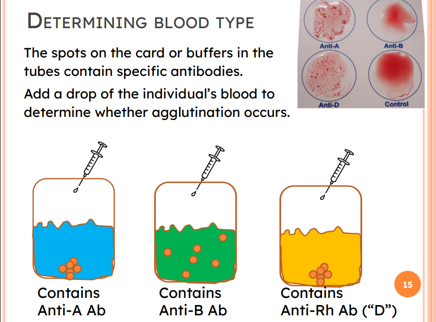
DETERMINING BLOOD TYPE
The spots on the card or buffers in the tubes contain specific antibodies.
If Ag exist → Ag-Ab reaction → agglutination
TRANSFUSIONS: RBC VS WHOLE BLOOD
Packed red blood cells transfusion
= plasma, platelets, and WBCs removed, RBCs transfused to restore oxygen-carrying capacity of receiver
Whole blood transfusion
= donor antibodies included in plasma
Transfusion of incompatible blood can be fatal
PACKED RBC
No plasma = no donor antibodies!
Blood type compatibility: life or death!
WHAT ABOUT RH+/- ??
Rh Factor (Rhesus factor)
“D” antigen = + = Rh positive
Anti-D Antibodies not present at birth
Require a second exposure to Rh+ blood → equivalent to a typical transfusion reaction
due to IgG developed by Rh- individual

MORE BLOOD GROUPS
Other blood groups (MNS, Duffy, Kell, and Lewis) usually weak agglutinogens
Antigens of ABO and Rh blood groups cause vigorous transfusion reactions
THINK PAIR SHARE
What blood types could Malfoy receive?
⇒ Packed RBC transfusion.
Malfoy’s Antigens on RBC: A indicates there are A antigens; + indicates there are Rh antigens.
Malfoy’s Antibodies: anti-B antibodies.
INCOMPATIBLE TRANSFUSION REACTIONS (MISMATCHED BLOOD)
Donor's cells
Attacked by recipient's plasma agglutinins (Ab)
Agglutinate and clog small vessels RBC lysis and release hemoglobin into bloodstream
Result in
Diminished oxygen-carrying capacity
Diminished blood flow beyond blocked vessels
Hemoglobin in kidney tubules → renal injury
HOMEOSTATIC IMBALANCE: HEMOLYTIC DISEASE OF THE NEWBORN
Also called erythroblastosis fetalis
Only occurs in Rh– mom carrying Rh+ fetus
Anti-Rh Ab (=anti-D) do not exist until exposure to Rh+ blood.
Rh– mom exposed to Rh+ blood of fetus during delivery of first baby
baby healthy
Mother’s immune response is activated against Rh+ Ag (“D”)
Mother synthesizes anti-Rh antibodies and Memory B cells
Second pregnancy (= Re-exposure)
Mom's memory B cells are activated → ⍺-Rh IgG
anti-Rh IgG cross placenta and destroy RBCs of Rh+ baby
NOTE: Anti-A and anti-B Abs (IgM) appear early on in infancy; too large to pass the placenta barrier
ERYTHROBLASTOSIS FETALIS
To the mother’s immune system: Rh antigen is a potential threat → Immune response → anti-Rh Ab
During healthy pregnancy, mother’s and fetus’ bloods do not mix. → Increased risk at birth: placenta delivery, miscarriage, ectopic pregnancies, others
2nd pregnancy: Memory B cells of mother are activated and produce anti-Rh IgG antibodies that pass the placental barrier →
anti-Rh IgG of mother agglutinate Rh+ RBC of the fetus!

PREVENTING ERYTHROBLASTOSIS FETALIS
If mother Rh– , and possibility of Rh+ fetus:
Administer anti-D antibodies to prevent the mother’s IS to build an initial IR during pregnancy/ delivery! ⇒
Who develops anti-D Ab to donate?

INCOMPATIBLE TRANSFUSION
Incompatible transfusion
• Decreased oxygen-carrying capacity
• Blocked vessels
• Renal failure
CONCEPT REVIEW - Blood 4
RBC carry Ag at their surface
Ag can elicit an immune response that produces Ab specific to the Ag.
Ab against the A and B Ag of the ABO group are IgM and do not pass the placental barrier. They are present early in life.
eg, Anti-A IgM agglutinate Type A RBC.
Determination of blood type and matching blood transfusions is essential to save lives.
A person that carries a specific Ag on their RBC does not carry Ab against that Ag.
Anti-Rh (anti-D) IgG are present after sensitization by exposure of a Rh– person to Rh+ RBC.
Erythroblastosis fetalis can lead to miscarriage, stillbirths.
FUNCTIONS OF THE LYMPHATIC SYSTEM
Drains excess interstitial fluid (ISF) → return to blood
maintains circulating blood volume → blood pressure -
Transports dietary lipids
lipids absorbed by gastrointestinal tract
fat-soluble vitamins (A, D, E, K)
Carries out immune responses
includes lymphoid tissue, lymphoid organs
contains most lymphocytes (B, T)
THE LYMPHATIC SYSTEM
Exudation → rush of self and foreign material into lymphatic vessels
Lymphatic system
Recovers fluid in ISF leaked from blood vessels, back to blood (approx 3L/day!)
Immune system: Main structures include primary organs (red bone marrow, thymus) and secondary organs (spleen, tonsils, lymph nodes and vessels).

LYMPHOID TISSUE CELLS
Lymphocytes Produced in red bone marrow Mature into one of two main varieties
T lymphocytes → Thymus
B lymphocytes → bone marrow
Other cells of lymphatic system
Macrophages → phagocytosis
Dendritic cells → phagocytosis
Epithelial cells → help select lymphocytes
Reticular cells produce reticular fibers
Lymph node (SEM): Reticular cells (brown) make up a connective tissue mesh of reticular fibers. White blood cells (lymphocytes, yellow), red blood cells (red) and macrophages (pink).
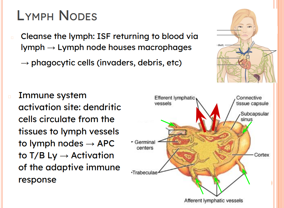
LYMPH NODES
Cleanse the lymph: ISF returning to blood via lymph → Lymph node houses macrophages → phagocytic cells (invaders, debris, etc)
Immune system
activation site: dendritic cells circulate from the tissues to lymph vessels to lymph nodes → APC to T/B Ly → Activation of the adaptive immune response
LYMPHATIC SYSTEM
Consists of three parts
1) Network of lymphatic vessels (lymphatics)
2. Lymph – fluid in vessels
3. Lymph nodes – cleanse lymph (⌀ 1-25mm)
Lymph journey: ISF → ly capillaries → ly vessels → ly trunks → ly ducts → [cisterna chyli] → thoracic & right lymphatic ducts join veins → blood (systemic circulation)
![<p>Consists of three parts</p><p>1) Network of lymphatic vessels (lymphatics)</p><p> 2. <strong>Lymph </strong>– fluid in vessels </p><p>3<strong>. Lymph nodes</strong> – cleanse lymph (⌀ 1-25mm)</p><ul><li><p>Lymph journey: ISF → ly capillaries → ly vessels → ly trunks → ly ducts → [cisterna chyli] → thoracic & right lymphatic ducts join veins → blood (systemic circulation)</p></li></ul><p></p>](https://assets.knowt.com/user-attachments/fbc15e76-0637-478b-b238-eafc121af819.png)
WHY DOES ISF FLOW TO THE LYMPHATIC CAPILLARIES? Why not directly back to the systemic venous capillaries?
Out of 20 L/day that leave the capillaries, 17L return to the venous circulation. → 3 L in lymphatic vessels
WHY DOES ISF FLOW TO THE LYMPHATIC CAPILLARIES? Why not directly back to the systemic venous capillaries?
Out of 20 L/day that leave the capillaries, 17L return to the venous circulation. → 3 L in lymphatic vessels
NO backflow:
○ Endothelial flaps orientation and
○ backflow prevention flaps ○
Smooth muscle
LYMPHATIC CAPILLARIES AND RETURN TO BLOOD
Pressure gradients flow fluid from ISF to lymph.
Hydrostatic pressure: HPISF > HPLV
Oncotic (colloid osmotic) pressure: large soluble proteins in lymph → Pulls fluid into lymph: OP LV > OPISF
NO backflow: orientation of endothelial flaps and unidirectional valves.
LYMPHATIC CAPILLARIES AND RETURN TO BLOOD
HPISF > HPLV
OPLV > OPISF
OP: pressure exerted by the presence of proteins that pulls fluid into the compartment
HP: pressure applied by the fluid on the walls of the container
Greatest HP pushes water out Greatest OP pulls water in

HYDROSTATIC PRESSURE & ONCOTIC PRESSURE
Hydrostatic pressure (HP): Force applied by fluid on the walls of its compartment → pushes fluid out of the compartment.
Oncotic pressure (OP, π): Force drawing fluid into the compartment to balance the concentrations of large solutes that cannot cross the walls of the compartment

CONCEPT REVIEW LYMPHATIC SYSTEM - session 5
Lymphoid tissue
houses lymphocytes and provides a site for maturation, activation and proliferation
allows for organism’s surveillance (infection and filtration) through lymphocytes and macrophages
Lymphoid organs
produce, mature, and differentiate immune cells
remove pathogens and aged red blood cells
Lymphatic vessels
Return excess ISF as lymph to systemic blood circulation
Pressure gradients determine the flow of fluid between the blood vessels, ISF, lymphatic vessels I
Important impact on blood pressure and blood volume (next)
IMMUNITY
Resistance to disease
Extrinsic (infection, injury)
Intrinsic (cancer cells)
Immune system
Two main systems
Innate (nonspecific) defense system
Adaptive (acquired, specific) defense system
Innate defense system has two lines of defense
First - external body membranes (skin and mucosae)
Second - antimicrobial proteins, phagocytes, and other cells, chemical mediators
Prevent infection,
Inhibit spread of invaders
Inflammation process
Clean dead tissue/cells
“Quick and dirty”
Adaptive defense system
Third line of defense attacks specifically a particular foreign substance
Takes longer to react than innate system
It is specific and highly effective “Slow and targeted”
OVERVIEW OF INNATE AND ADAPTIVE DEFENSES

INNATE DEFENSES: 1ST LINE
Surface barriers ward off invading pathogens
Skin, mucous membranes, and their secretions
Physical barrier to most microorganisms
Acid secretion
Phagocytes & dendritic cells
Enzymes - lysozyme of saliva, respiratory mucus, and lacrimal fluid
Antimicrobial secretions: Defensins
SURFACE BARRIERS BREACHED …
→ second line of defense must protect other/deeper tissues
INNATE DEFENSES: 2ND LINE
Internal Defenses: Cells and Chemicals
Leukocytes:
Phagocytic cells (Neutrophils, macrophages [“tissue monocytes”])
Natural killer (NK) cells (type of Lymphocyte)
Additional cells that promote immune response (basophil, eosinophils)
Antimicrobial proteins (interferons and complement proteins, defensins, Antimicrobial Peptides)
Fever
Inflammatory response
LEUKOCYTES: TWO CATEGORIES
Granulocytes (PMN)– Visible cytoplasmic granules
Neutrophils, eosinophils, basophils, NKC
Agranulocytes – No visible cytoplasmic granules
Lymphocytes*, monocytes
GRANULOCYTES
Neutrophils
aka Polymorphonuclear leukocytes (PMNs or polys)
Very phagocytic
Eosinophils
Lysosome-like granules
Allergies, asthma, and general immune response
Basophils
Granules contain histamine
Histamine = inflammatory chemical → vasodilator → ↑ WBCs to inflamed sites
Natural Killer Cells
Large granular cytotoxic LY
attack virus-infected, cancerous cells
AGRANULOCYTES
These cells do not have visible granules
Lymphocytes
Mostly in lymphoid tissue few circulate in blood
T lymphocytes (T cells) against virus-infected cells and tumor cells
B lymphocytes (B cells) → antibody producing cells
Monocytes
Leave circulation → enter tissues and differentiate into macrophages
Actively phagocytic cells that present antigens
Activate lymphocytes

PHAGOCYTIC CELLS
Cells that detect and ingest harmful foreign particles
Neutrophils most abundant but die fighting
Become phagocytic on exposure to infectious material
Phagocytize 1 bacterium and die
Macrophages differentiate from monocytes – chief phagocytic cells – robust cells
Can phagocytize up to 200 bacteria
Free macrophages wander through tissue spaces
Fixed macrophages permanent residents of some organs (eg, Langerhans cells (skin), Microglia (CNS))