Reflex Physiology and General senses
0.0(0)
Studied by 0 people Call Kai
 Learn
Learn Practice Test
 Spaced Repetition
 Match
Match Flashcards
 Knowt Play
Card Sorting
1/50

Earn XP
Description and Tags
Last updated 4:50 AM on 11/17/22
Name | Mastery | Learn | Test | Matching | Spaced | Call with Kai | Chat |
|---|
No analytics yet
Send a link to your students to track their progress
51 Terms
1
New cards
What are the 5 steps of the reflex arc
Receptor, Sensory Relay, Integration, Motor Command Relay, Effector
2
New cards
Reflex
Simple automatic response to a stimulus
3
New cards
Most neural reflexes are designed to what?
Maintain balance and posture, protect from danger or injury, carry out routine activities like chewing or walking
4
New cards
Monosynaptic vs Polysynaptic
based on the number of neurons involved
5
New cards
Monosynaptic reflexes
Faster and integrated in the spinal cord ex, knee jerk
6
New cards
Polysynaptic reflex
contain at least one interneuron, most are polysynaptic
7
New cards
Somatic
Voluntary, involves skeletal muscles
8
New cards
Autonomic
Involuntary, involves smooth muscles, glands, or visceral glands
9
New cards
acquired reflexes
learned through experience, driving, walking
10
New cards
General Stimuli
temperature, pH, pressure
11
New cards
Classifications of General stimuli
Exteroceptors, Interoceptors
12
New cards
Exteroceptors monitor what?
Temperature, touch, pain, pressure
13
New cards
Interoceptors monitor what?
internal conditions, Ex. chemoreceptors
14
New cards
Free (naked) nerve endings
Dendrites terminate in epidermis/dermis junction of skin and mucosal epithelium, detect pain
15
New cards
Merkel cells
Located in the stratum germinitivum, detect light touch
16
New cards
Encapsulated Receptors
Meissner's Corpuscles, Pacinian Corpuscles
17
New cards
Meissner's corpuscles
more superficial, detects light touch
18
New cards
Pacinian Corpuscles
More deep, detects forceful pressure, vibrations
19
New cards
Free nerve endings vs Merkel cells
Free nerve endings terminate basically at the surface of skin or mucosa and detect pain, Merkel cells terminate at the deepest layer of the epidermis and detect touch
20
New cards
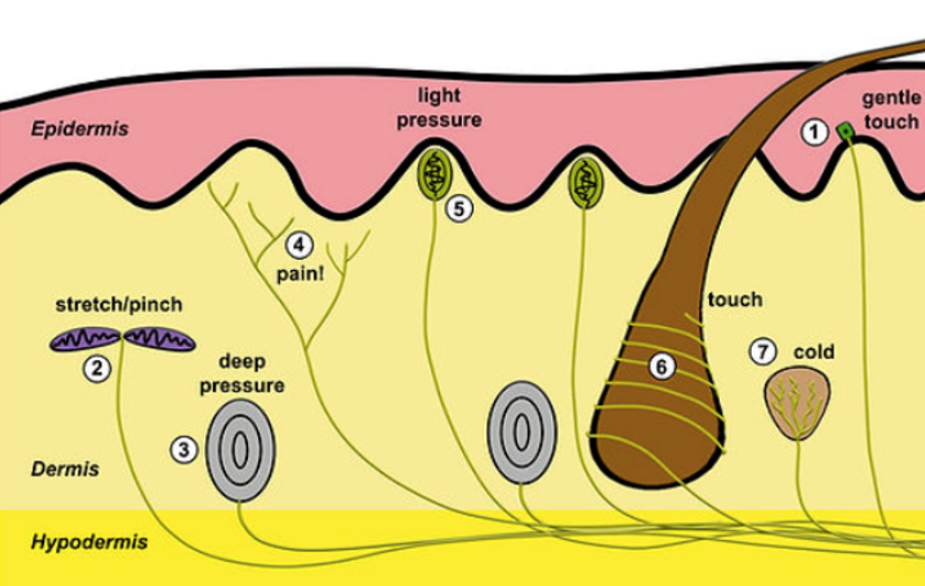
Label number 1 in the illustration
Merkel disk
21
New cards
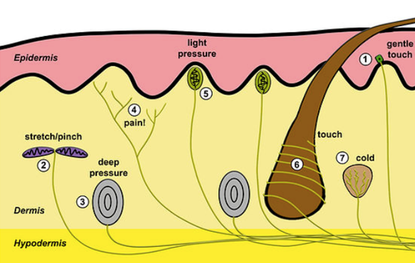
Label number 2 in the illustration
Ruffini corpuscle
22
New cards
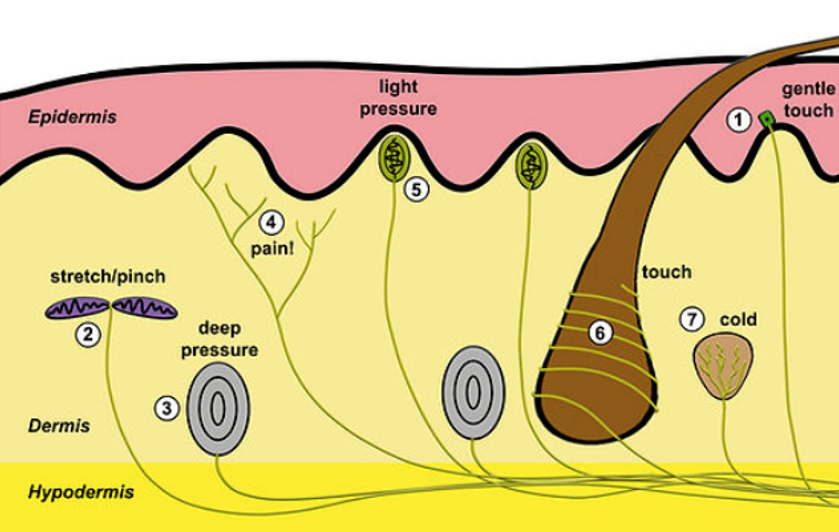
Label number 3 in the illustration
Pacinian corpuscle
23
New cards
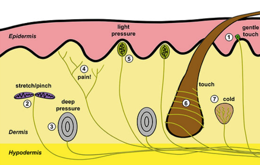
Label number 4 in the illustration
nociceptor (free nerve endings)
24
New cards
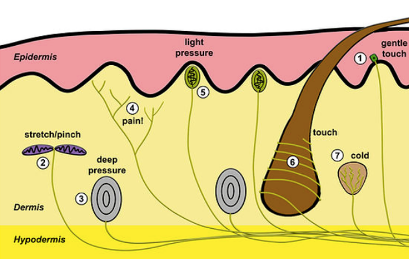
Label number 5 in the illustration
Meissner's corpuscle
25
New cards
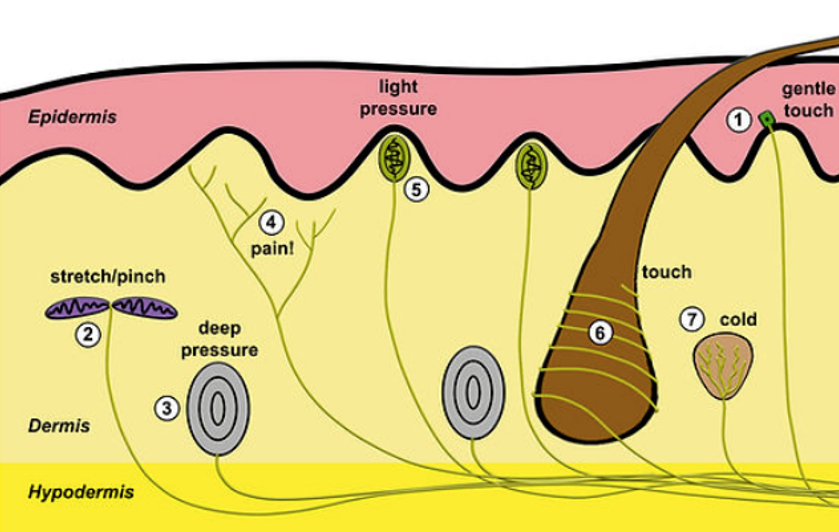
Label number 6 in the illustration
Root hair plexus
26
New cards
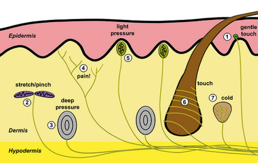
Label number 7 in the illustration
Krause and bulb
27
New cards
Muscle spindle purpose
monitor muscle position, ensures that muscles are not overstretched
28
New cards
α-motor neurons
innervate contractile region of external fibers
29
New cards
γ-motor neurons
innervate contractile region of intrafusal fiber
30
New cards
Sensory neuron sends what to what back to what via what
continual impulses, CNS, extrafusal fibers, α-motor neurons
31
New cards
Willful muscle contraction
Brain sends impulse to γ-motor neurons in order to contract the intrafusal fibers which relieves compression of spindle
32
New cards
Receptors are what
Transducers
33
New cards
Transducers
Convert stimuli to action potentials
34
New cards
Action potentials are distinguished as unique by what?
Their destination in the brain
35
New cards
Receptor Fields
The area within, if stimulated by two separate stimuli, is translated as a single stimulation
36
New cards
What is adaptation?
Desensitization to constant stimuli
37
New cards
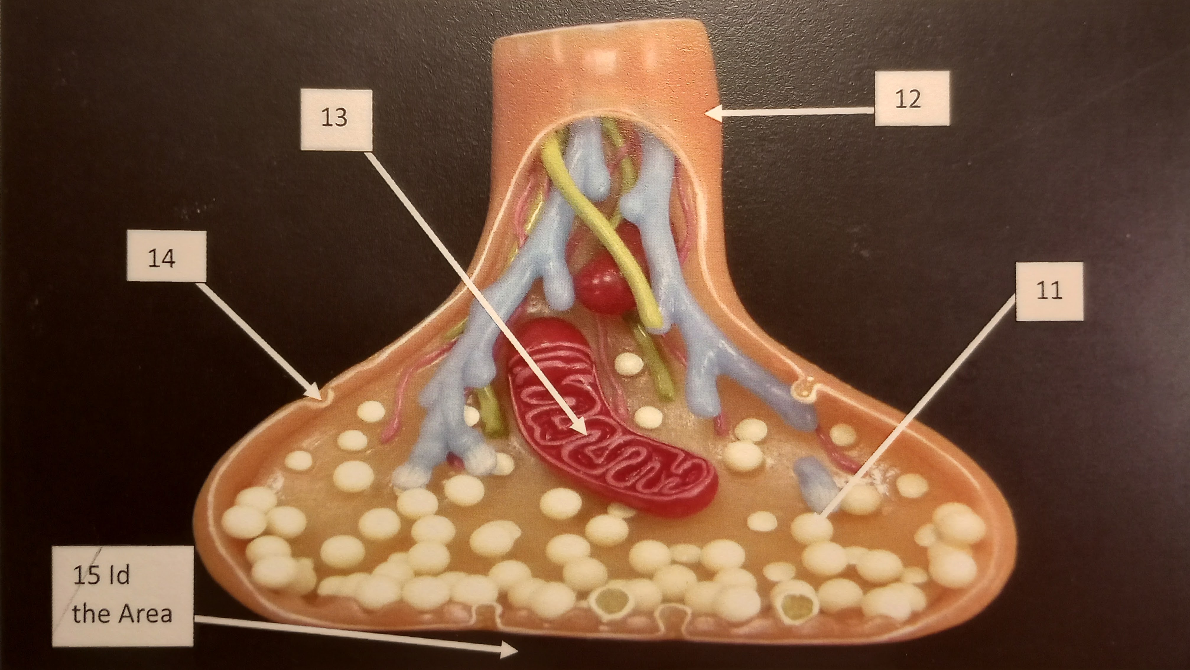
Identify number 11 in the illustration
Neurotransmitter vesicle
38
New cards
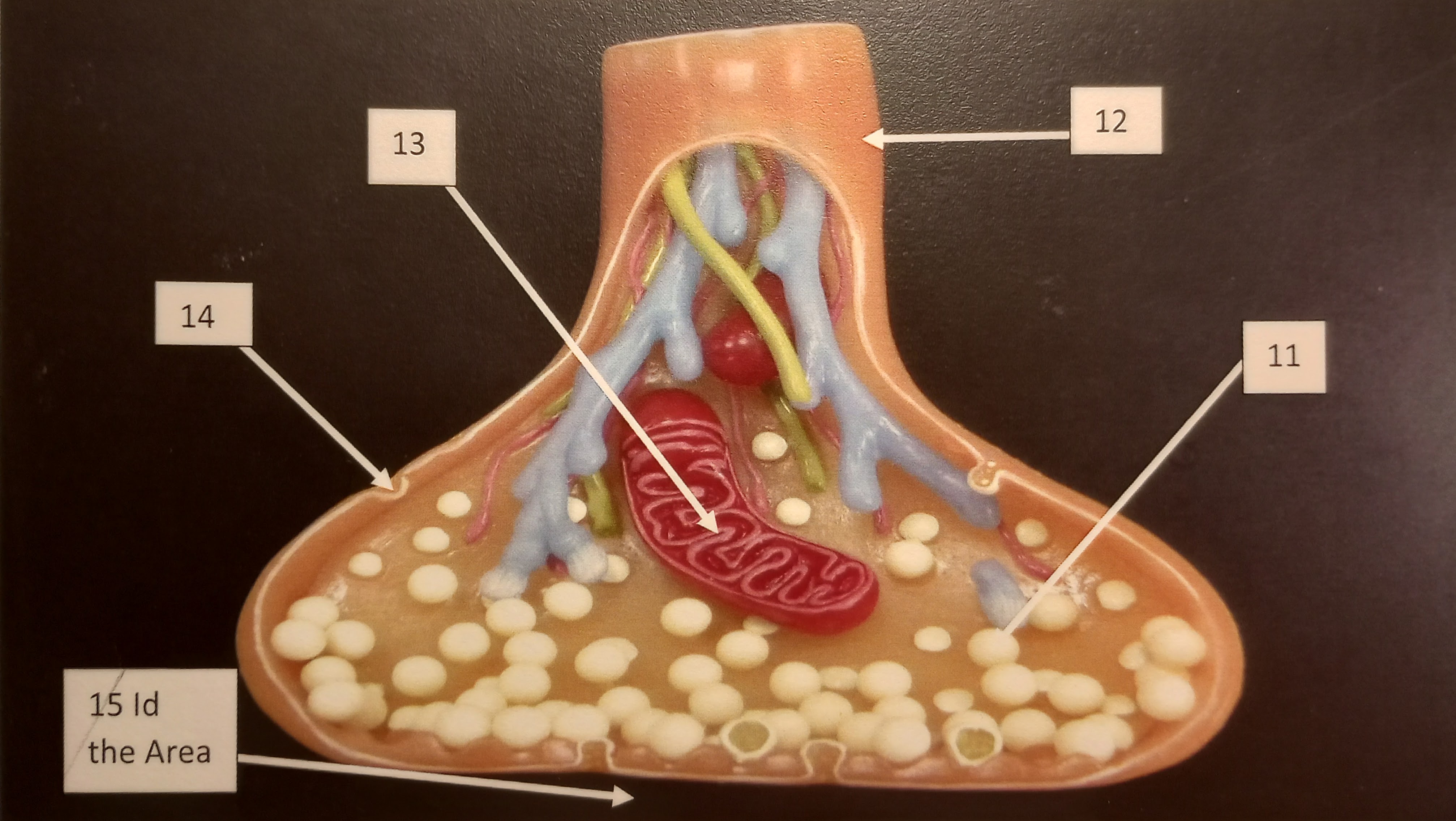
identify number 12 in the illustration
Axon Hillox
39
New cards
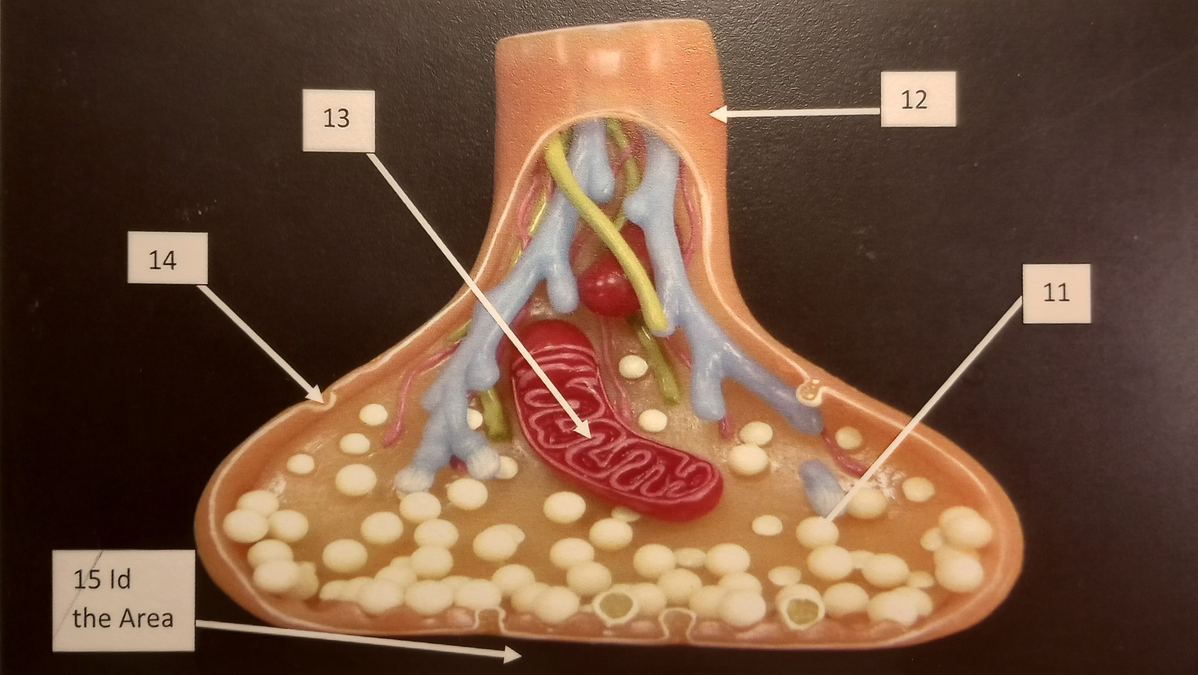
Identify number 13 in the illustration
Mitochondria
40
New cards
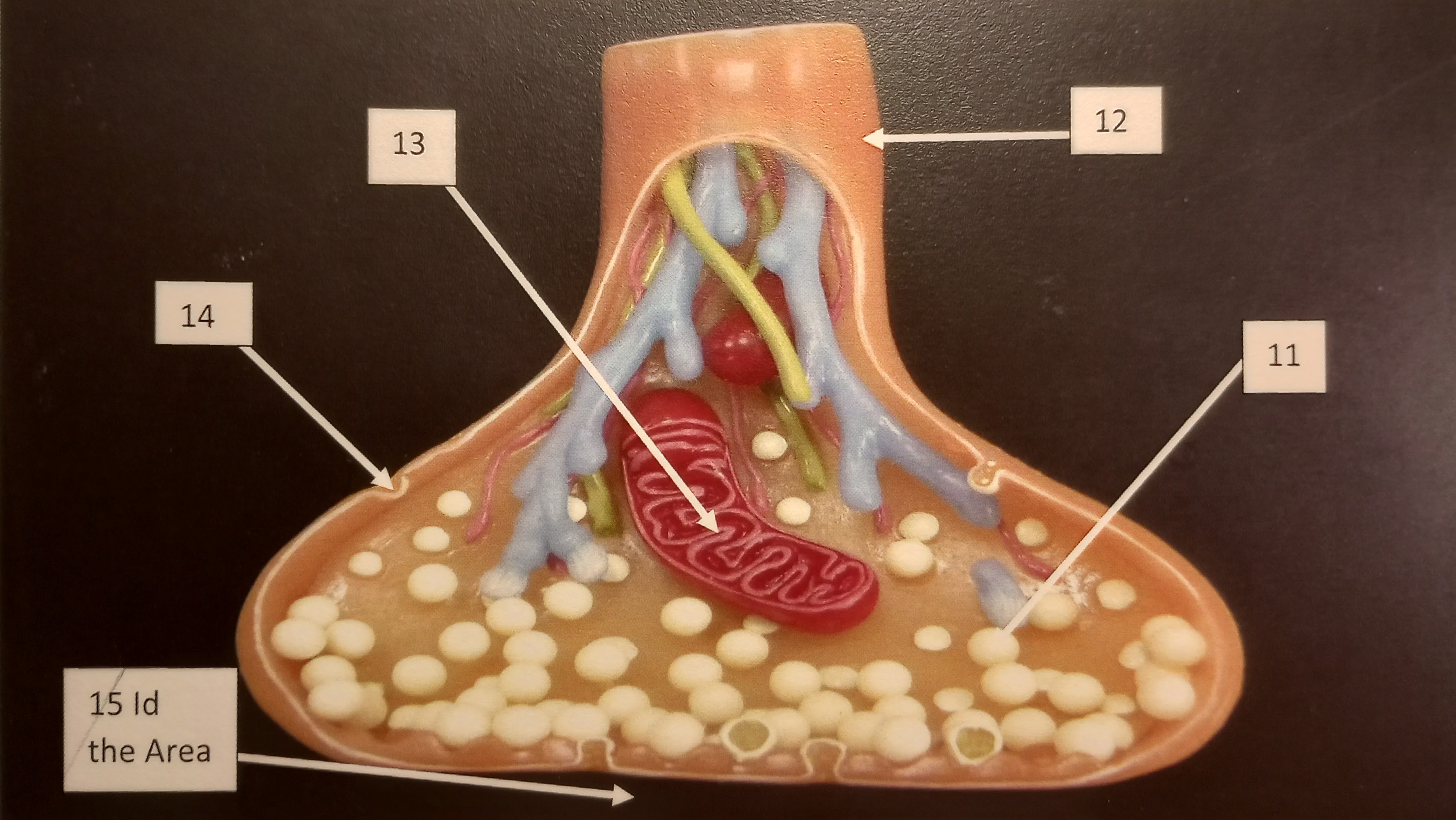
Identify the number 14 in the illustration
Calcium channel
41
New cards
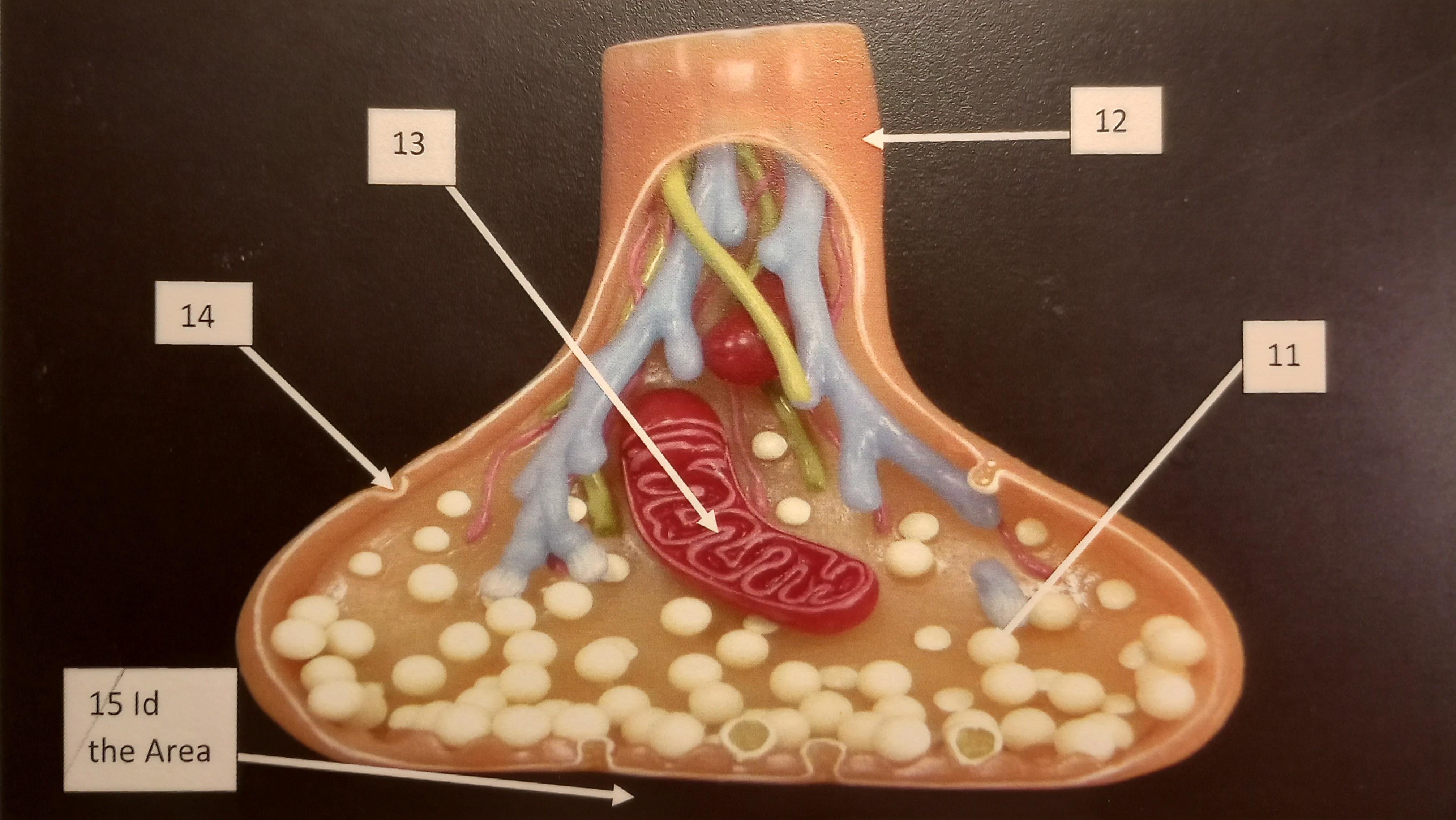
identify the number 15 in the illustration
Motor end plate
42
New cards
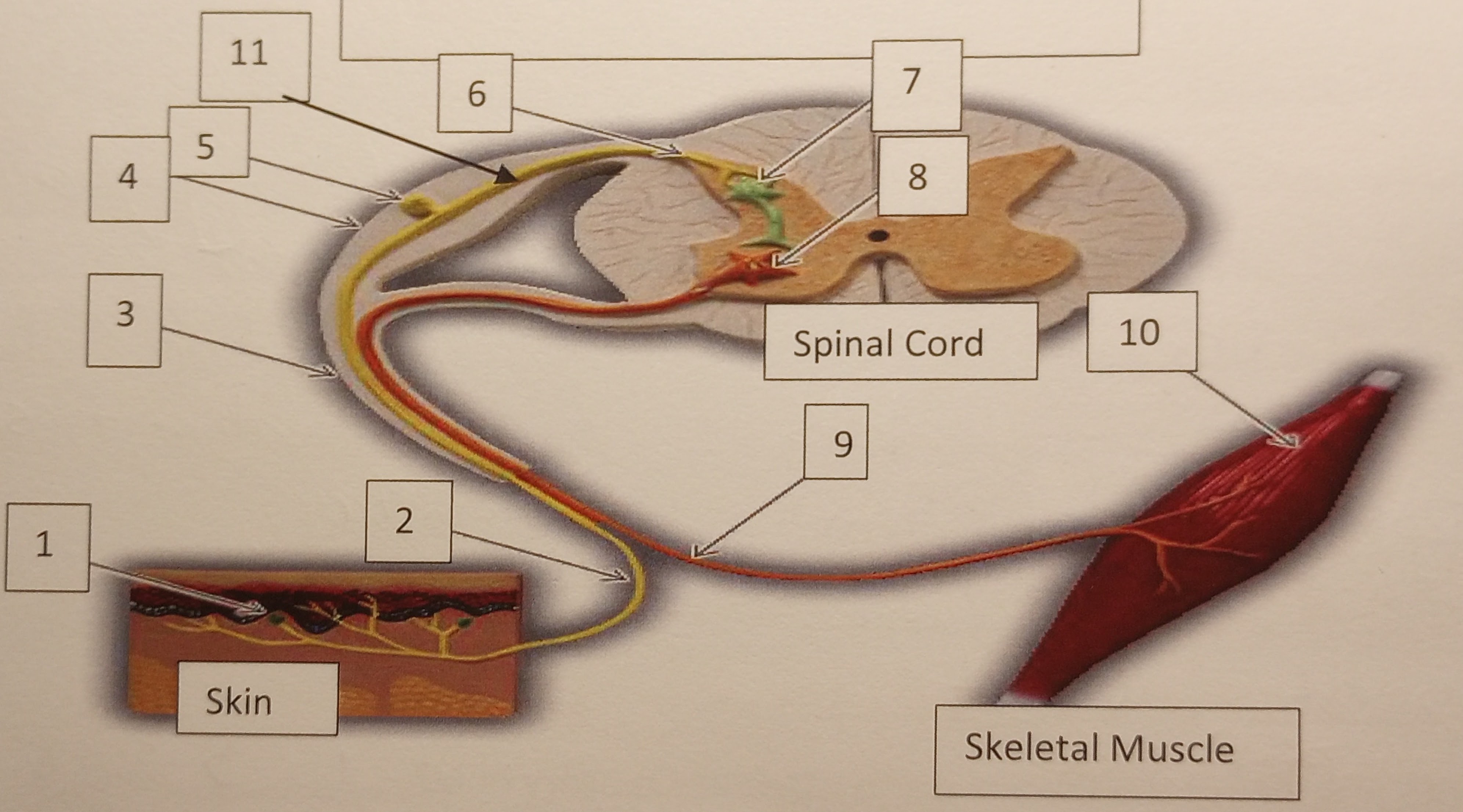
Identify the number 1 in the illustration
sensory receptor
43
New cards
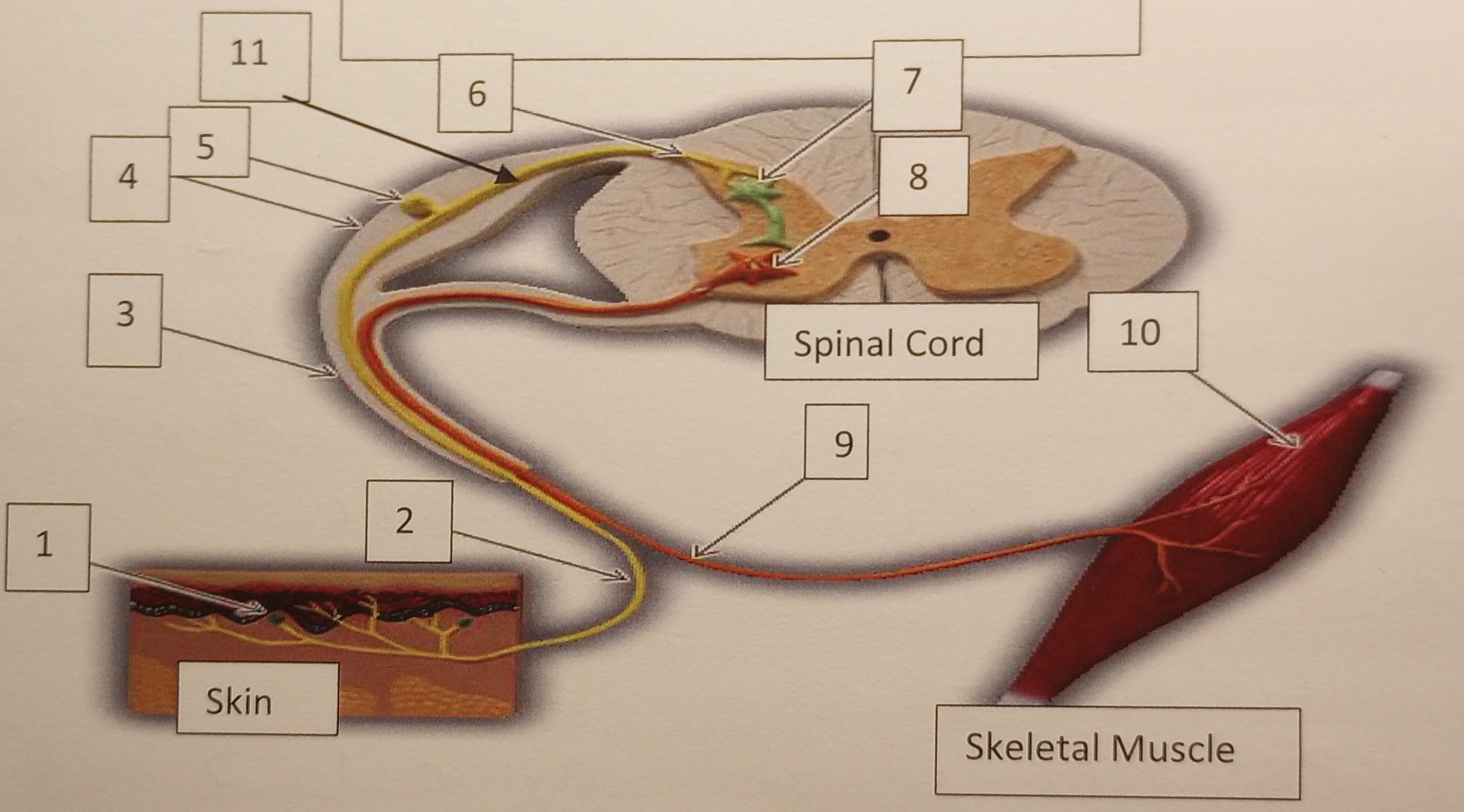
Identify the number 2 in the illustration
Sensory nerve (Afferent)
44
New cards
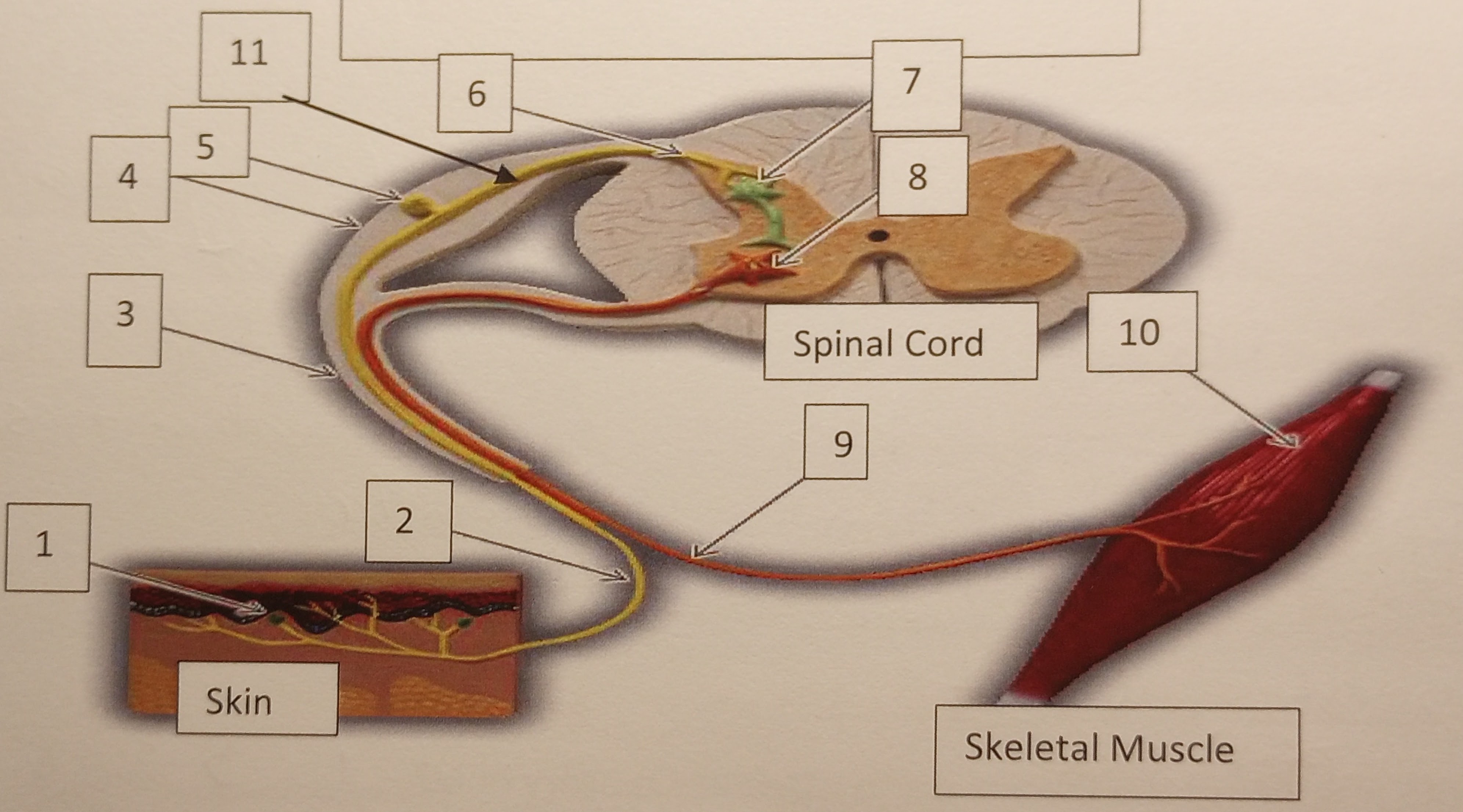
Identify the number 3 in the illustration
Spinal nerve
45
New cards
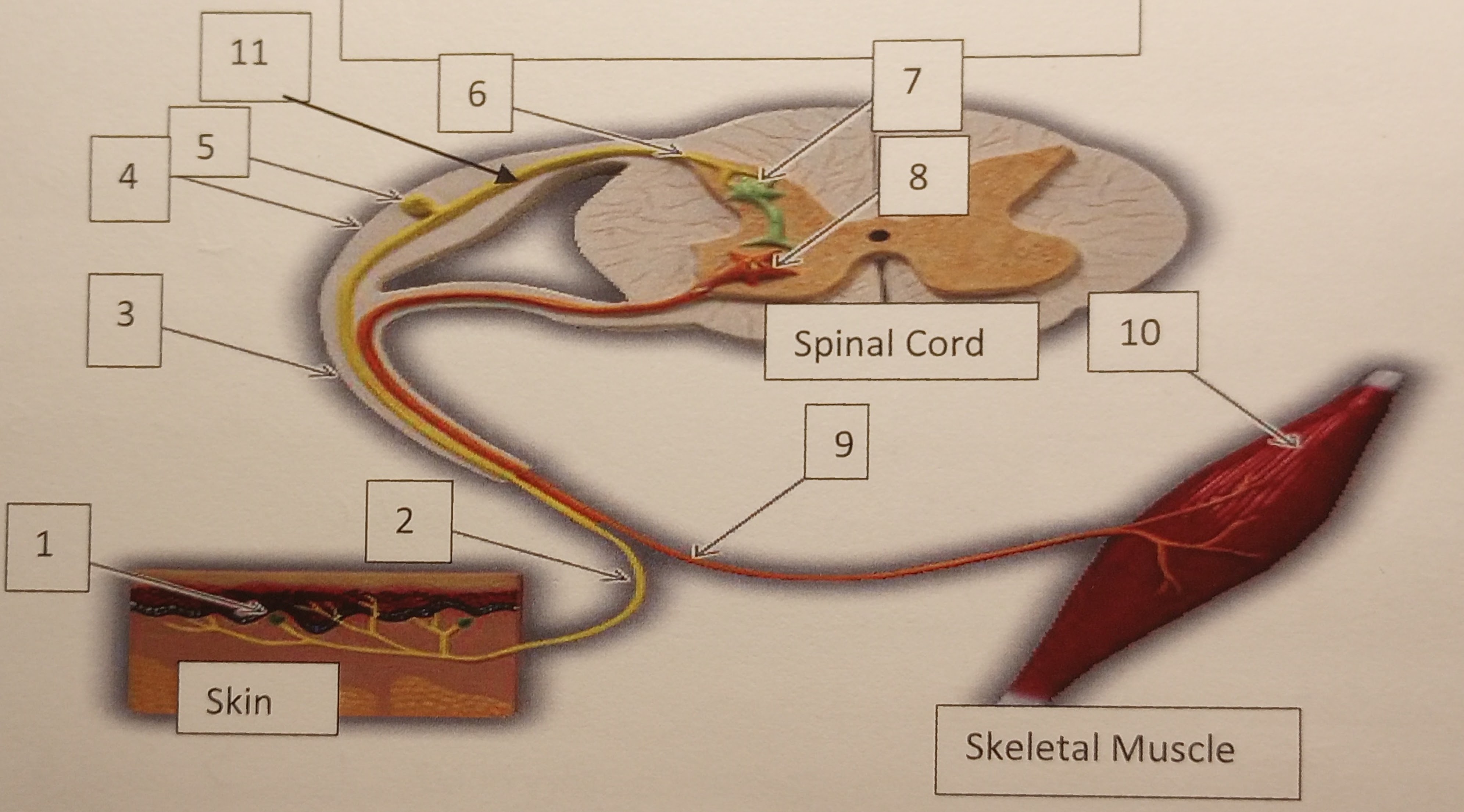
Identify the number 4 in the illustration
Dorsal root
46
New cards
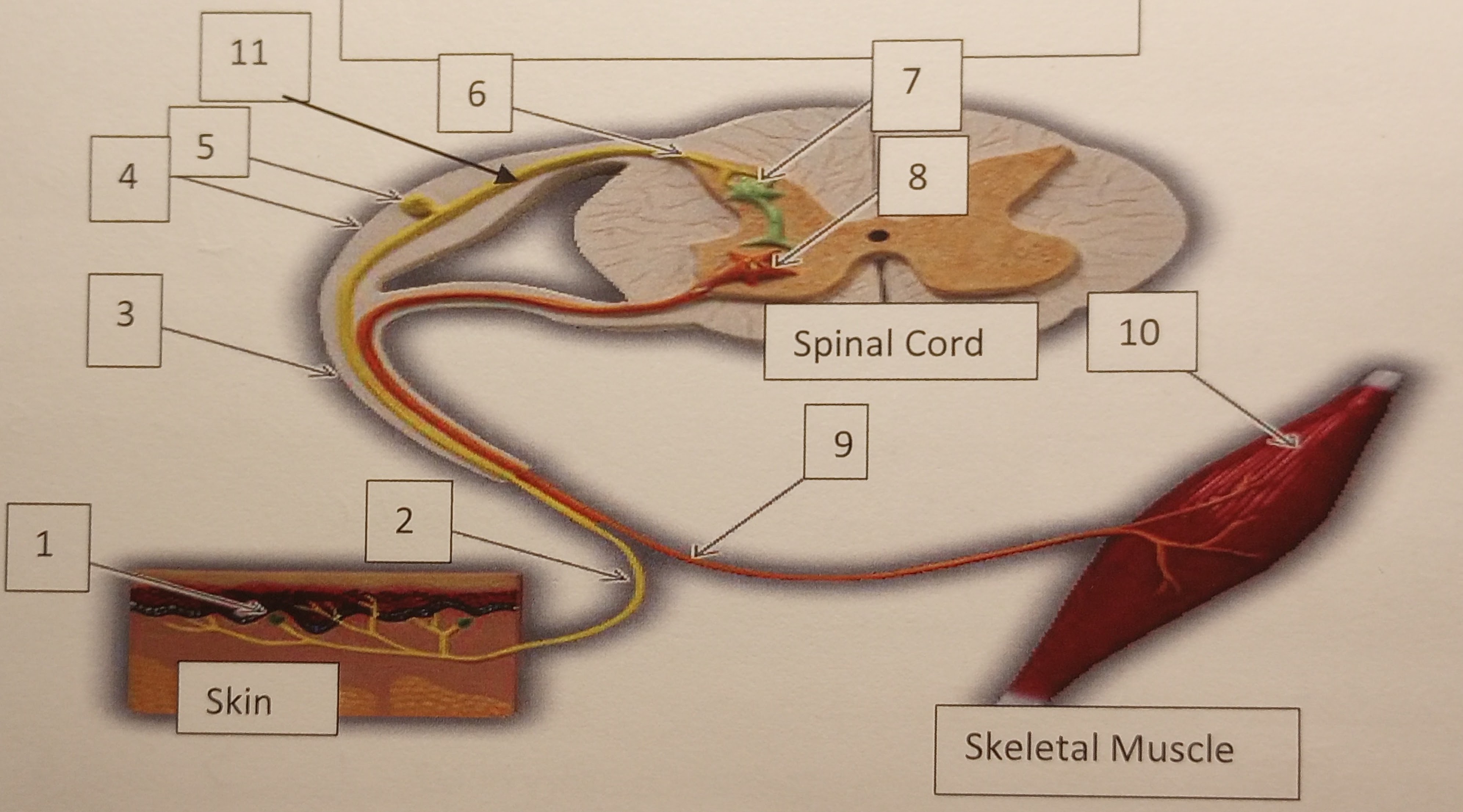
Identify the number 5 in the illustration
dorsal root ganglion
47
New cards
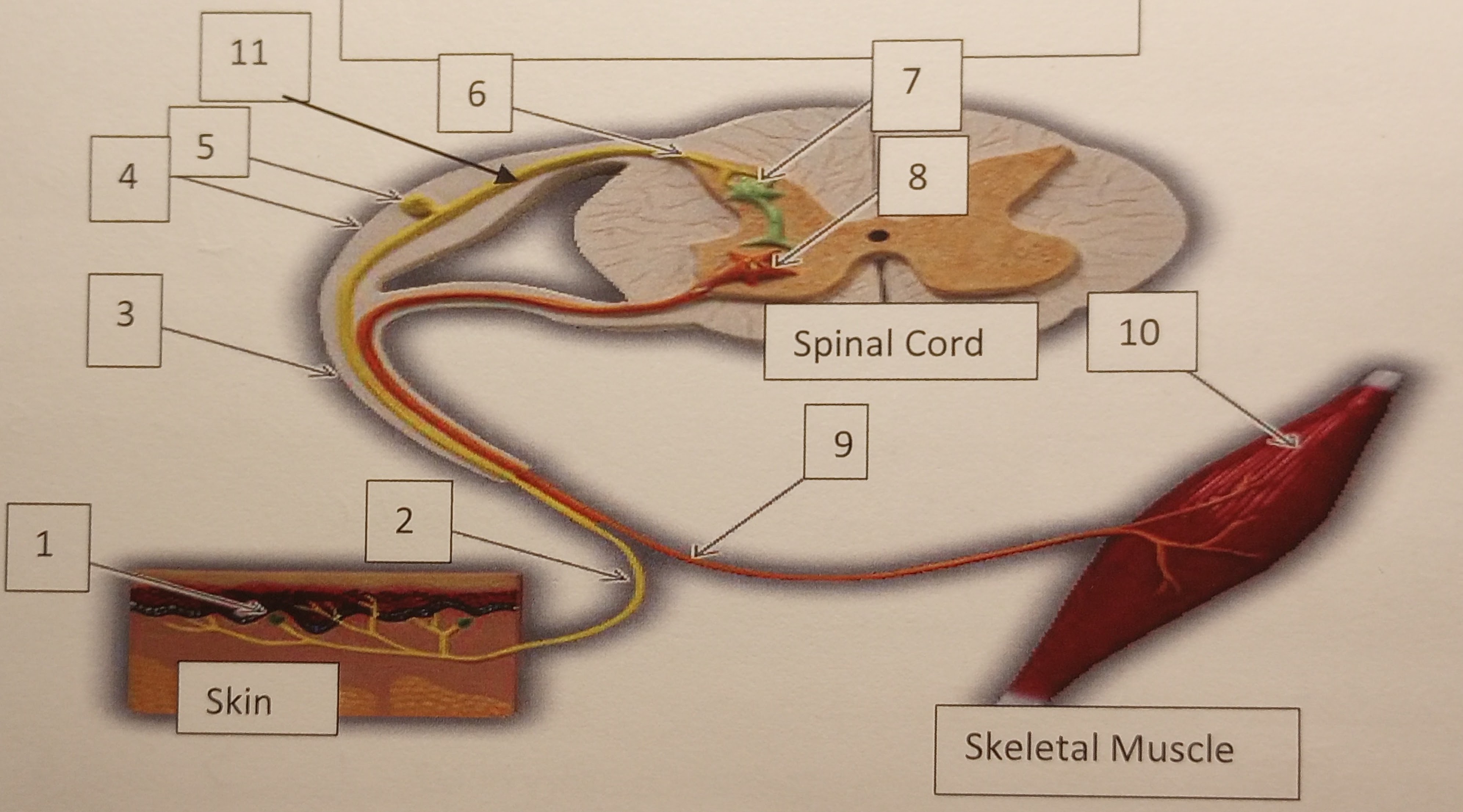
Identify the number 6 in the illustration
Dorsal horn
48
New cards
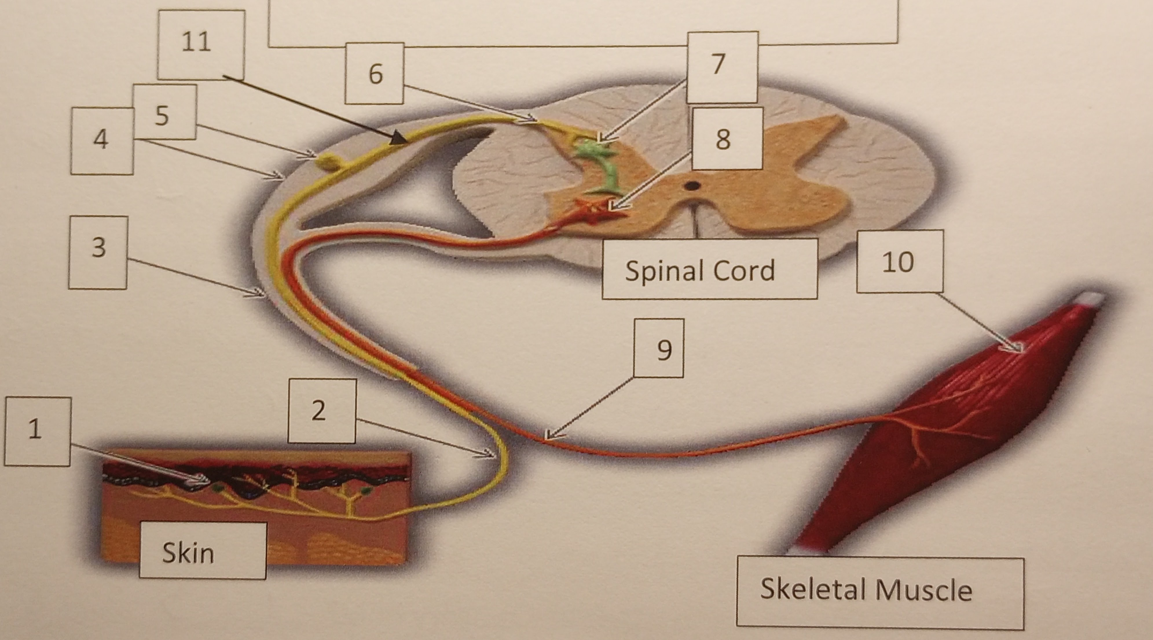
Identify the number 7 in the illustration
interneuron
49
New cards
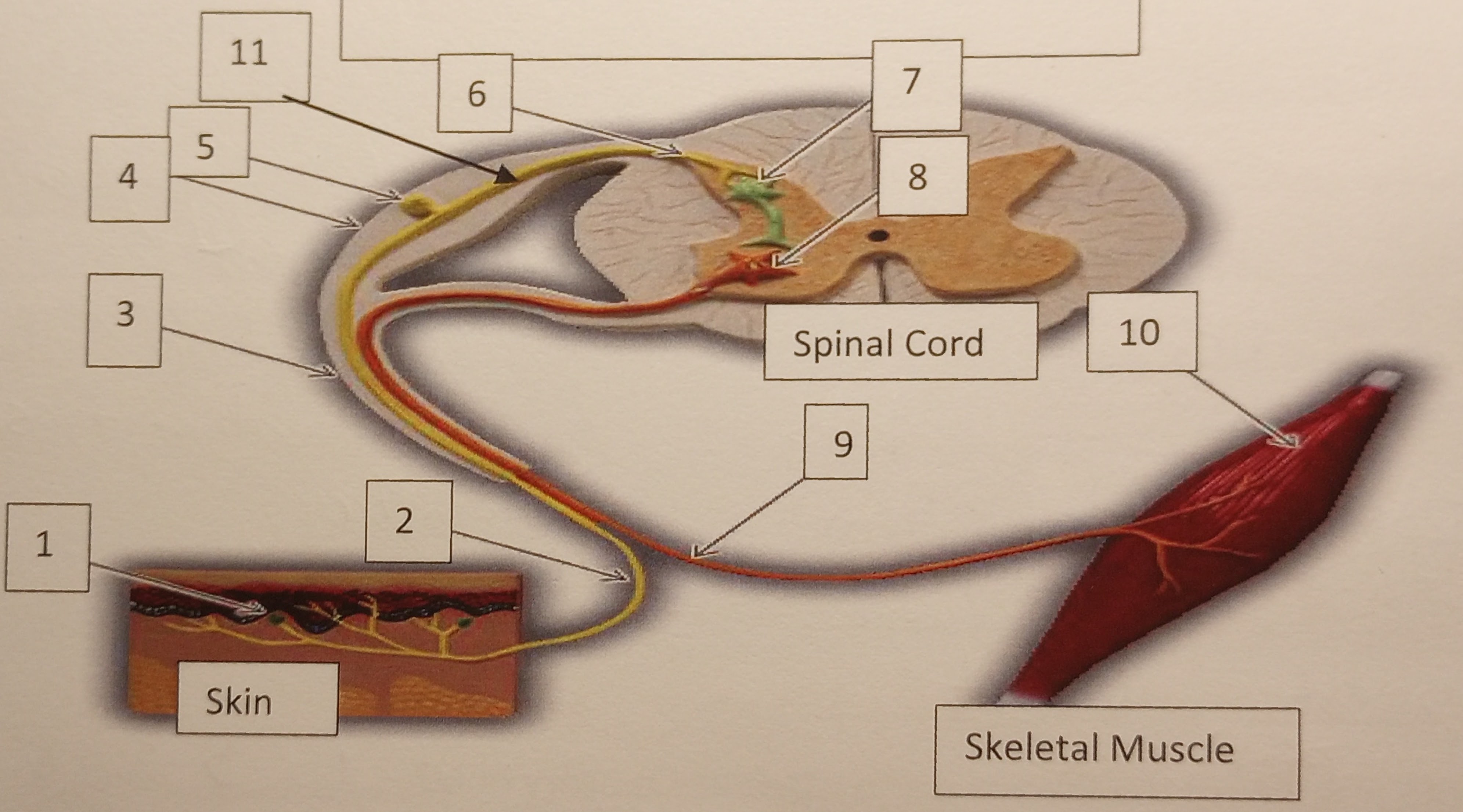
Identify the number 8 in the illustration
Motor neuron
50
New cards
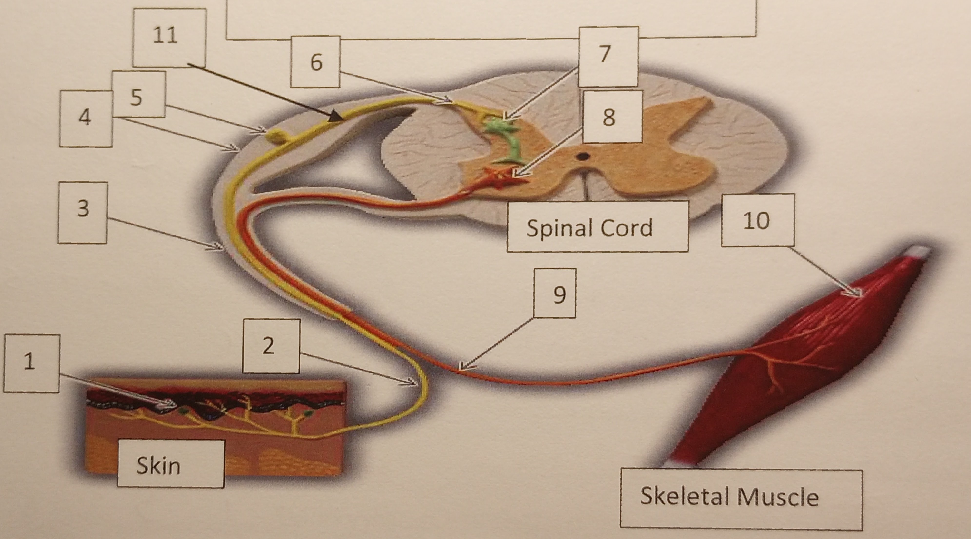
Identify the number 9 in the illustration
Motor nerve (efferent)
51
New cards
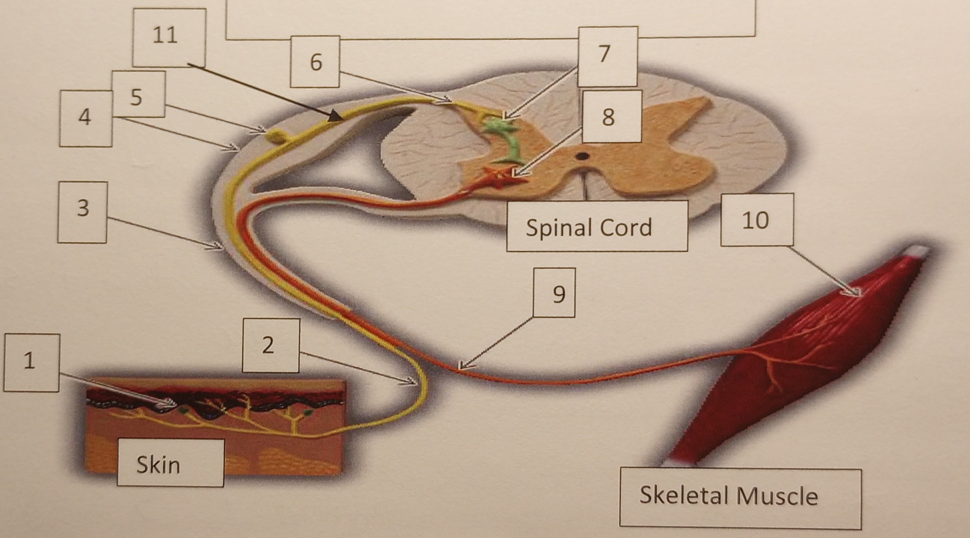
Identify the number 10 in the illustration
Muscle cell