WBC Lecture 2: BM, Non-Malignant WBC Disorders
 Call Kai
 Learn
Learn Practice Test
 Spaced Repetition
 Match
Match Flashcards
 Knowt Play
1/67

There's no tags or description
Looks like no tags are added yet.
Name | Mastery | Learn | Test | Matching | Spaced | Call with Kai | Chat |
|---|
No analytics yet
Send a link to your students to track their progress
68 Terms
Spicule
(BM eval) Relatively intact marrow consisting of hematopoietic and stromal cells (observed under low power -10x)
x100 magnification
Count 300-500 nucleated cells
Determine M:E ratio
How to perform an aspirate differential (3)
1.5:1 - 3.3:1
Normal M:E ratio of BM
Absent
Decreased
Adequate
Moderately increased
Markedly increased
BM iron is reported as… (5)
2-10/lpf
Hypoplasia or poor quality aspirate
Hyperplasia
(——) is the normal range of megakaryocytes in the BM, while a low amount would indicate (—2—) and a cluster would indicate (——)
Assess peripheral blood dilution
Find bony spicules and areas of clear cell morphology
Fat:Marrow ratio (estimate cellularity)
Search for tumor cells in clusters
Examine/estimate megakaryocytes
With a bone marrow aspirate, what is observed at low power (10x)? (5)
Observe myelocytic and erythrocytic maturation
Distinguish abnormal distribution of cells or cell maturation stages
Perform differential count on 300 to 1000 cells
Compute myeloid-to-erythroid ratio
With a bone marrow aspirate, what is observed at high power (50x and 100x)? (4)
50% (1:1)
100 - age ± 10
Increased, decreased, normal
BM cellularity in adults is approximately (——), but the general rule to estimate normal cellularity for a certain age is (——). Cellularity is reported as… (—3—)
Normal
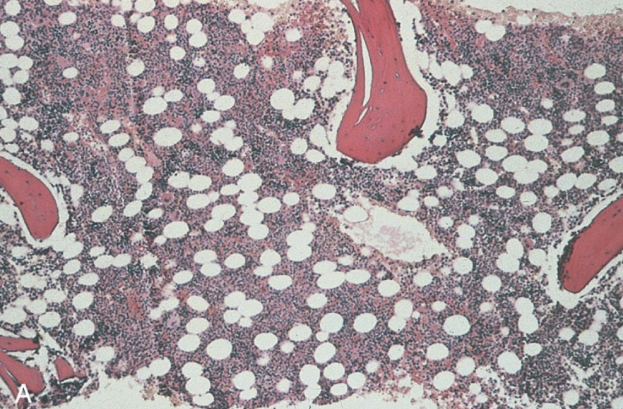
Evaluate the BM cellularity
Hypercellular/increased
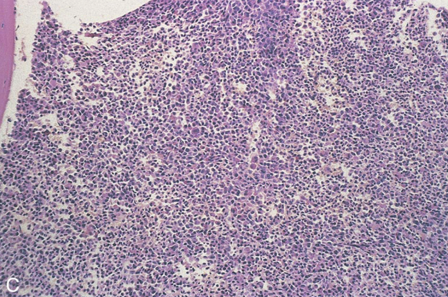
Evaluate the bone marrow cellularity
Hypocellular/decreased
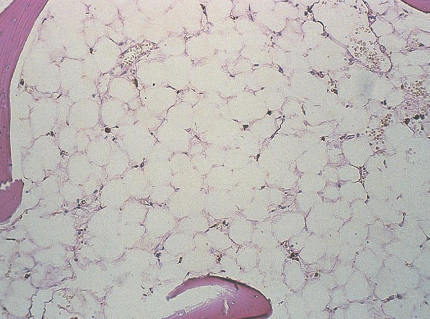
Evaluate the bone marrow cellularity
Pt history, age, sex, etc.
Description of samples received
PB: CBC, ESR, retic, manual diff, PBS
BM diff
Description of cellularity, M:E ratio
Iron stores
Description of histological sections
Diagnostic conclusion
What is included in a bone marrow report? (8)
Spicule
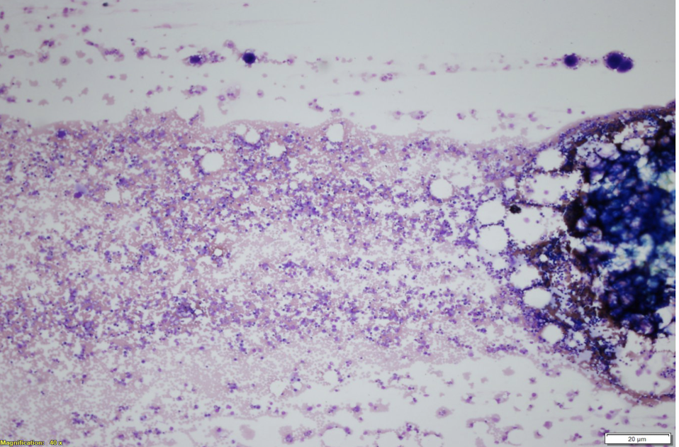
The current field in a BM smear is a good example of a…
Plasma cells
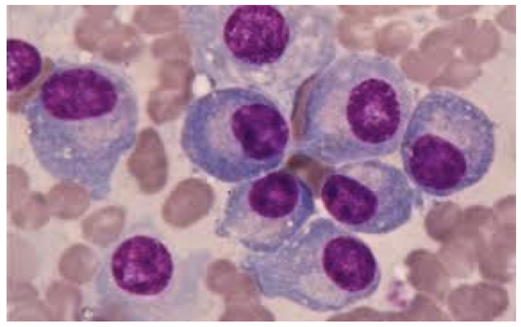
Identify the cells shown in the BM aspirate
Shift in neutrophils from marginal to circulatory pool
Increase in BM production
Release of neutrophils from storage to PB
Seen in infections
Characteristics of pathological absolute neutrophilia (4)
Infection
Storage pool
Neutrophilia and toxic changes
Left shift describes as increased number of immature cells as an indicator of (——) with increase in bands, metamyelocytes, and myelocytes. It involves an increase in release from (——) and is usually seen alongside (—2—)
> 50×103/µL
Neutrophilia
Marked left shift (band, meta, myelo)
Characteristics of leukemoid reaction (3)
Severe/chronic infection (i.e., TB, pneumonia)
Metabolic disease
Inflammation
Response to malignancy
Leukemoid reactions are mostly a result of… (4)
LR: Normal count and morph
CML: Elevated, maybe mixed granulation
Difference between leukemoid reactions and chronic myeloid leukemia regarding eosinophils and basophils
LR: toxic gran and Dohle bodies
CML: maybe psuedo-Pelger Huet
Difference between leukemoid reactions and chronic myeloid leukemia regarding neutrophil morphology
LR: normal
CML: Elevated early, decreased late
Difference between leukemoid reactions and chronic myeloid leukemia regarding platelet count
LR: Normal
CML: Possible giant, hypogranular, and/or bizarre forms
Difference between leukemoid reactions and chronic myeloid leukemia regarding platelet morphology
LR: Elevated
CML: Decreased
(RI: 15-170)
Difference between leukemoid reactions and chronic myeloid leukemia regarding LAP score
LR: Normal
CML: Positive for t(9;22); BCR-ABL
Difference between leukemoid reactions and chronic myeloid leukemia regarding genetics
Immature neutrophils
nRBCs
Dacrocytes
Neutrophilia often (not always)
Leukoerythroblastic picture (4)
Metastatic tumors
Fibrosis
Lymphoma
Leukemia
Primary myelofibrosis
Leukoerythroblastic reactions point to the possibility of a space-occupying lesion in the bone marrow, such as… (5)
Hyposegmentation
Hypersegmentation
Pyknotic and necrobiotic forms
Qualitative (acquired) granulocyte disorders in the nucleus (3)
Toxic granulation
Degranulation
Vacuolization (with/without engulfed matter)
Dohle bodies
Qualitative (acquired) granulocyte disorders in the cytoplasm (4)
Chronic infection
Megaloblastic anemia
Drugs
Hypersegmentation is usually seen in… (3)
Myelodysplastic syndromes
Asynchrony of nuclear maturation (→ clumped chromatin, no segmentation)
Hyposegmentation is usually seen in… (2)
Pyknotic
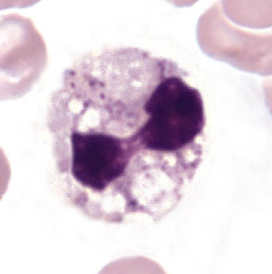
Dying cell with a very dark/dense nucleus; filaments still visible
Necrotic
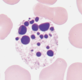
Dead cell, no filaments
Toxic granulation
Dohle bodies
Cytoplasmic vacuoles
Toxic changes in neutrophils include… (3)
Inflammation
Infection
Administration of G-CSF
Toxic granulation is associated with… (3)
Bacterial infection
Sepsis
Pregnancy
Dohle bodies are associated with… (3)
Septicemia (or other infection)
Autophagocytosis secondary to drug ingenstion
Acute alcoholism
storage artifact
Cytoplasmic vacuolization of neutrophils are associated with… (4)
Neutrophils
Ribosomal RNA
Dohle bodies are only seen in (——), and they are remnants of (——-)
Platelet satellitism
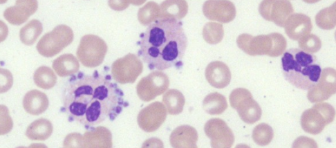
Identify what is happening in this picture
Yeast
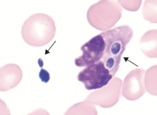
Identify the inclusion in the WBC
Bacilli
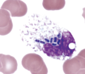
Identify the inclusion in the WBC
Histoplasma capsulatum
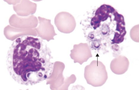
Identify the inclusion in the WBC
Autosomal dominant
Inheritance pattern of Pelger Huet Anomaly
Heterozygous state
Identify the form of Pelger Huet anomaly according the the following characteristics:
Bi-lobed nucleus
Dense heterochromatin
Incomplete nuclear segmentation
Dumbbell or pince-nez morphology
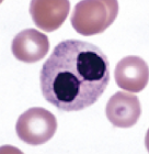
Homozygous state
Identify the form of Pelger Huet anomaly according the the following characteristics:
Nucleus is round or oval
No segmentation
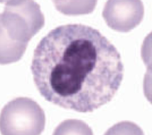
70-90%
In true Pelger-Huet Anomaly, (——) of neutrophils are affected
Drugs
HIV, TB, Mycoplasma pneumonia, severe bacterial infection
Myelodysplastic syndromes, acute leukemia, chronic myeloproliferative disorders
What can cause pseudo-Pelger-Huet anomaly (8)
Autosomal recessive
Alder Reilly Anomaly inheritance pattern
Alder-Reilly Anomaly
Abnormally large metachromatic granules in the cytoplasm of granulocytes, monocytes, and lymphocytes; leukocyte function is unaffected
Mucopolysaccharides
Alder Reilly anomaly involves the incomplete degradation of (——) deposits in WBCs
Autosomal dominant
May Hegglin Anomaly inheritance pattern
Giant, bizarre PLTs (maybe hypogran)
Leukopenia w/ large basophilic inclusions
Variable thrombocytopenia
Usually asymptomatic or mild bleeding disorder
May Hegglin Anomaly characteristics (4)
Autosomal recessive
Chediak Higashi Syndrome inheritance pattern
Chediak Higashi Syndrome
Characterized by abnormal fusion of granules in most cells that contain granules (including hematopoietic cells); fused granules are large and mostly dysfunctional; disease manifestations can be found in hair, skin, adrenal/pituitary glands, and nerves
Dysfunctional phagocytosis and ineffective bacterial destruction
How does Chediak Higashi affect WBCs
Dense PLT granules → tendency to bleed
How does Chediak Higashi affect PLTs
Albinism (affects melanocytes)
Neurological complications through childhood (peripheral neuropathy)
Other effects of Chediak-Higashi (not found in blood) (2)
Rare X linked or Autosomal Recessive
Chronic granulomatous disease inheritance pattern
NADPH oxidase
Normal or with toxic changes
Chronic granulomatous disease involves the decreaed ability of phagocytes to produce (——) (kills microorganisms); neutrophil morphology is usually (——)
Granulomas
In chronic granulomatous disease, bacteria are not killed upon ingestion; instead, they are encased in (——) to keep them localized, but they may rupture
Nitroblue tetrazolium (NBT) reduction test
Flow cytometry
What can be used to diagnose chronic granulomatous disease (2)
Nuclear contortion
Inc. cytoplasmic volume
Inc. # cytoplasmic granules
Evidence of phagocytic activity (vacuoles, debris, irregular border)
(Qualitative Changes) Reactive changes during infection, recovery from BM aplasia or post GM-CSF include… (4)
Macrophages and monocytes in BM and spleen
Lysosomal storage diseases are enzyme deficiencies where phagocytized material accumulate in cells due to degredation. It is typically seen in…
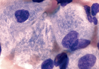
Gaucher Disease
Most common lysosomal lipid storage disease; unmetabolized lipid accumulates in the macrophages throughout the body; BM contains macrophages that have abundant fibrillar blue gray cytoplasm with wrinkled apeparance
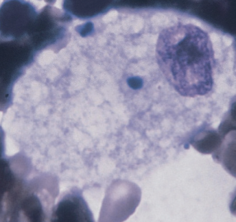
Niemann Pick Disease
Lipid buildup in macrophages; BM contains macrophages that contain foamy cytoplasm packed with lipid-filled lysosomes that appear as vacuoles after staining
Epstein Barr Virus (EBV)
Infectious mononucleosis is caused by the (——); this virus infects >90% of the world population, usually with young children
CD21
EBV preferentially infects B lymphocytes by attaching to (——) receptors
Heterophile antibodies
Infectious mononucleosis involves the production of (——) in serum, which reacts with RBCs from horse or sheep → principle of Mono test
Reactive lymphs
Elevated WBC (10-30×109/L)
Absolute lymphocytosis
Wide variation in lymphocyte morph
Infectious mononucleosis PBS characteristics (4)