(N)Chapter 8 Cells of the Nervous System単語カード | Quizlet
1/131
There's no tags or description
Looks like no tags are added yet.
Name | Mastery | Learn | Test | Matching | Spaced | Call with Kai |
|---|
No study sessions yet.
132 Terms
Brain and spinal cord
function: integration, processing, sending info out
CNS components and function
Nerves
sensory component: afferent (sends info towards CNS)
motor component: effect (info is sent away from CNS)
PNS components and function
PNS
breaks down to
A. Sensory or B. Motor
1. Somatic motor division (volun.)
2. Autosomatic motor division (invol.)
a. parasympathetic
b. sympathetic
What is the break down of the PNS?
somatic motor division
Division of PNS
Carries signals to skeletal muscles, voluntary control
autonomic motor division
Division of the PNS that regulates involuntary responses such as heart rate
1. parasympathetic
2. sympathetic
What are two branches of the autonomic motor division?
parasympathetic nervous system
the division of the autonomic nervous system that calms the body, conserving its energy
rest and digest
sympathetic nervous system
the division of the autonomic nervous system that arouses the body, mobilizing its energy in stressful situations
fight or flight
enteric nervous system
autonomous control and autonomic division control
under the influence of autonomic but can also control itself
ex: GI tract
neurons & glial cells
The nervous system is composed primarily of two cells; what are they?
neurons
basic signaling units of nervous systems
dendrites
receive incoming signals
increases surface area of a neuron, allowing it to communicate with multiple other neurons
some nerve cells can have 1 or more of these
receive incoming info and transfer it to an integrating region within the neuron
What is the function of dendrites in the PNS?
sends signals back and forth w/ other neurons in the brain
What is the function of dendrites in the CNS?
axons
sends/carry outgoing information
can be myelinated or unmyelinated
have collaterals (branching)
axon terminals: where you synaspe
somatic motor division
can have varicosities (pooling/swelling along the neuron where the NT is released)
- transmit outgoing electrical signals from soma (integration center) of neuron to the target cells at the end of the axon
- @ distal end of axon, the electrical signal usually causes a secretion of a NT (passing through gap functions in some CNS neurons)
What is the primary function of an axon?
presynaptic neuron
synaptic cleft
postsynaptic neuron
What makes up a synapse?
Axodendritic
Axoaxonic
Axosomatic
dendodendritic
neuroeffector junction
What are some of the variations of synaspes?
cell body/soma
contains the nucleus and axon hillock
axon hillock
The conical region of a neuron's axon where it joins the cell body; typically the region where nerve signals is generated.
1. pseudounipolar (most common)
2. bipolar (in the eye and nasal)
What are two types of sensory (afferent) neurons?
interneurons
Central nervous system neurons that internally communicate and intervene between the sensory inputs and motor outputs
anaxonic - no apparent axon
multipolar - highly branched but lack long extensions
multipolar
What are mostly motor (efferent) neurons?
efferent neurons
Multipolar: PNS
has 5-7 dendrites, each branching 4-6 times.
a single long axon may branch several times and
the region where an axon terminal meets it's target cell
What is a synapse?
presynaptic cell
the neuron that delivers a signal to the synapse
postsynaptic cell
neuron that receives the signal
synpatic cleft
a narrow space between two cells where information is transfered
chemical
The vast majority of synapses in the body are what kind of synapse?
chemical synapses
a presynaptic cell releasing a chemical signal that diffuses across a synaptic cleft and binds to a membrane receptor on the postsynaptic cell
one direction
what kind of synapse is this?
electrical synapses
a presynaptic and postsynaptic cell are connected by gap junctions channels
the channels allow electrical current to flow directly from cell to cell
communication is bidirectional and faster than chemical
direct transfer of ATP
what kind of synapse is this?
ribosomes and ER
The axon cytoplasm is filled with many types of fibers and filaments, but what does it lack?
nucleus, ER and packaged by Golgi
Where are the proteins that are made for the axon and axon terminal synthesized?
slow axonal transport
materials are moved by cytoplasmic streaming in the axon from soma to axon terminal
used for components like: structural proteins, enzymes, cytoskeleton proteins
fast axonal transport
walks vesicles and mitrochondria down along with microtubules network (cytoskeleton)
used for transport of NT and vesicles
can go in two directions:
anterograde - vesicles move down axon
retrograde - empty vesicles move back up axon (recycling)
cell body
nucleus, ER, golgi: protein synthesis
Where are NT produced, transported, stored?
communicate with neurons and provide important biochemical support
basically assists the neurons
function of glial cells?
Schwann cells
Satellite cells
What are two types of glial cells in the PNS?
schwann cells
support and insulate axons by forming myelin
found in PNS
1 neuron axon associates w/ many schwann cells
1 schwann cell associates with 1 neuron axon
myelin sheath
A layer of fatty tissue segmentally encasing the fibers of many neurons;
enables vastly greater transmission speed of neural impulses
nodes of ranvier
gaps between schwann cells/neurofibril nodes
segments of myelin sheath
allow for saltatory conduction
saltatory conduction
Rapid transmission of a nerve impulse along an axon, resulting from the action potential "hopping" from one node of Ranvier to another
satellite cells
non-myelinating cells in the PNS
multiple cells surround cell bodies in PNS ganglia
support PNS cell bodies by providing nutrients, clean stuff up
Oligodendrocytes
astrocytes
microglia
ependymal cells
What are the glial cells in the CNS?
Guillain-Barre syndrome
inflammatory response leading to loss of PNS myelin
- demyelination
can cause paralysis and loss of sensations
recoverable for most, given time
astrocytes
communication network via gap junctions
blood-brain barrier
support neurons by providing nutrients
regulate ions in ISF
uptake and release of chemicals at synapses
microglia
immune cells - phagocytosis
release reactive oxygen species (ROS) - form free radicals
ependymal cells
neural stem cells: differentiate into neurons and glial cells
line ventricles & central canal
- with capillaries form choroid plexus
PRODUCES CSF
selectively permeable endothelial layer
Yes
If the cell body dies, does the entire neuron die?
the neuron survives
the damaged part of the axon will degrade
But the Schwann cells may form a scaffolding for re-growth of the axon in the PNS
If the cell body survives, but the axon is damaged, what happens?
rarely
still experimental
In the CNS, can undifferentiated ependymal cells be stimulated to help damaged/dying neurons?
1. the uneven distribution of ions across the cell membrane. Normally, Na, Cl, and Ca are more concentrated in the ECF than in the cytosol. K is more concentrated in the cytosol than the ECF.
2. Differing membrane permeability to those ions. The resting cell membrane is much more permeable to K than to Na or Ca. This makes K the major ion contributing to the resting membrane potential
What two factors influence the membrane potential?
-90 mV
What is the equilibrium potential (Eion) for K?
-70 mV
What is the resting membrane potential for neurons?
by adding the Ek + Ena + Ecl + E ca together inside the cell
How is -70 mV the resting membrane potential?
1. K+ conc. gradient
2. cell's resting permeability to K, Na, Cl.
*** a change in either the K conc. gradient or ion permeabilities changes the membrane potential.
What are key factors that determine resting membrane potential?
electrochemical gradient and ion channels
ions movement is based on what?
Correct - if the membrane suddenly increases its Na permeability, Na enters the cell, moving down its electrochemical gradient
At rest, the cell membrane of a neuron is only slightly permeable to Na.
depolarizes the cell membrane (less negative)
creating an electrical signal
The addition of positive Na ions entering the cell's ICF does what to the cell's membrane and creates what?
membrane potential graph

If the cell membrane suddenly becomes more permeable to K, positive charge is lost from inside the cell and the cell becomes more negative (hyperpolarize)
K+ channels open
a cell may also hyperpolarize if negatively charged ions, such as Cl, enter the cell from the ECF.
How does a cell membrane become hyperpolarized?
very FEW
the ICF and ECF concentrations of ions remain essentially unchanged even when the membrane potential changes a lot.
A significant change in membrane potential occurs with the movement of ______ ____ ions
1. Na channels
2. K channels
3. Ca channels
4. Cl channels
What are the four major types of selective ion channels in the neuron?
leak channels
spend most of their time in the open state
- the K leak channels are the major determinant of the resting membrane potential
1. mechanically gated ion channels
2. chemically gated ion channels
3. voltage gated ion channels
What are the 3 types of gated channels?
mechanically gated ion channels
found in sensory neurons and open in response to physical forces like pressure or stretch
chemically gated ion channels
in most neurons, they respond to a variety of ligands, such as extracellular NT and neuromodulators or intracellular signal molecules
voltage-gated ion channels
opens and closes in response to changes in the cell's membrane potential.
Voltage-gated Na and K channels play an important role in the initiation and conduction of electrical signals along the axon.
some "leak" channels may actually be voltage gated channels that remain open in the voltage range of the resting membrane potential
Different voltage gated channels open at different voltages.
Give an example about leak* channels.
Na and K channels of axons are both activated by cell depolarization
Na channels open rapidly, K channels are slower to open.
The result is an initial flow of Na across the membrane, followed later by K flow.
Different channels open at different speeds, give an example
many channels that open in response to depolarization only close when the cell repolarizes.
the gating portion of the channel protein has an electrical charge that moves the gate between open and closed positions as the membrane potential changes.
open or closed positions: K
open, inactivated, closed positions: Na
**they must go through these cycles before they can open again
Different channels behave differently during repolarization. Examples?'
(different conformations)
out of
K+ flows ______ the cell.
into
Na, Cl, Ca flow ______ the cell.
- tend to occur in dendrites and cell body
- can depolarize or hyperpolarize
- vary in strength (strong/weak)
- spreads towards trigger zone but is not propagated or conducted
- decremental (becomes weaker as it travels throughout cell)
Characteristics of graded potentials
chemically (ligand) gated channels
mechanically gated channels
occasional voltage gated
What channels can initiate a graded potential?
graded potential
variable strength signals that travel over short distances and lose strength as they travel through the cell.
Used for short distance communication
IF a depolarizing graded potential is strong enough when it reaches an integrating region within a neuron, the graded potential initiates action potential.
action potentials
very brief, large depolarizations that travel for long distances through a neuron without losing strength. Their function is rapid signaling over long distances, like from toe to brain.
input
What type of signal is a graded potential?
entry of ions through gated channels
What can initiate a graded potential signal?
no
Is there a minimum level required to initiate a graded potential?
If a graded potential is strong enough, it will reach the trigger zone.
- located at the axon hillock or initial segment
What is the trigger zone? (graded potential)
subthreshold graded potential
a graded potential that is below threshold by the time it reaches the trigger zone
suprathreshold graded potential
a graded potential that is strong enough to cause an action potential when it reach the trigger zone ( -55 mV)
-55 mV
What is threshold that must be reached to initiate an action potential?
- occurs at the trigger zone (axon hillock or initial segment)
- ONLY depolarizing
- uses voltage gated ion channels
- strength: ALL or none; cannot be summed
- propagated current flow (new action potential is initiated in each segment of axon)
- non-decremental
Characteristics of Action Potential
suprathreshold graded potential at the trigger zone opens ion channels
What initiates an action potential signal?
because of the refractory period.
Absolute refractory period - cannot initiate another action potential. What is there must be all or none
Why can't two signals coming close together in time cannot sum during an action potential?
conduction of action potential
spreads towards axon terminal
new action potential initiated at each segment of axon
propagated in one direction
non decremental
What is propagated current flow - action potential?
- limits the # of action potentials
- one way propagation
What does a refractory period do?
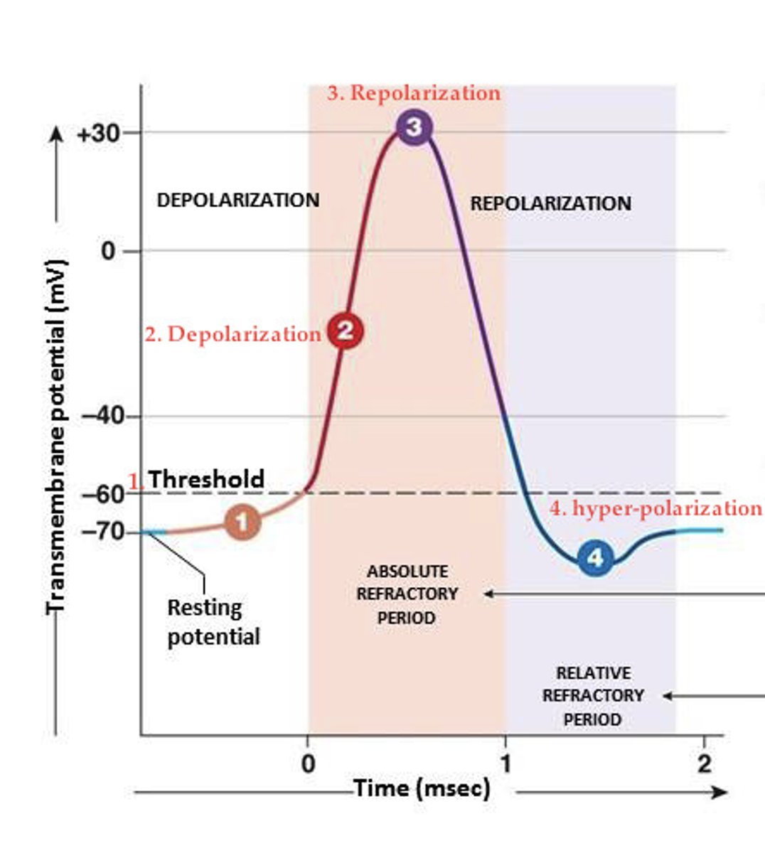
1. resting membrane potential (-70 mV)
2. depolarizing stimulus
3. membrane depolarizes to threshold. (-55mV) Voltage-gated Na and K channels begin to open.
4. rapid Na entry depolarizes cell (flows into the cell down its conc. gradient)
(overshoot: reach ENa, Na channels inactivate and K open)
5. Na channels close and slower K channels open
6. K moves from cell to extracellular fluid
7. K channels remain open and additional K leaves cell, hyperpolarizing it. (after hyperpolarization - undershoot)
8. Voltage gated K channels close, less K leaks out of the cell.
9. cell returns to resting ion permeability and resting membrane potential
Explain the steps in an action potential.
refractory period
once an action potential has begun, a second action potential cannot be triggered for about 1-2 msec, no matter how large the stimulus.
The delay is called the absolute refractory period:
the time required for the Na channel gates to reset to their resting positions.
absolute refractory period
time during which another action potential is impossible; limits maximal firing rate
Na channel gates must reset to their resting positions
relative refractory period
follows the absolute refractory period.
some but not all Na channel gates have rest to their original positions, in addition K channels are still open
*** it is possible to initiate another action potential but it all depends on:
1. how many channels close
2. how strong the stimulus is
Action potential propagation
trigger zone/initial segment
- suprathreshold graded potential
- voltage gated Na channels open
- stimulation of adjacent area
Adjacent area:
- voltage gated Na channels
- depolarization
- nondecremental
- stimulation of adjacent area
Previous area:
- absolute refractory period
- one directional flow
larger axons, less resistance, faster conduction
Rates of conduction: diameter
unmyelinated and small: slower conduction
- 0.5-2 m/s
myelinated: faster conduction
120 m/s, large and myelinated
Rates of conduction: myelination
salatory
myelinated axons have what type of conduction?
continuous
unmyelinated axons have what type of conduction?
concentration of voltage gated Na channels
saltatory conduction
efficiency: saves time and space
metabolically efficient
-fewer ions to move back
What type of conduction do Nodes of Ranvier have?
K balance is important - key ion in determining resting membrane potential
hyperkalemia
hypokalemia
What is the importance of ECF ion concentrations?