Derm E2: need to know
1/139
Earn XP
Name | Mastery | Learn | Test | Matching | Spaced |
|---|
No study sessions yet.
140 Terms
What are precancerous lesions?
actinic keratosis, cutaneous horns, keratoacanthoma, congenital melanocytic nevus, dysplastic nevus
What are cancerous lesions?
Basal cell carcinoma, squamous cell carcinoma, Merkel cell, Melanoma, mycosis fungoides
What precancerous lesion is a precursor lesion to SCC?
actinic keratosis
What is a precursor to malignant melanoma?
dysplastic nevus
What is the diagnostic workup for Actinic Keratosis?
clinical or dermoscopy; bx if uncertain
What is the tx for Actinic Keratosis?
topical fluorouracil
severe: cryo
resistant: curettage
suspicion of SCC: excision
What is the dx & tx for a Cutaneous Horn?
excisional biopsy
What is the diagnostic workup for Keratoacanthoma?
biopsy
What is the tx for Keratoacanthoma?
surgical excision or Mohs (for face lesions)
What is the diagnostic workup for a Congenital Melanocytic Nevus?
clinical or dermoscopy
What is the tx for a Congenital Melanocytic Nevus?
can remove when old enough to tolerate anesthesia
What is the diagnostic workup for a Dysplastic Nevus?
digitalized dermoscopy
What is the tx for a Dysplastic Nevus?
surgical excision if suspicious
What is the diagnostic workup for Basal Cell Carcinoma?
skin biopsy
What is the tx for BCC?
excision (Mohs)
What is the diagnostic workup for Squamous Cell Carcinoma?
skin biopsy or dermoscopy
What is the tx for SCC?
surgical excision (Alt: Mohs)
What is the diagnostic workup for Merkel Cell Carcinoma?
skin biopsy, get imaging for staging & to check for metastasis
What is the diagnostic workup for Melanoma?
thorough history, ABCDEs, dermoscopy, excisional biopsy, find Clark level (1-5)
What is staging 1 &2?
negative lymph node (lymph mapping, CXR, labs)
What is staging III?
positive lymph node (lymph mapping, CXR, labs, CT, PET)
What is staging IV?
metastasis (lymph mapping, CXR, labs, chest CT, MRI brain, bone scan, GI series)
What is the tx for Melanoma?
early: excisional biopsy
stage 4: chemo
What are the different types of Melanoma?
Superficial Spreading: MC
Nodular Melanoma
Lentigo Maligna Melanoma
Acral Lentiginous Melanoma (MC in darker skins)
Amelanotic Melanoma (no pigment)
What is the diagnostic workup for Mycosis Fungoides?
skin biopsy
What is the tx for Mycosis Fungoides?
UVA phototherapy, steroids, topical chemo, radiation
What is the cause behind Erythema Nodosum?
delayed hypersensitivity rxn caused by drugs
What is the cause behind Erythema Multiforme?
acute immune mediated rxn; caused by bacterial, fungal, or viral infx (especially HSV & mycoplasma), or by malignancy or meds
What is the cause behind Erythema Migrans?
lyme disease (black deer tick)
What is the clinical presentation of Erythema Nodosum?
erythematous tender nodules on shins
What is the clinical presentation of Erythema Multiforme?
target-like lesions
Minor: little/no mucosal involvement, extremities & spreads inward, HSV
Major: mucous membrane involved, caused by drug rx
What is the clinical presentation of Erythema Migrans?
bull’s eye (similar to multiforme), fever, chills, HA
What conditions are associated w/ Erythema Nodosum?
sarcoidosis, GI disease, strep, malignancy
What conditions are associated w/ Erythema Multiforme?
infections, malignancy, sarcoidosis, hormone issues, COVID
What conditions cause hyperpigmentation?
Melasma, Acanthosis Nigricans, Post inflammatory hyperpigmentation
What conditions cause Hypopigmentation?
Vitiligo, Albinism, Post inflammatory hypopigmentation
What causes Post inflammatory hyperpigmentation?
acne, lichen planus, dermatitis → hyperpigmented macules or papules
What causes Melasma?
hyperfunctional melanocytes
What causes Acanthosis Nigricans?
elevated glucose or metabolic syndrome → thick velvety hyperpigmented plaques
What causes Post inflammatory hypopigmentation?
chemical leukoderma, drug induced leukoderma, halo nevus, psoriasis, pityriasis versicolor → hypopigmented macules & patches
What causes Vitiligo?
usually thyroid or DM related, loss of melanocytes in epidermis → sharply marginated depigmented macules & patches
What causes Albinism?
congenital reduction or absence of melanin → hypopigmented macules/patches, usually have eye symptoms too
What is the criteria of a 1st degree burn?
superficial epidermis only, pain, erythema, heals w/in 3-6 days
What is the criteria of a 2nd degree burn?
partial (epidermis & dermis), painful, weeping, blistering, can take 3 weeks to heal
What is the criteria of a 3rd degree burn?
full (epidermis, dermis, & SC), no pain, waxy, gray/white, need surgical tx
What is the criteria of a 4th degree burn?
full (SC, muscle, fascia, bone), no pain, life threatening, need surgical tx

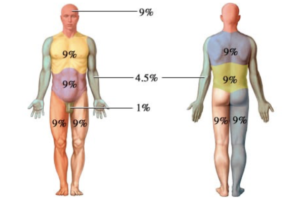
Know how to calculate % TBSA w/ Rule of 9s
What is the burn center criteria?
partial burns >10% TBSA
burns on face, hands, feet, genitalia, major joints
3rd degree +
electrical & chemical burns
burned children in hospitals w/o qualified personnel
burn in pts who require special social, emotional, or rehab intervention
What is the clinical presentation of Acute Cutaneous Lupus?
malar or butterfly rash, morbilliform rash (red eruption on sun exposed areas)
What is the clinical presentation of Subacute Cutaneous Lupus?
papules, psoriasiform, or annular plaques
What is the clinical presentation of Chronic Discoid Cutaneous Lupus?
(MC type) indurated plaques w/ well formed scale in hair follicle, plaques heal & leave scars & pigmentation
What is the clinical presentation of Chronic Lupus Panniculitis?
painful nodules → scarring
What is the prevention and tx for Cutaneous Lupus?
SPF, steroids (topical, IL or oral); antimalarial drugs if refractory
What forms of Lupus can include vascular manifestations?
all of them
What is the clinical presentation of Behcet Syndrome?
recurrent apthous & genital ulcers, cutaneous lesions
What would the lab findings of Behcet Syndrome?
>3 oral apthae in 1 yr + genital ulcers, eye lesions, skin lesions, can do pathergy test (would be +)
What is the tx for Behcet Syndrome?
aphthous and genital ulcers
topical glucocorticoids +/- topical sucralfate
Recurrent ulcers
Colchicine 1-2 mg/day divided twice daily
Refractory lesions
Prednisone 15 mg/day and taper down
or Azathioprine 50 mg daily
What is the clinical presentation of Dermatomyositis?
proximal muscle weakness, Gottron’s papules, Heliotrope eruption (periorbital skin), Shawl sign (on neck/shoulders)
What would the lab findings of Dermatomyositis show?
CK, aldolase, LDH, ANA, Anti-Mi-2, Anti-Jo antibodies
What is the tx for Dermatomyositis?
Hydroxychloroquine + Methotrexate
What would the lab findings of Cutaneous Lupus show?
ANA positive
What is the clinical presentation of Scleroderma?
pruritus, edema, thickening skin, raynauds, painful ulcer in DIP & PIP, sclerodactyly
What would the lab findings of Scleroderma show?
ANA +, ACA & Anti-Scl-70 Ab sometimes
What is the tx for Scleroderma?
Methotrexate
What is the clinical presentation of Raynauds?
digital color changes, numbness, pain
What is the tx for Raynauds?
CCBs (amlodipine)
What is the clinical presentation of IgA Vasculitis?
palpable purpura, arthritis, arthralgia, abd pain
What would the lab findings of IgA Vasculitis?
leukocytoclastic vasculitis in postcapillary venules w/ IgA deposition
What is the tx for IgA Vasculitis?
supportive analgesics; hospital if Sx are severe
What is the clinical presentation of Polyarteritis Nodosa?
purpura, painful nodules, ulcers, necrosed skin
What would the lab findings of Polyarteritis Nodosa?
ANCA, ANA, complement PTNs
What is the tx for Polyarteritis Nodosa?
prednisone, severe: azathioprine or MTX
What is the clinical presentation of Kawasaki Disease?
children, fever, erythema & edema w/ desquamation, bilateral conjunctivitis, cracked cherry red lips, cervical LAD
What would the lab findings of Kawasaki Disease show?
high LFTs/ESR, leukocytosis, thrombocytosis, anemia, pyuria
What is the tx for Kawasaki Disease?
IVIG infusion (slowly) and aspirin
What is the clinical presentation of Sarcoidosis?
papules, plaques, lupus pernio (violet infiltrate on nose), erythema nodosum
What would the lab findings of Sarcoidosis show?
apple jelly semi translucent on diascopy, noncaseating granuloma
What is the tx for Sarcoidosis?
intralesional or topical steroids; if systemic → oral steroids; refractory MTX or hydroxychloroquine
What is the clinical presentation of Granulomatosis w/ Polyangiitis?
necrotizing granuloma in URT/lungs, vasculitis, glomerulitis
What would the lab findings of Granulomatosis w/ Polyangiitis show?
anemia, leukocytosis, high ESR, impaired renal function, RBC casts, anti-neutrophil cytoplasmic antibodies
What is the tx for Granulomatosis w/ Polyangiitis?
cyclophosphamide or rituximab & prednisone, followed by maintenance w/ MTX or rituximab or azathioprine
What is the clinical presentation of Bullous Pemphigoid?
bulla, pruritus
What would the lab findings of Bullous Pemphigoid show?
none, just do biopsy
What is the tx for Bullous Pemphigoid?
clobetasol or PO steroid or doxy
What is the tx for Erythema Nodosum?
if moderate NSAID, if severe PO steroid
How do you dx Pityriasis Rosea?
clinical dx: look for Herald patch or Christmas tree pattern of scaly oval pink/hyperpigmentated papulosquamous lesions
What is the tx for Pityriasis Rosea?
sx relief w/ med potency topical steroids for pruritis
How do you dx Granuloma annulare?
dx w/ biopsy to confirm (can do KOH to r/o tinea)
What is the tx for Granuloma Annulare?
topical or IL steroids
How do you dx Lichen Planus?
+Koebner, dermoscopy, or biopsy; look for Wickham Striae & 6 Ps (pruritic, purple, polygonal, planar, papules, plaques)
What is the tx for Lichen Planus?
super high potency top steroids
How do you dx Erythema multiforme?
w/ biopsy; minor will NOT have mucous involvement, major will
What is the tx for Erythema Multiforme?
Sx: antihistamines, analgesics, top med potency steroids, acyclovir if HSV
How do you dx Melasma?
clinical dx
What is the tx for Melasma?
Hydroquinone or Retin-A, can also do chemical peels or oral tranexamic acid
How do you dx Vitiligo?
biopsy, do labs to r/o other conditions
What is the tx for Vitiligo?
localized: top steroids or calcineurin inhibitors
refractory/disseminated: UVB phototherapy
How do you dx Albinism?
clinical dx
What is the tx for Albinism?
prevent w/ sunscreen & eye protection
What is a stage 1 ulcer?
intact skin, impending ulceration