module 1: review of basic extra-oral and intra-oral anatomy
 Call Kai
 Learn
Learn Practice Test
 Spaced Repetition
 Match
Match Flashcards
 Knowt Play
1/93

There's no tags or description
Looks like no tags are added yet.
Name | Mastery | Learn | Test | Matching | Spaced | Call with Kai | Chat |
|---|
No analytics yet
Send a link to your students to track their progress
94 Terms

radiolucent structures are structures that what?
less dense and allow radiant energy to pass through
what does the term radiolucent mean?
appears dark or black on an x-ray
radiolucent structures are LESS dense because what?
x-rays can pass through them easily
because more x-rays reach the image receptor/sensor, those areas appear what?
dark
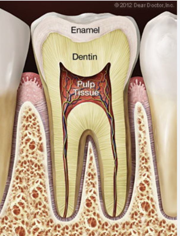
pulp
soft tissue containing nerves and blood vessels inside the tooth

periodontal ligament space (PDL)
the thin space surrounding the tooth root

soft tissue
less dense than teeth and bone

air spaces
allow x-rays to pass through very easily and therefore appear very dark

when something is radiopaque that means it appears what?
white or light on an x-ray
radiopaque structures are what?
dense because x-rays have trouble passing them
in radiopaque x-rays what happens?
fewer x-rays reach the image receptor behind these structures so they appear white or light on the radiograph
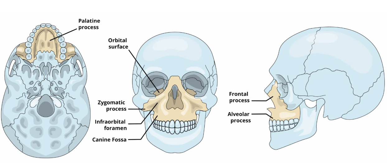
what is the maxilla?
it is two bones that form the upper jaw and part of the palate its joined together by the maxillary suture
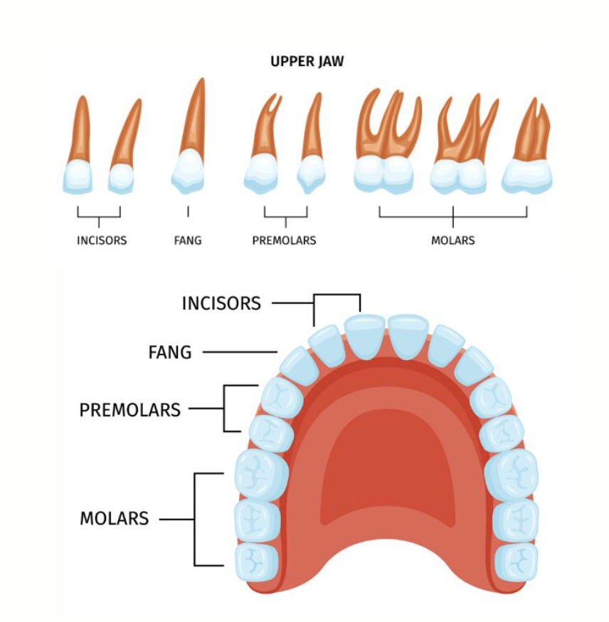
where are the upper teeth located?
they are located in the alveolar process of the maxilla
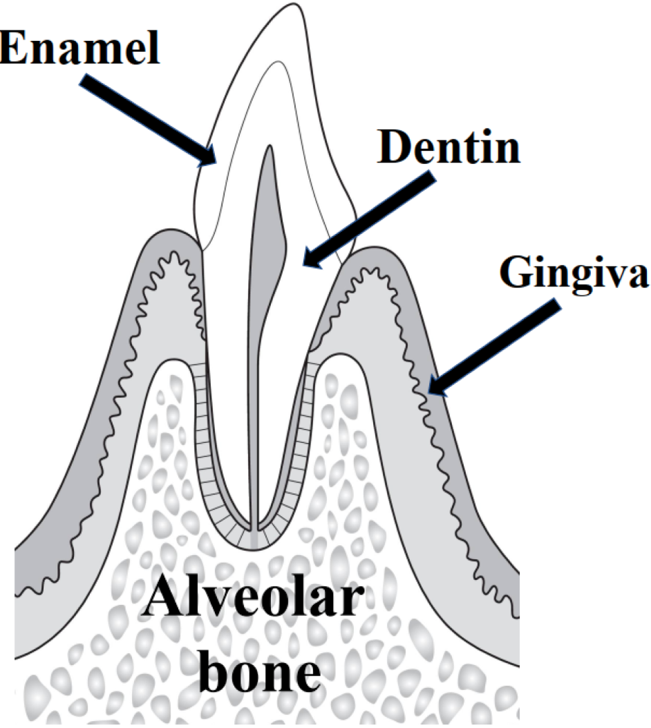
what is the alveolar bone?
The part of the jawbone that holds and supports the teeth in their sockets.
the maxilla appears WHAT on radiographs and digital images?
radiopaque
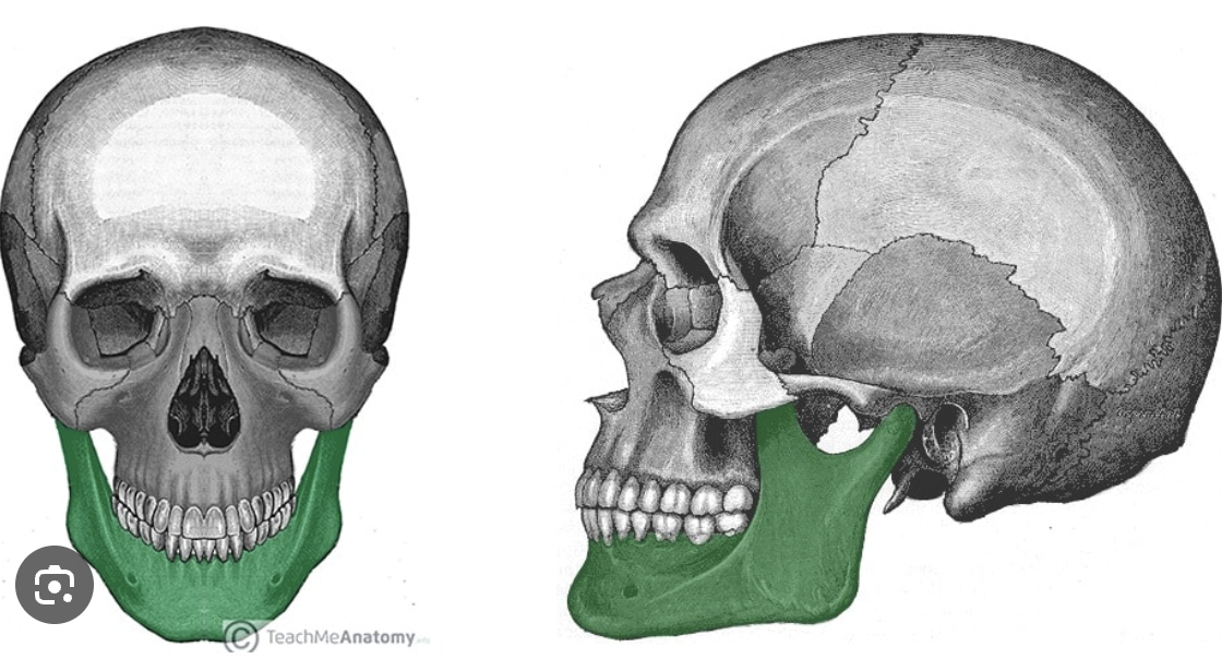
what is the mandible?
it’s a single bone that makes up the lower jaw and is the only movable bone in the skull
the mandible is made up of what?
the condyloid process, the coronoid process, the ramus, the body of the mandible, the alveolar process and the mental protuberance

what is the condyloid process?
The rounded upper part of the lower jaw (mandible) that connects the jaw to the skull and allows the jaw to move.
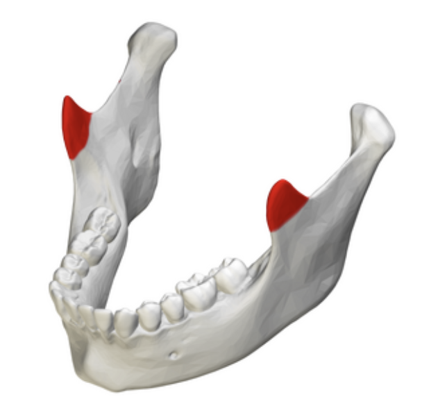
what is the coronoid process?
The pointed upper part of the lower jaw (mandible) where a major chewing muscle attaches. It helps you chew and move your jaw.

what is the ramus?
The vertical part of the lower jaw (mandible) that connects the body of the jaw to the upper parts of the mandible.
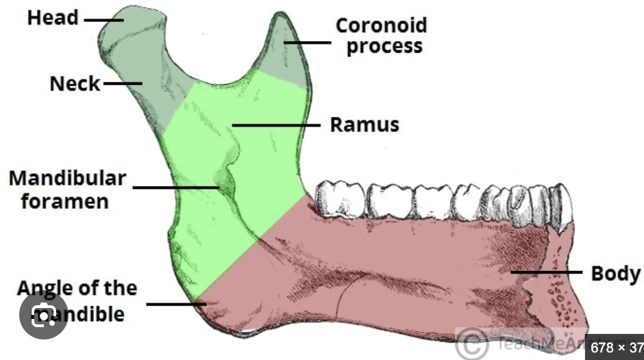
what is the body of the mandible?
the curved, horizontal, horseshoe-shaped front and side portion of the lower jawbone that holds the lower teeth and forms the lower jawline and chin.
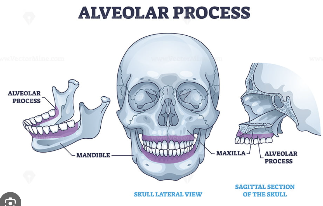
what is the alveolar process?
The part of the jawbone that contains the tooth sockets and holds the teeth in place.
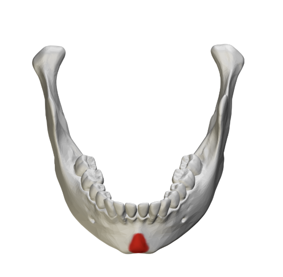
what is the mental protuberance?
The bony prominence on the front of the lower jaw (mandible) that forms the chin.
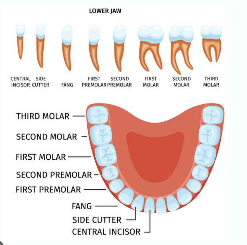
where is the lower teeth located?
in the alveolar process of the mandible
the mandible appears WHAT on radiographs and digital images?
radiopaque
the condyle of the mandible appears WHAT under radiographs and digital images?
radiopaque
the condyle of the mandible is a
ball like structure that forms the hinge between the temporal bone and mandible its a part of the temporomandibular joint
the condyle of the mandible appears on
panoramic, cephalometric and computed tomography (CT) radiographs and digital images.
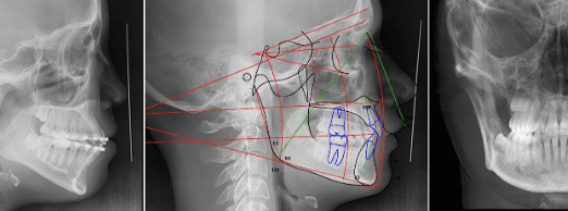
what is a cephalometric x-ray?
it is a a specialized side-profile image of the face, jaw, and skull
the ramus appears as WHAT on radiographs and digital images?
radiopaque
the body of the mandible appears as WHAT on radiographs and digital images?
radiopaque
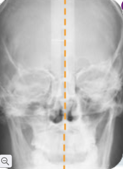
what is the midsagittal plane?
it is the imaginary vertical plane that equally separates the body into right and left halves
the midsagittal plane is also known as what?
the midline
what is the orbit?
it is the eye cavity
where does the orbit appear?
it appears on panoramic and cephalometric radiographs and digital images
the orbit appears as WHAT on radiographs and digital images?
radiolucent

what is the frankfort plane?
it is the imaginary line from the bottom of the eye socket to the auditory meatus ( ear canal)
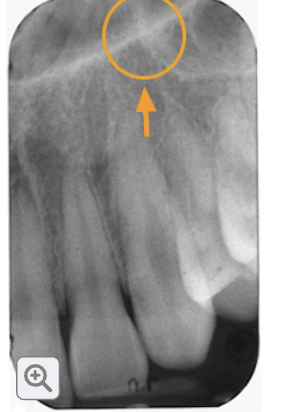
what is the inverted y?
it is the junction of the lateral wall of the nasal fossa and the wall of the maxillary sinus
where is the inverted y located?
it is located above the maxillary canine in the canine pre molar region
the inverted y appears WHAT on maxillary canine periapical images?
radiopaque
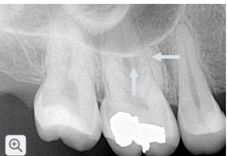
what are the maxillary sinus?
large air filled cavities located above (superior) to the maxillary pre molar and molar teeth
the maxillary sinus appear how?
they appear radiolucent on maxillary premolar and molar periapical images
the outline of denser tissue (the floor of the sinus) appears WHAT?
radiopaque
the roots of the maxillary first molar almost always appear to WHAT?
to penetrate into the maxillary sinuses but they rarely actually penetrate into the sinuses
nutrient canals are what?
they are small passageways through bone that contain blood vessels, nerve tissue, and lymphatic tissues that supply the teeth
nutrient canals appear as WHAT on periapical images?
they appear as vertical radiolucent lines

the nutrient canals are called WHAT once they enter the tooth?
pulp tissue
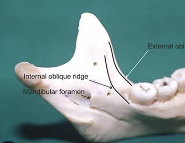
internal oblique ridge is what?
a raised line of bone on the inside of the lower jaw (mandible) near the molar teeth
where is the internal oblique ridge located?
it is located near the apices of the molars, extending down and forward from the ramus to either the third molar region or to the mylohoid ridge
how does the internal oblique ridge appear?
they appear as a radiopaque band that may run parallel to or appear superimposed on the external oblique ridge on mandibular periapical images
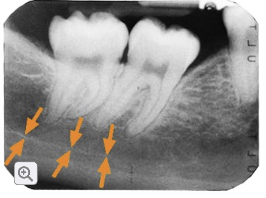
what is the mandibular canal?
long tunnel inside the lower jawbone (mandible) that carries a nerve and blood vessels to the lower teeth
what does the mandibular canal carry?
the inferior alveolar nerve and blood vessels
the inferior alveolar nerve provides what?
feeling and sensation to the lower teeth
what does the mandibular canal look like on an x-ray?
the mandibular canal looks like a radiolucent band with two thin radiopaque lines along its border canal itself= radiolucent / borders of canal= radiopaque
cortical bone is what?
its dense and lines the outside layers of the maxilla and mandible
cancellous bone is what?
soft spongy bone inside the jawbone
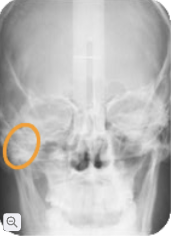
zygoma
the cheekbone. it appears as a radiopaque band on maxillary posterior periapical radiographs and digital images
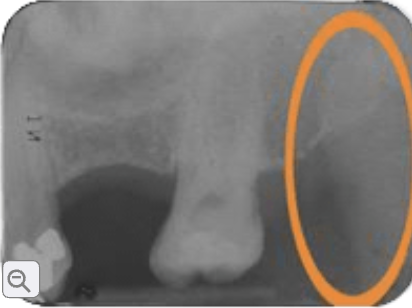
coronoid process
a pointed piece of bone at the top/front of the lower jaw although it can sometimes appear on an x-ray taken of the upper molars because the structures can overlap. it usually appears as a triangular shaped radiopaque.

zygomatic process of the maxilla
a projection of bone from the upper jaw that helps form the cheekbone. it is located above the upper molar teeth. on a dental x-ray it usually looks J shaped or U shaped radiopaque area

maxillary tuberosity
a rounded area of bone located just behind the last upper molar. on an x-ray it appears radiopaque because it is bone
what does the maxillary tuberosity do?
blood vessels and nerves can enter the upper jaw through this area
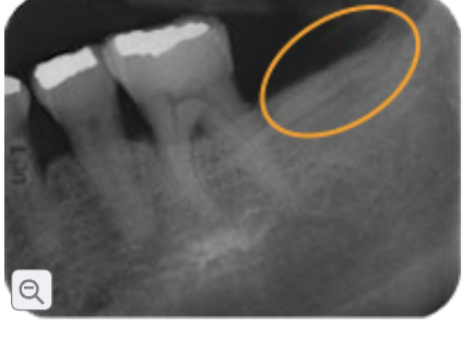
external oblique ridge/ external oblique line
a raised line of bone on the outside of the lower jaw (mandible) near the back molars. it starts near the ramus of the mandible and runs downward and forward toward the molars. on the x-ray it appears radiopaque because its a dense bone
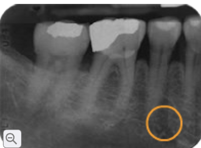
mental foramen
a small opening in the lower jawbone where nerves and blood vessels pass through to supply the lower lip and chin. its located near the roots of the lower premolars especially around the second pre molar. on an x-ray it appears like a small round/oval area it is radiolucent

mental process or ridge
a raised line of bone on the front part of the mandible near the chin just below the lower front teeth. on the x-ray it appears as a thick white line or band and it may look like an upside down V. it is radiopaque
mental
chin area
ridge
raised line of bone

alveolar process
the part of the jawbone that contains the tooth sockets and holds the teeth in place. it is found in both jaws (maxilla and mandible) on the x-ray it looks radiopaque although the spongy bone within it has a mixed pattern
alveolar ridge
the bony ridge of the jaw that contains the tooth sockets and supports the teeth on the x-ray it appears radiopaque
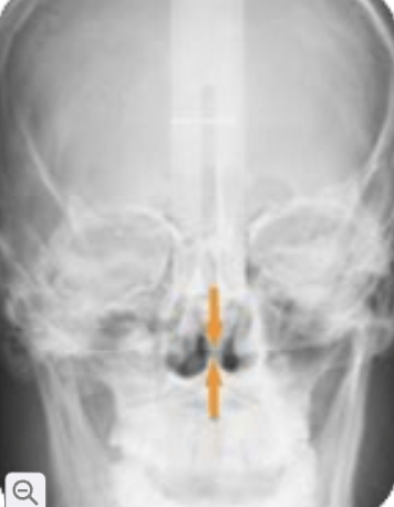
nasal septum
he bony structure that separates the right and left nasal cavities. It appears as a radiopaque (white) line between the two dark nasal cavities on an X-ray.
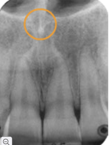
anterior nasal spine
A V-shaped bony projection at the bottom of the nasal cavity, above the upper front teeth. It appears radiopaque (white) on an X-ray.
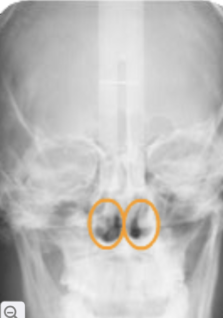
nasal fossa (nasal cavity)
Two large air-filled spaces above the maxilla
Separated by the nasal septum
Located above the roots of the maxillary incisors/canines
Appears radiolucent (dark) on X-rays
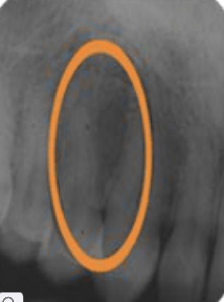
lateral fossa/canine fossa
A shallow depression in the maxilla near the lateral incisor and canine that appears radiolucent (dark) on X-rays.
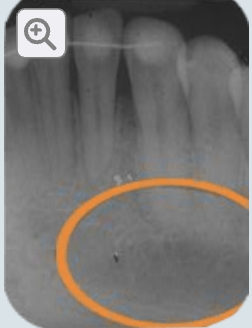
mental fossa
A depression on the external surface of the anterior mandible that appears radiolucent (dark) above the mental ridge on mandibular incisor X-rays.
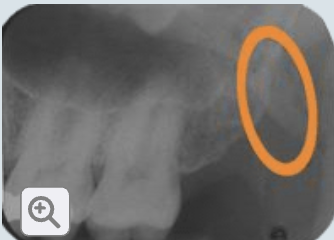
What is the hamulus (hamular process)?
A small hook-shaped projection of the sphenoid bone located behind the maxillary tuberosity. It appears radiopaque (white) on maxillary molar X-rays.
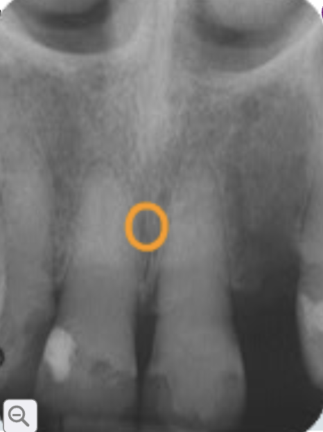
incisive foramen
A round/pear-shaped opening in the anterior maxilla, located between/behind the maxillary central incisors. The nasopalatine nerve and vessels pass through it. It appears radiolucent (dark) on X-rays.

mylohoid ridge
A raised line of bone on the internal mandible where the mylohyoid muscle attaches. It appears as a radiopaque (white) band on X-rays.
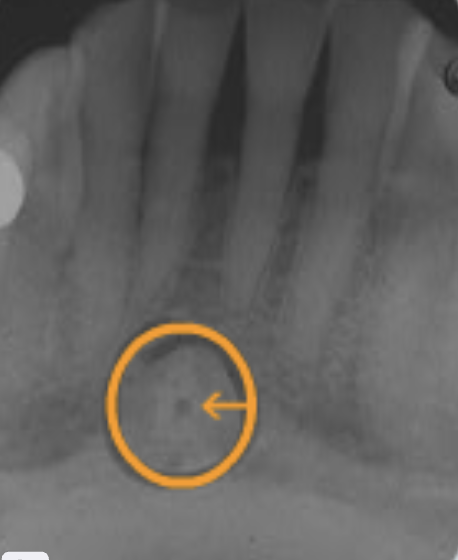
lingual foramen
A small opening at the midline of the internal mandible that appears as a small radiolucent (dark) dot, often within the genial tubercles.
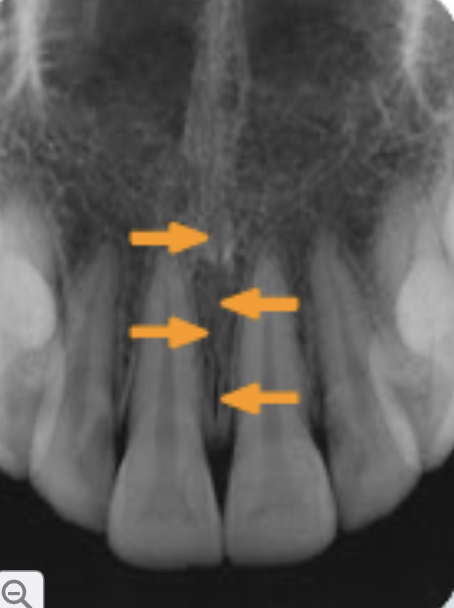
median palatal suture
The line of fusion between the right and left palatal processes of the maxilla. It appears as a thin radiolucent vertical line between the upper central incisors.
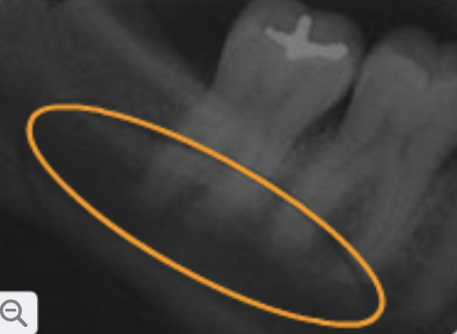
submandibular fossa
A depression on the lingual side of the mandible that contains the submandibular salivary gland. It appears radiolucent (dark) below the mandibular molar roots.
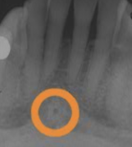
genial tubercles
Small bony projections on the internal midline of the mandible that surround the lingual foramen. They appear as a radiopaque (white), donut-shaped ring.
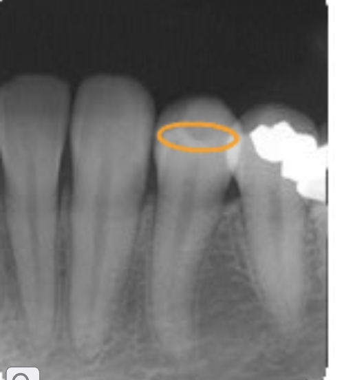
enamel
The hard outer covering of the tooth crown. It is the hardest tissue in the body and appears very radiopaque (white).

dentin
The mineralized tissue that forms the main body of the tooth. It is less radiopaque than enamel.

dentinoenamel junction
The line where enamel meets dentin.
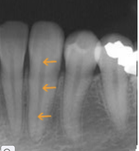
pulp
The soft inner portion of the tooth containing nerves, blood vessels, and lymphatic tissue. It appears radiolucent (dark).

periodontal ligament
Connective tissue that supports and suspends the tooth in its socket. It appears as a thin radiolucent line around the root.
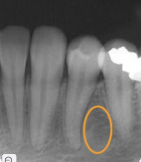
alveolar bone
The bone surrounding and supporting the teeth and forming their sockets. It appears generally radiopaque.
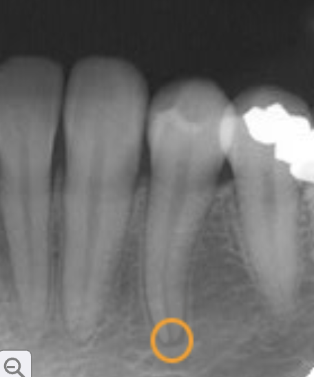
apical foramen
A small opening at or near the apex (root tip) through which nerves and blood vessels enter and exit the tooth. It is usually difficult to see radiographically.
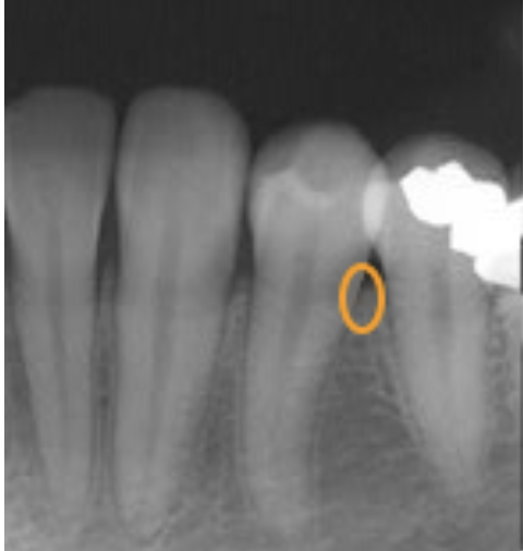
cementoenamel junction
The area where the enamel of the crown meets the cementum of the root. It marks the crown-root boundary and is usually not clearly visible radiographically.
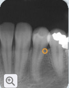
alveolar crest
he most coronal portion of alveolar bone between teeth. It appears radiopaque and is normally about 1–2 mm below the CEJ.
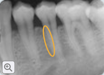
cementum
Calcified tissue that covers the tooth root and provides attachment for periodontal ligament fibers. It is usually not separately visible on X-rays.
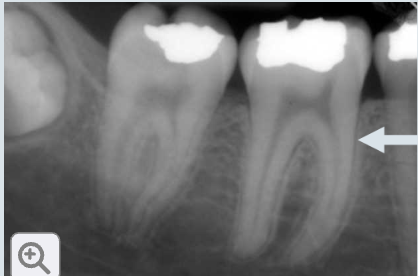
lamina dura
The dense bony wall of the tooth socket. It appears as a thin radiopaque (white) line around the root.

apex or apices
The tip or end of the tooth root.