Lesson 7 - Interpreting of Abdominal Radiographs I
 Call Kai
 Learn
Learn Practice Test
 Spaced Repetition
 Match
Match Flashcards
 Knowt Play
1/118

There's no tags or description
Looks like no tags are added yet.
Name | Mastery | Learn | Test | Matching | Spaced | Call with Kai | Chat |
|---|
No analytics yet
Send a link to your students to track their progress
119 Terms
right lateral
The fundus of the stomach can be seen in which view of the abdomen?
left lateral
The pylorus of the stomach can be seen in which view of the abdomen?
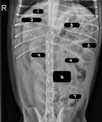
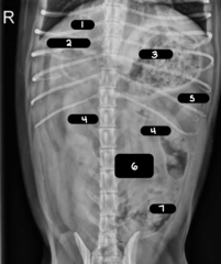
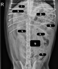
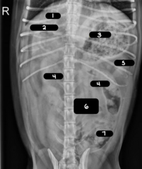
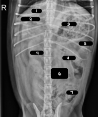
stomach
1
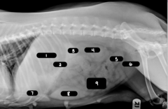
colon
2
spleen
3
kidney
4
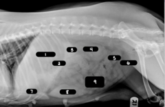
colon
5
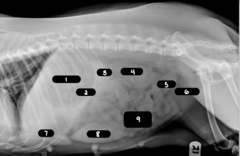
bladder
6
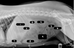
liver
7
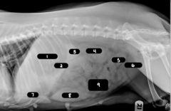
spleen
8
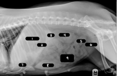
small intestine
9
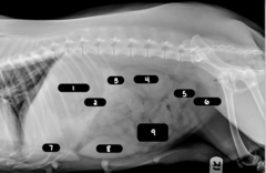
liver
1
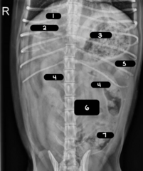
duodenum
2
stomach
3
kidneys
4
spleen
5
small intestine
6
colon
7
1. liver
2. spleen
3. stomach
4. duodenum (+/-)
5. small intestine
6. cecum (+/-)
7. colon
8. kidneys
9. urinary bladder
10. prostate (+/-)
Which structures can normally be seen on abdominal radiographs?
1. pancreas
2. adrenal glands
3. lymph nodes
4. ureters
5. ovaries/uterus
Which structures can not normally be seen on abdominal radiographs?
1. number
2. size
3. shape
4. position
5. opacity
What are the Roentgen signs?
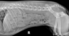
normal right lateral view of a dog
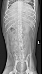
normal VD view of a dog
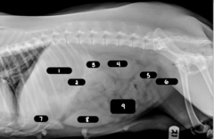
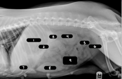
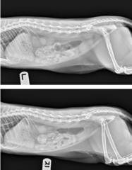
normal right and left lateral views of a cat
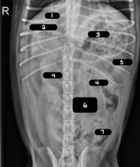
normal VD view of a cat
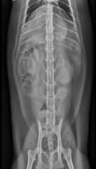
extra-abdominal constipation in a canine patient
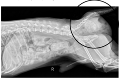
extra-abdominal mass
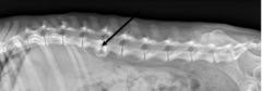
spondylosis deformans
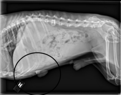
hernia
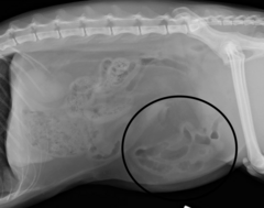
diaphragmatic hernia
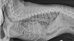
inguinal hernia
This is a 3-years-old DSH presenting after being HBC, with a swelling on Rt. Inguinal region. What is the radiographic finding?
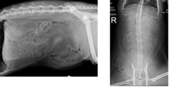
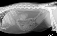
right lateral view of an obese feline
VD of an obese feline
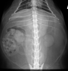
moderate to large fat deposits in the retroperitoneal space and peritoneum (especially in the falciform ligament)
What can be seen on radiographs of an obese feline patient?
difficulty seeing the contours of individual organs
What is reduced/lost serosal detail?
1. neonatal (brown fat)
2. emaciation (loss of fat)
Which body conditions can cause reduced/loss serosal detail?
1. free fluid
2. peritonitis (steatitis)
3. carcinomatosis
4. mass effect
5. post op changes (can persist for weeks)
Which pathalogical conditions can cause reduced/loss serosal detail?
neonates and juveniles up to about 6 months old
Which animals have minimal intraperitoneal fat?
brown fat
Which type of fat has a higher water content and results in poor serosal detail?
1. peritonitis
2. small amount can cause focal loss of detail
3. large amount can cause an overall increase in opacity
4. fat stranding
5. displacement of structures
What can free fluid in the abdomen cause?
peritoneal effusion
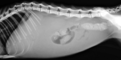
peritoneal effusion
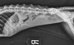
peritoneal effusion with fat stranding/whispy soft tissue opacities
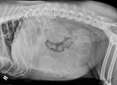
inflammation of fat in cats from vitamin E deficiency, chronic pancreatitis, high fish diet
What is steatitis?
steatitis
What can cause loss of serosal definition in cats?
pancreatitis
What can cause loss of serosal definition in dogs?
metastatic seeding of neoplasms throughout the peritoneum
What is carcinomatosis?
hazy loss of detail, free fluid, and potential nodular appearance of serosal margins
What can be seen on radiographs in patients with carcinomatosis?
peritonitis/FIP
What can carcinomatosis not be differentiated from radiographically?
carcinomatosis
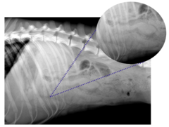
decreased serosal definition due to a mass effect
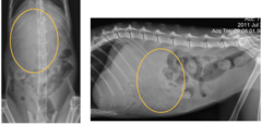
pneumoperitoneum
What can cause increased contrast of serosal definition?
free gas within the peritoneal cavity most common secondary to surgery (can persist for a month)
What is a pneumoperitoneum?
1. penetrating trauma
2. rupture of a hollow viscus (GI tract- emergency)
3. necrotic neoplasia/abscess
What can cause a pneumoperitoneum?
1. Highlighted serosal surfaces
2. Geometric-shaped gas opacities
3. Look near diaphragm, liver and cranial to the bladder
4. May see focal loss of serosal detail
5. Positional views (horizontal beam)
What are the rdiographic signs of a pneumoperitoneum?
pneumoperitoneum due to perforation/abcess
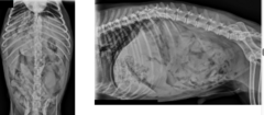
good serosal definition
1 year old DSH, anorexia for 3 days, what is your describtion for the serosal definition?
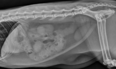
enhanced serosal definition/gas opacity
1 year old DSH that was HBC presenting with marked abdominal distension what is the radiographic finding?
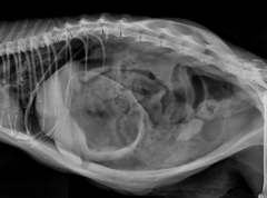
dystrophic mineralization/tissue death that can affect serosal definition
What is intrabdominal mineralization?
1. metabolic disorders (Cushing's)
2. inflammation
3. neoplasia
4. mesenteric cyst/Bates body
What can cause intrabdominal mineralization?
bates body (incidental finding in cats)
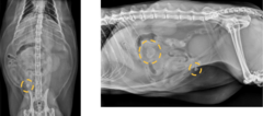
1. extraperitoneal space
2. situated ventral to the vertebrae and paraspinal muscles
3. extends along the length of the entire abdomen from the diaphragm to the anus
4. the lateral boundaries are the abdominal and pelvic walls
5. the ventral boundary is the dorsal parietal peritoneum
What are the characteristics of the retroperitoneum?
kidneys
What retroperitoneal structures can be seen on a radiograph?
1. effusion/fluid
2. mass
3. gas
What can cause decreased/loss of definition in the retroperitoneum?
ventral displacement of colon by retroperitoneal effusion
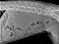
1. neoplasia
2. mineralization of adrenal glands
3. lymphadenopathy
4. bone mass
What are they types of masses seen in the retroperitoneum?
retroperitoneal mass
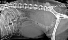
bone tumor at L4
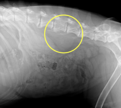
a decrease in overall opacity with clear depiction of structures from gas in the peritoneum
What is a pneumoperitoneum?
1. extension of pneumomediastinum
2. abscess
3. gas producing bacteria
What can cause a pneumoperitoneum?
pneumoretroperitoneum
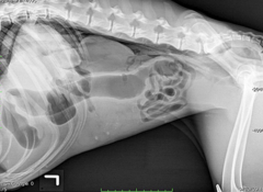
1. cranio-ventral on lateral
2. caudal to diaphragm
3. cranial to stomach
4. within the ribcage
5. soft tissue opacity
6. triangle-wedge, sharp caudal ventral contour (hepatic angle)
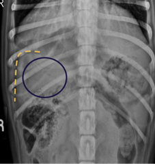
What are the characteristics of the liver on a radiograph?
falciform fat
What can displace the liver in obese cats?
canine liver
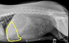
feline liver
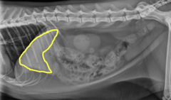
1. rounding or blunting of the caudoventral liver margin
2. extension beyond the costal arch
3. caudal (+- medial) displacement of the gastric axis
4. may include caudal displacement of right kidney, transverse colon, and cranial duodenal flexure
What can be seen on a lateral radiograph with hepatomegaly?
an increased opacity may be present in the right cranial abdominal quadrant, extend beyond the gastric fundus
What can be seen on a VD radiograph with hepatomegaly?
normal liver with normal hepatic angle
What is seen on the left?
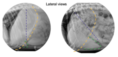
liver with hepatomegaly
What is seen on the right?
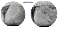
hepatomegaly
What liver pathology is being shown?
1. nodular regeneration/extramedullary hematopoiesis
2. passive congestion from right congestive heart failure
3. endocrinopathies like hyperadrenocorticism, hypothyroidism, diabetes mellitus
4. neoplasia
5. lipidosis (cats)
What can cause hepatomegaly?
lesions located on the caudal surface of the liver
What focal liver lesions are seen more clearly?
liver-increase in size, focal
1. normal, breed/age variation
2. neoplasia, large mass (hepatocellular and bile duct carcinoma) in dogs
3. biliary cystadenoma/adenocarcinoma (cats)
4. metastatic disease
5. benign nodular hyperplasia
6. bulge at the mid-ventral aspect of the liver may be the gallbladder (cats)
What are the causes of focal hepatomegaly?
microhepatia
What pathology is being shown?
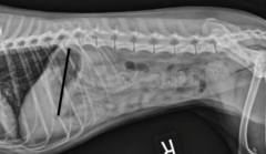
cranially displaced gastric axis
What is being shown with the black line?
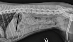
1. Normal variant
2. Porto-systemic shunt (younger)
3. Microvascular dysplasia
4. Cirrhosis (older)
What causes microhepatia?
intrahepatic gas from pneumobilia, hepatic abscess, or emphysematous cholecystitis in diabetic patients
What can cause a decrease in opacity of the liver?
mineralization from chronic inflammation, abscess, necrosis, or branching of the biliary system
What can cause an increase in opacity of the liver?
incidental finding of mineral opacities in the biliary tree
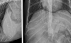
gallbladder mineralization (feline)
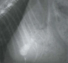
intrahepatic gas from an abscess (left) vs emphysematous cholecystitis (right)
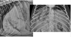
pneumobilia
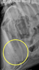
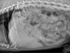
increased opacity and cranial displacement of the liver
13 Y- F- Golden retriever, rummaged through the trash (several chicken and fish bones) No vomiting, no changes in parameters or physical examination, except tachypenia. What is/are the Roentgen sign/s?
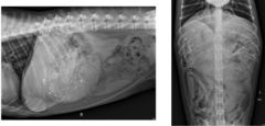
typically not seen
How does the pancreas appear on dog radiographs?
left lobe may be seen in obese cats
How does the pancreas appear on cat radiographs?
1. loss of serosal definition,
2. ground glass appearance in the pancreatic area
3. focal mineral opacitiies
4. mass effect with organ displacement of the descending duodenum, cranial displacement of the stomach, caudal displacement of the transverse colon
5. persistent gas dilation of regional intestinal segments
6. thickening, corrugation, or spasticity of the duodenum
What are the radiographic signs of pancreatitis?
normal angle of the pylorus duodenal junction with a normal sized pancreas
What is the orange line showing?
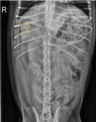
widened angle of the pylorus duodenal junction with an enlarged pancreas from pancreatitis
What is the orange line showing?
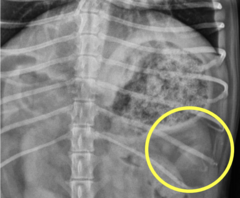
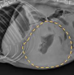
head of the spleen in a dog
What is in the yellow circle?