Radiology Chapters 38-42
1/297
There's no tags or description
Looks like no tags are added yet.
Name | Mastery | Learn | Test | Matching | Spaced | Call with Kai |
|---|
No analytics yet
Send a link to your students to track their progress
298 Terms
Angulation
Alignment of central ray of x-ray beam in horizontal and vertical planes
ALARA Concept
As low as reasonably achievable
Ampere
A unit of electric current
Anode
Positive electrode in the x-ray tube
Artifact
Blemish or unintended radiographic image that is not present in the actual structure
Atom
Basic unit of matter
Automatic processor
Machine that automates all film-processing steps
Beam alignment device
Assists in the positioning of the position indicator device
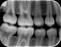
Bitewing
Image view that shows the crowns of both arches on one film
Bisecting (bisecting of the angle) technique
Intraoral technique of exposing dental images
Bitewing
Type of image used for interproximal examination
Bremsstrahlung radiation
“braking radiation”; the sudden deceleration of electrons as they interact with highly positively changed nuclei
Calcium tungstate
Common type of phosphor
Cathode
Negative electrode in the x-ray tube
Cassette
Contains extraoral films during exposure
Central ray
X-rays at the center of the beam
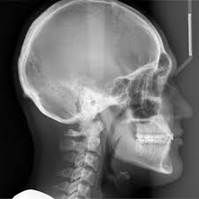
Cephalometric film
Shows the bony and soft tissue areas of the facial profile
Cephalostat
Special device that allows the operator to easily position both film and patient
Charged coupled-device (CCD)
Solid-state image sensor used in intraoral digital imaging
Computer tomography (CT)
Radiographic technique the produces images or “slices” of one layer or section of the specific area; also referred to as computed axial tomography
Cone beam computed tomography (CBCT)
Three-dimensional digital imaging method that uses a cone-shaped beam of radiation that rotates around the patient
Confidentiality
Never revealing any personal information about the patient
Contact area
Area of the mesial or distal surfaces of a tooth that touches an adjacent tooth in the same arch
Contrast
Differences in degrees of blackness on an image
Control panel
Portion of the x-ray unit that contains the master switch, the indicator light, the selector buttons, and the exposure button
Crestal bone
Coronal portion of alveolar bone found between the teeth
Density
Overall darkness or blackness of an image
Dental radiography
Process of recording images of the teeth and adjacent structures to x-radiation
Developmental disability
Impairment of mental or physical functioning that usually occurs before adulthood and lasts indefinitely
Diagnostic quality
Referring to images with the proper structures and necessary density, contrast, definition, and detail for diagnostic purposes
Digital image
Electronic signals captured by sensors and displayed on computer monitors
Digital imaging
Filmless method of capturing an image and displaying it by using an image receptor, an electronic signal, and a computer to process and store the image
Digital panoramic units
Filmless method of recording a panoramic image and displaying it by using an electronic sensor and a computer to process and store the image
Digitize
Scanning of tradition film-based radiographs into a digital image
Disclosure
Process of informing the patient about a procedure, for example, the procedure of taking x-rays
Distortion
The disproportionate charge in the size of images that is caused by excessive or insufficient vertical angulation
Dosimeter
Used to measure the amount of occupational exposure to ionizing radiation
Duplicating film
Film designed for use in a film duplicating machines
Emulsion
Coating on the x-ray film that contains energy-sensitive crystals
Extraoral film
Film designed for use in cassettes
Extraoral images
Images taken when large areas of the skull or jaw must be examined
Extraoral imaging
Image of the tooth and bones made by placing the film or cassette against the face or the head and projecting the x-rays from the opposite side
Film composition
Fast film speeds result in less sharp detail because of the large crystal size
Film speed
Sensitivity of the emulsion on the film to radiation
Field of view
Area that can be shown when performing imaging procedures
Focal trough (trof)
Imaginary three-dimensional horseshoe-shaped zone used to focus panoramic radiographs
Focal spot size
A machine with a small focal spot produces a sharper image than a machine with a larger focal spot size
Frankfort plane
Imaginary plane that passes through the top of the ear canal and the bottom of the eye socket
Image Receptor
A recording medium; examples include x-ray film, phosphor plate, or digital sensor
Informed consent
Permission granted by a patient after being informed about the risks, benefits, and alternatives of a procedure
Intensifying screen
Part inside an extraoral cassette that converts x-ray energy into visible light, which in turn exposes screen film
Interproximal
Between two adjacent surfaces
Intersecting
Cutting across or through
Intraoral film
Film designed for placement in the patient’s mouth
Ionization radiation
Harmful and produces biologic changes on living tissues
Kilovoltage
Highest voltage of x-ray tube used during an exposure
Label side
Colored side of the film that faces the tongue
Latent
Time between exposure to ionizing radiation and appearance of symptoms
Latent image
invisible image on the x-ray film after exposure but before processing
Lead apron
Device used to protect the reproductive and blood-forming tissues from scatter radiation
Liability
Accountability or legal responsibility
Long axis of the tooth
Imaginary line dividing the tooth longitudinally(vertically) into two equal halves
Magnification
Proportional enlargement of an image
Malpractice
Professional negligence
Matter
Anything that occupies space and has form or shape
Midsagittal plane
Imaginary line that divides the patient’s face into right and left side
Milliampere (MA)
One one-thousandth (1/1000) of an ampere; a unit of measurement used to describe the intensity of an electrical current
Movement
Any movement of the patient or image receptor (the film or sensor), no matter how slight, will degrade the sharpness of the image
Negligence
Failure to provide a proper or reasonable level of care
Occlusal
Radiographic view that shows larger areas of the maxilla or mandible
Occlusal technique
Used to examine large areas of the upper or lower jaw
Panoramic film
Used in cassettes to provide a wide-view of both the upper and lower jaws
Parallel
Moving or lying in the same plane, always separated by the same distance
Paralleling technique
Intraoral technique of exposing periapical and bitewing images
Penumbra
Blurred or indistinct area that surrounds an image
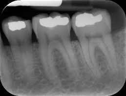
Periapical
Radiographic view that shows the crown, root tip, and surrounding structures
Perpendicular
Intersescting at or forming a right angle
Personal radiation monitoring badge
Device that measures exposure of personnel to ionizing radiation by measuring the intensity of visible light emitted from a crystal in the detector when heated; the intensity of light emitted depends on the radiation exposure
Photon
Minute (tiny) bundle of pure energy that has no weight or mass
Phosphor storage plate (PSP)
Reusable film-sized plated coated with phosphor as the image receptor
Physical disability
Impairment in certain function(s) of the body, such as vision, hearing, or mobility
Positioning instruments (device)
intraoral devices used to position and hold the film, sensor, or PSP
Primary beam
Most penetrating beam produced at the target of the anode
Primary radiation
Is made up of x-rays that come from the target of the x-ray tube. Primary radiation is often referred to as the useful beam , or primary beam
Quality (of x-ray beam)
Mean energy or penetrating ability of the x-ray beam
Quality assurance
Plan to ensure that the dental office produces consistent, high-quality images with a minimum of exposure to patients and personnel
Quality control tests
Specific tests used to ensure quality in dental x-ray equipment, supplies, and film processing
Quantity (of x-ray beam)
Number of x-rays produced in the dental unit; the quantity of x-rays produced is controlled by mA
Radiation
Forms of waves of energy emission through space or material
Radiograph
Image produced on photosensitive film by exposing the film to radiation and then processing it
Radiology
The science of study of radiation as used in medicine; a branch of medical science that deals with the therapeutic use of x-rays, radioactive substances, and other forms of radiant energy
Right angle
Angle of 90 degrees formed by two lines perpendicular to each other
Risk management
Policies and procedures that will reduce the chance that a malpractice lawsuit will be brought against the dentist; key areas of risk management include patient informed consent, patient records, confidentiality, liability issues, and patient education
Scatter radiation
Form of secondary radiation that occurs when an x-ray beam has been deflected from its path by interaction with matter
Secondary radiation
X-radiation that is created when the primary beam interacts with matter
Sensor
Solid-state image receptor that contains a silicon chip with an electric circuit
Sharpness
Measure of how well an image reproduces the fine details or outline of an object
Somatic effects
Effects of radiation that cause illness and are responsible for poor health (such as cancer, leukemia, and cataracts) but are not passed on to offspring
Standard of care
Level of knowledge, skill, and care comparable with that of other dentists who are treating similar patients under similar conditions
Stepwedge
Device constructed of layered aluminum steps to demonstrate film densities and contrasts