MLS 2213 Exam 1
 Call Kai
 Learn
Learn Practice Test
 Spaced Repetition
 Match
Match Flashcards
 Knowt Play
1/135

There's no tags or description
Looks like no tags are added yet.
Name | Mastery | Learn | Test | Matching | Spaced | Call with Kai | Chat |
|---|
No analytics yet
Send a link to your students to track their progress
136 Terms
Explain the parts of an amino acid
All have an Alpha Carbon, to which is bonded:
An Amine Group (---NH2)
A Carboxy Group (----COOH)
A Hydrogen
Another Thingy (H or something else) This Thingy is what makes each Amino Acid unique.
explain characteristics common to all amino acids
An Amine Group (---NH2)
A Carboxy Group (----COOH)
A Hydrogen
explain how the amino acids differ from each other
Another Thingy (H or something else) This Thingy is what makes each Amino Acid unique.
If the Thingy is:
H or just a Hydrocarbon, it is uncharged.
Contains another Carboxy Group, it is acidic and is negatively charged.
Contains another Amino or Nitrogen group, it is alkaline and is positively charged
explain the differences and similarities among single amino acids, tri-peptides, polypeptides, and proteins.
One all by it’s lonesome - single AA
Three bonded together = Tri-peptide
Many bonded together, but total molecular weight less than 10,000* are Polypeptides
More than one Polypeptide bonded together, or polypeptides > 10,000 make a Protein
Define primary structure proteins
Sequence of amino acids
Define secondary structure of proteins
Coiling of amino acids
Explain tertiary structure of protein
coiling causes one part to bond with another part
Explain quaternary structure of protein
Molecule consists of two or more different proteins
Explain the characteristics of albumins
True proteins, but consist entirely of amino acid chains.
Explain the characteristics of globulin proteins
AA chains forming true proteins, but contain non-AA constituents.
Not as soluble as albumins
Explain the characteristics of total proteins
Measuring all Albumins and Globulin proteins together.
Give an example of the different types of globulin proteins
AA Chains plus -----
Carbohydrates = Glycoproteins
Lipids = Lipoproteins
Metals = Metaloproteins
Funky Amine-Carbohydrates = Mucoprotei
What is the equation for A/G ratio
Albumin/globulin
What is the general chromatography theory
Separation of individual analytes from a mixture of two or more analytes.
Separation made between affinity for a stationary phase vs. a mobile phase.
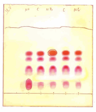
What is thin-layer chromatography
A technique used to separate non-volatile mixtures based on the adsorption of compounds to a stationary phase and their solubility in a mobile phase
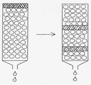
What is Column (liquid-liquid) chromatography
A chromatographic technique used for separating compounds in a liquid phase, where the stationary phase is a liquid coated on a solid support and the mobile phase is a liquid that carries the analytes through the column.
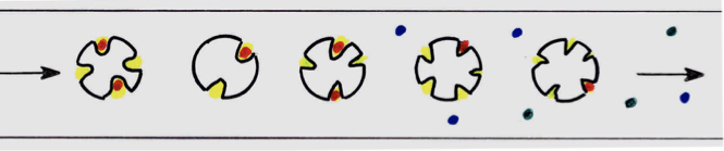
What is Gel filtration (gel permeation, molecular sieving, steric exclusion) chromatography
A technique used to separate molecules based on size, where smaller molecules are retained in the pores of the stationary phase while larger molecules pass through more quickly.
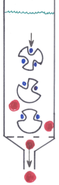
What is ion-exchange chromatography
A chromatographic method that separates ions and polar molecules based on their charge. In this technique, charged particles are exchanged between an ion-exchange resin and the solution being analyzed.
What is High Performance Liquid Chromatography (HPLC) (aka High Pressure Liquid Chromatography)
Like Liquid-Liquid chromatography, but performed under extremely high pressure.
Elution time is very fast (seconds to minute).
Once measured, analyte is recoverable.
Works for volatile and non-volatile analytes.
What amino acids are elevated in PKU
Totally lacking the enzyme that converts Phenylalanine (Phe) to Tyrosine (Phenylalanine Hydroxylase).
Blood levels of Phenylalanine build up and become toxic.
What are the symptoms of PKU
Phe blocks CNS and Nerve receptors used by Glutamic Acid
Both blood and urine high in Phe, Phenylpyruvic Acid and Phenlylactic Acid.
Causes severe seizures and mental retardation.
Treatment is to withhold Phe from diet for first 3 years (or longer) to avoid retardation and CNS damage.
What are the diagnostic tests used to diagnose PKU
Urine screened with Ferric Chloride test
turns blue/green
2,4-DNPH = turbid
Confirmatory is blood and urine quantitation or semi-quantitation by TLC and/or HPLC
At birth screening is the Guthrie Test
What amino acid is elevated in tyrosinosis and what enzyme is missing
Type 1: Lacking the enzyme to convert Tyrosine to it’s Pyruvate form (keto). or
Type 2:Lacking the enzyme to convert the Pyruvate form to Homogentisic Acid.
Type 1: Tyrosine Transaminase, or
Type 2:P-Hydroxy-Phenylpyruvate Hydroxylase
What are the symptoms of tyrosinosis
Deficiency 1 is the most severe, and causes severe Liver damage, enlarged Spleen. Usually fatal to infants.
What are the diagnostic tests used to diagnose tyrosinosis
The urine and blood have elevations in Tyrosine, p-OH Phenylpyruvic Acid and p-OH Phenyl-lactic Acid.
Urine is Ferric Chloride Positive, but with a very quick-fading green (Dude, like within a few seconds).
What are the amino acids that are elevated in maple syrup urine disease
Deficiency is Branched-Chain Keto Acid Decarboxylase
Leucine, isoleucine, and valine
What are the symptoms of maple syrup urine disease
The urine of these infants has an odor similar to burnt sugar or maple syrup.
Symptoms appear during first week of life
severe neurological developmental problems,
severe Mental Retardation,
Early Infant Death.
What are the diagnostic tests used to diagnose maple syrup urine disease
Urine is Ferric Chloride Positive, producing a Blue color.
(DNPH positive also).
What are the amino acids elevated in alkaptonuria
Deficiency is Homogentisic Acid Oxidase,
can’t convert HGA to Acetoacetic Acid and Fumaric Acid.
What are the symptoms of alkaptonuria
This doesn’t cause symptoms until age 20 to 40
causes arthritis, and black pigmentation of tissues containing large amounts of cartilage (ears, nose, etc.).
Very unusual finding is that the urine Darkens upon standing in light.
There are two conditions associated with urine that darkens with light:
Alkaptonuria or Elevated Porphobilinogen (PBG) (porphyrias, such as Acute Intermittent Porphyria)
What are the diagnostic tests used to diagnose alkaptonuria
Urine is Ferric Chloride Positive, producing a Transient Blue color. (DNPH positive also).
What amino acids are elevated in cystinuria
Cysteine, Cystine, Lysine, and Arginine (and Ornithine, but not an amino acid)
What are the symptoms of cystinuria
Hallmark is a baby that develops Kidney Stones.
What are the tests used to diagnose cystinuria
Urine Ferric Chloride test is Negative
no keto forms, because blood levels of AA are normal.
Urine Cyanide-Nitroprusside test is Positive.
What amino acid is elevated in Histidinemia
Histidase enzyme absent or low
can’t properly metabolize Histidine.
Histidine elevated in blood and urine, as well as Imidazole, Pyruvate and Lactate forms of Histidine.
What are the symptoms of Histidinemia
Symptoms variable,
normal patient,
speech defects, and/or
possible mental retardation.
What are the diagnostic tests used to diagnose Histidinemia
Urine Ferric Chloride Positive, and DNPH Positive.
What amino acid is elevated in Non-ketotic Hyperglycemia
Block in Glycine cleavage enzyme system.
Can’t properly metabolize Glycine.
What are symptoms of Non-ketotic Hyperglycemia
Patient suffers Seizures, Mental Retardation, Hypotonia.
Blood has low Osmotic Pressure, therefore cells of tissues swell, causing CNS and nerve damage.
Glucose elevates, but patient does not go into ketosis.
What diagnostic tests are used to diagnose Non-ketotic Hyperglycemia
Glycine and pyruvate/lactate forms elevate in blood, urine, & CSF.
What amino acid is elevated in Methylmalonic Acidemia
Deficiency of Methylmalonyl CoA Synthetase (partial or complete)
Glycine and Methylmalonic Acid elevated in Blood, Urine, and CSF, as are the pyruvate and lactate forms of Glycine.
Name the reagents used, the principles involved, and the clinical utility of the following protein-related test methods: Ninhydrin
Stains all amino acids some hue of pink to purple. Useful with TLC to visualize AA separations.
Most amino acids stain about the same red color, but some stain slightly different and are distinguishable by color
For example, Aspartic Acid is more Purple
Name the reagents used, the principles involved, and the clinical utility of the following protein-related test methods: Bromsresol green method (dye binding)
Most commonly-used serum Albumin reagent.
Albumin will bind BCG Dye. The complex is Green Colored.
Will not bind any globulin proteins.
Bromcresol Purple is a similar reagent.
Name the reagents used, the principles involved, and the clinical utility of the following protein-related test methods: Biuret method
Copper Sulfate (cupric form) in NaOH, KI (to keep in cupric form) and gobs of Na, K-Tartrate.
Cupric ion interacts with two adjacent amide bonds of a protein. There is an excess of Cu2SO4, so reaction is influenced by the number of amide bonds available (protein concentration).
Only acceptable for serum, will react with tri peptides and larger, will not react with single amino acids
Name the reagents used, the principles involved, and the clinical utility of the following protein-related test methods: Refractive index
Proteins influence the Refractive Index of light.
Special Protein ocular can be obtained and used with a refractometer, just like doing urine specific gravities.
Slow, manual method makes this not a popular method.
Name the reagents used, the principles involved, and the clinical utility of the following protein-related test methods: TCA method
Protein in small amounts, such as Urine and CSF, can be quantitated photometrically by precipitating them.
The amount of cloudiness from precipitation indicates protein concentration. Generally have linear ranges from 10 – 150 mg/dL.
TCA is trichloroacetic acid which is very caustic and used at a 13.5% solution
Name the reagents used, the principles involved, and the clinical utility of the following protein-related test methods: Sulfosalicylic acid method
Protein in small amounts, such as Urine and CSF, can be quantitated photometrically by precipitating them.
The amount of cloudiness from precipitation indicates protein concentration. Generally have linear ranges from 10 – 150 mg/dL.
SSA is sulfosalicylic acid which is less caustic with a pH around 3 usually used in a 10% solution
Name the reagents used, the principles involved, and the clinical utility of the following protein-related test methods: Coomassie Blue (dye binding)
Gets it’s name because the reagent comes in concentrated form, and it’s Red! Actually, the working reagent is faintly Blue-Yellow and turns Blue when protein binds it (albumin and globulins).
This is our most sensitive method of measuring extremely low levels of protein, especially in urine (for Microalbuminuria testing).
Linear range can go down to 5 or 3 mg/dL.
Why can you not use Bieret method for protein urine or CSF?
not suitable for urine or CSF protein quantitation because it requires a higher protein concentration than typically found in these fluids.
What three methods of assay can be used to test protein in CSF and urine
SSA, TCA, coomassie blue
What causes elevated protein in CSF
Meningitis
Encephalitis
MS
SAH
What can cause elevated protein in Urine
Acute glomerulonephritis
Acute pyelonephritis
Nephrotic syndrome (primary)
Multiple myeloma
What can cause microalbuminuria
Diabetes
What protein and glucose levels would you expect to see in bacterial meningitis
High protein low glucose
What protein and glucose levels would you expect to see in viral meningitis
High protein normal glucose
What protein and glucose levels would you expect to see in chemical meningitis
CSF Protein slightly elevated with a normal CSF Glucose – must rule-out viral causes.
What is electrophoresis
Separation of individual molecules from a mixture in an electrical field.
Separation is made by migration according to Size and Electrical Charge.
outline steps of cellulose Acetate Electrophoresis
A strip of cellulose is soaked in buffer pH 8.6 - 8.8
sample is applied
an electric current is applied, the proteins move towards anode
proteins separate by size and charge
Summarize the following in serum protein Electrophoresis: The buffer used and the pH.
Verinal buffer (buffered barbutyric acid)
pH - 8.6 to 8.8
Summarize the following in serum protein Electrophoresis: The qualitative electrical charge of the proteins.
If the buffer is added then the proteins become negatively charged
Summarize the following in serum protein Electrophoresis: The direction of protein migration.
The proteins migrate towards the anode due to their negative charge.
Summarize the following in serum protein Electrophoresis: The protein fraction that migrates the fastest.
Albumin is the fastest migrating protein fraction in serum protein electrophoresis.
Explain the advantages and/or disadvantages of the following support media, as compared to cellulose acetate protein electrophoresis: Agarose Gel Electrophoresis:
most commonly used, able to be scanned. Gel has limited stability, it will dry out and is fragile
Explain the advantages and/or disadvantages of the following support media, as compared to cellulose acetate protein electrophoresis: Polyacrylamide Gel Electrophoresis:
used for extremely high resolution for things like HIV antibodies and antigens. Media is colored, and can’t be scanned
Explain the advantages and/or disadvantages of the following support media, as compared to cellulose acetate protein electrophoresis: Capillary Electrophoresis
newest of the EP techniques. Capillary is made of fused Silica. Capillaries are 20 – 200 um (up to 0.2 mm) thick, and 10 – 200 cm long.
Empty capillaries are negatively charged. Sample is added at the cathode. Both ends of the capillary are immersed in buffer chamber.
Separation made under very high voltage, and takes only a minute to complete. Heat buildup is not a problem.
Explain the advantages and/or disadvantages of the following support media, as compared to cellulose acetate protein electrophoresis: Immunoelectrophoresis (IEP)
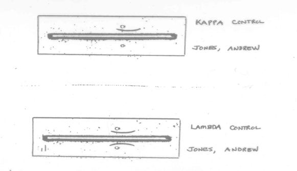
An old method (since the 1950’s), but gives very good qualitative separation of proteins. Possible to isolate each individual protein.
A trough is cut out of the agar between the patient sample and control. Anti-sera is added to the trough and allowed to diffuse into the agar. When antibody diffuses to protein and reacts with it, a visible precipitin band forms. in heaven, every precipitin band is unique to every protein – to the trained eye (with a Voodoo Doll!).
Explain the advantages and/or disadvantages of the following support media, as compared to cellulose acetate protein electrophoresis: Immunofixation Electrophoresis (IFE)
Sorta like the second-generation IEP, and is much faster (4 hours vs. 24 hours).
Support media is buffered gel. Samples and controls are applied to sample wells at cathode end of plate.
Proteins migrate towards anode in the electrical field.
Then, anti-sera is poured on top of gel, and precipitin reactions happen within an hour (no diffusion step). Like IEP, each protein has its own precipitation band.
Explain the advantages and/or disadvantages of the following support media, as compared to cellulose acetate protein electrophoresis: Isoelectric Focusing (IEF)
Substrate is unbuffered gel.
Patient serum is added to patient well at anode side of plate, and mixed with a gob of Ampholytes. (Poly-carboxy hydrocarbon chains). Current is applied.
The ampholytes move in the current faster than the patient proteins, and they establish a pH gradient.
The proteins migrate until they reach the pH that equals their pI, and then stop migrating (and focus into tight bands).
What are the five (5) major serum protein fractions obtained by Cellulose Acetate Electrophoresis (CAE), or Agarose Gel Electrophoresis (AGE),
albumin, alpha-1 globulins, alpha-2 globulins, beta globulins, and gamma globulins.
What proteins are alpha-1 globulins
A1Anti-Trypsin (A1AT) makes up 90% of this group, is a Protease Inhibitor, and one of the Acute Phase Proteins.
Alpha Lipoproteins (HDL)
A1Acid Glycoprotein (AAG) an Acute Phase Protein
Some other Glycoproteins and Mucoproteins
A1Fetoprotein when present (embryonic protein, can be a cancer marker).
What proteins are in alpha-2 globulins
A2 Macroglobulin - plasmin and protease inhibitor, an Acute Phase protein, It is among the largest proteins, and is the slowest to be lost in. Nephrotic Syndrome.
Haptoglobin -binds free Hgb, different protein after binding Hgb. is an acute phase protein, Decreased in Hemolytic conditions
Pre-Beta Lipoproteins -VLDL – carries mainly Triglycerides.
Ceruloplasmin - an acute phase protein, carries Copper. Decreased in Wilson’s Disease.
What proteins are beta globulins
Hemopexin - binds free heme groups
Transferrin - transport for Iron, NOT an acute phase protein, drops in inflammation.
B2-Microglobulin - Very small (~ 12,000 Daltons), Gets filtered at glomerulus, but reabsorbed by proximal tubule. in tubule problems, protein is lost in urine.
C-Reactive Protein (CRP) - An acute phase protein.
Complement C-3 and C-4.
Fibrinogen - only if plasma is used – absent in serum
Beta Lipoproteins - LDL – mainly carrying Cholesterol
What proteins are gamma globulins
Immunoglobulins (antibodies) I
gG – Most abundant antibody IgM – First antibody to be produced IgA – Usually a secretory antibody IgE – Elevated in allergies, internal parasites. IgD - ?????
Explain electrophoresis migration rate
The bigger, the slower moving.
The more charged, the faster moving.
The higher the buffer’s Ionic Strength, the slower moving. - High Ionic Strength opposes migration by causing Electroendosmosis.
What is wick-flow and how do you avoid it?
During electrophoresis, heat is built up.
Heat causes excessive evaporation, which will then cause Wick-Flow.
Wick-Flow screws up separation.
Ya gotta minimize the heat! Yup, we also use ice cubes or refrigerated cells, but we still get some heat problems.
What is electroendosmosis and how do you avoid it?
High Ionic Strength opposes migration by causing Electroendosmosis.
use the correct buffer
What is tailing and how is it avoided?
Low Ionic Strength causes migration to increase, but you loose ability to isolate tight bands of proteins.
High Ionic Strength reduces Tailing, but reduces migration
you don’t get good separation of 1 protein from another.
Good method has Ionic Strength optimized for specific sample type.
Explain the advantages of a Constant-Current power supply for electrophoresis, as compared to Constant-Voltage power supply.
Using Constant Voltage tends to create excessive heat (heat continually increases as current amperage increases).
Using Constant Current minimizes heat production. Current remains the same, resistance remains the same, heat production stabilizes – Recommended mode.
Explain how to identify each of the following electrophoresis problems, and describe the correct problem-solving steps for each of the following electrophoresis problems: improper buffer
if pH wrong, affects charge, and direction of migration.
If Ionic Strength wrong, affects migration and separation.
Explain how to identify each of the following electrophoresis problems, and describe the correct problem-solving steps for each of the following electrophoresis problems: unsatisfactory staining
If stain washes off during 5% Acetic Acid washes, the stain is too old.
If it fails to stain, too little sample was applied.
Explain how to identify each of the following electrophoresis problems, and describe the correct problem-solving steps for each of the following electrophoresis problems: improper sample application
If sample was over-applied, scanned Albumin will be Low, and Globulin fractions will be High.
If sample was under-applied, scanned Albumin will be High, and Globulin fractions will be Low.
Not priming with conditioning application is usual cause.
Explain how to identify each of the following electrophoresis problems, and describe the correct problem-solving steps for each of the following electrophoresis problems: discontinuities in sample application
Applicator wires bent = Bow Tie patterns
Applicator wires bent = Won’t pick-up correct amount of sample.
Explain how to identify each of the following electrophoresis problems, and describe the correct problem-solving steps for each of the following electrophoresis problems: distortion of protein zones
Dirt on Membrane.
Strip didn’t hydrate evenly.
Not sufficient blotting prior to applying sample.
Too much blotting dries out membrane.
Wiggling during sample application.
Explain how to identify each of the following electrophoresis problems, and describe the correct problem-solving steps for each of the following electrophoresis problems: artifacts
Buffer contaminated with bacteria.
Bent applicator wires
Membrane allowed to dry-out
Membrane blotted too much
Explain how to identify each of the following electrophoresis problems, and describe the correct problem-solving steps for each of the following electrophoresis problems: cellulose acetate or gel membranes wont clear
Typical of old membranes.
Jayson Clarke completely dehydrate the plates prior to clearing solution.
Didn’t use fresh Methanol in both dehydration steps.
Clearing solution too old.
Not long enough in the clearing solution
Explain what special sample preparation steps must be taken to perform Urine Protein electrophoresis and CSF protein electrophoresis.
Centrifuge to remove swimmies and floaties, and other artifacts.
Concentrate the sample using Osmosis chambers (about 50x - 100x concentration)
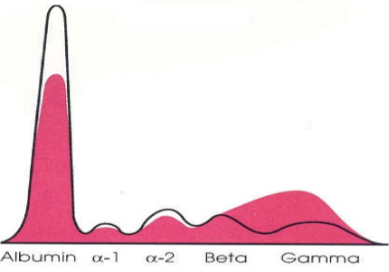
Explain what is meant by a Monoclonal and Polyclonal spike, with reference to serum protein electrophoresis.
Monoclonal Spike - One single protein is being produced in abundance. Typical of a few malignancies and Multiple Myeloma.
Polyclonal Spike - A broader spike that represents a bunch of proteins being produced at the same time (like a gob of different antibodies)
Evaluate serum protein electrophoresis patterns and differentiate from the normal pattern for the following diseases: Chronic liver disease or hepatic cirrhosis
Not making normal proteins
classic beta-gamma bridge
Evaluate serum protein electrophoresis patterns and differentiate from the normal pattern for the following diseases: Multiple Myeloma (heavy and light chain disease)
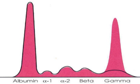
Evaluate serum protein electrophoresis patterns and differentiate from the normal pattern for the following diseases: Nephrotic syndrome or nephrosis
High alpha2 macroglobulin
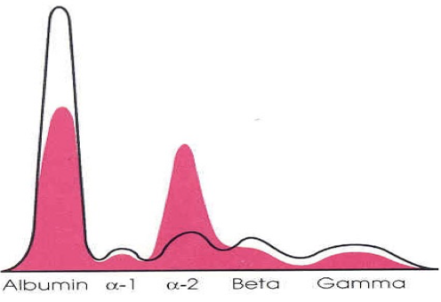
Evaluate serum protein electrophoresis patterns and differentiate from the normal pattern for the following diseases: acute inflamation
high AAT and haptoglobin
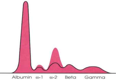
Evaluate serum protein electrophoresis patterns and differentiate from the normal pattern for the following diseases: chronic inflamation
under production of albumin
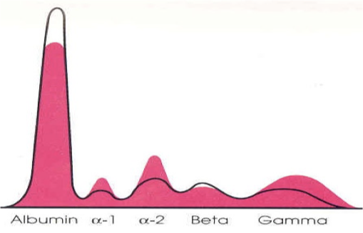
What serum proteins are acute phase proteins
Alpha 1 anti-trypsin
Alpha 2 macroglobulin
ceruloplasmin
crp
Alpha acid glycoprotein
haptoglobin
What is haptoglobin
A2Globulin that binds free Hemoglobin.
When bound to Hemoglobin, it is a whole different protein complex and migrates in the Beta region.
Jayson Clarke thinks that in Hemolytic conditions Haptoglobin is decreased. t’s still there, it just migrates somewhere else.
What diseases are assocated with an abnormal haptoglobin
Decreased in hemolytic conditions
What is alpha-1 antitrypsin
An acute phase alpha-1 protein
3 different alleles M,S and Z, 90% of people are MM. 1% have ZZ which makes undetectable levels of AAT
Levels are increased during inflammation
What diseases are assocated with an abnormal alpha-1 antitrypsin
Pre-mature emphysema
What is alpha-fetoprotein
Produced by fetal liver.
Should not be detectable 5 weeks after birth.
Detectable in the mother during pregnancy
What diseases are assocated with an abnormal alpha-fetoprotein
Primary Liver Cancer
Cirrhosis
Hepatitis B and C
Testicular carcinomas (some types)
Detectable in the mother during pregnancy.
Maternal blood tested at end of first trimester as a screen for Anencephaly, Spina Bifida, Myelocele, multiple fetuses.
What is ceruloplasmin
An acute phase A2 globulin that carries Copper.
Is an active catalase enzyme.
It increases in inflammation, and pregnancy.