eye movements II
1/18
There's no tags or description
Looks like no tags are added yet.
Name | Mastery | Learn | Test | Matching | Spaced | Call with Kai |
|---|
No analytics yet
Send a link to your students to track their progress
19 Terms
state the 6 extraocular muscles that eye movements are controlled by
–Medial and lateral rectus.
–Superior and inferior rectus.
–Superior and inferior oblique.
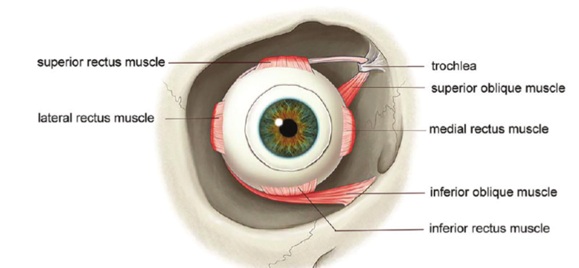
recti muscles include just the first 4 (recti = straight) / oblique muscles are the last 2 (oblique = angled)
where do the muscles attach to in the eye
The muscles attach at the apex of the orbit with the exception of the inferior oblique which arises anteriorly
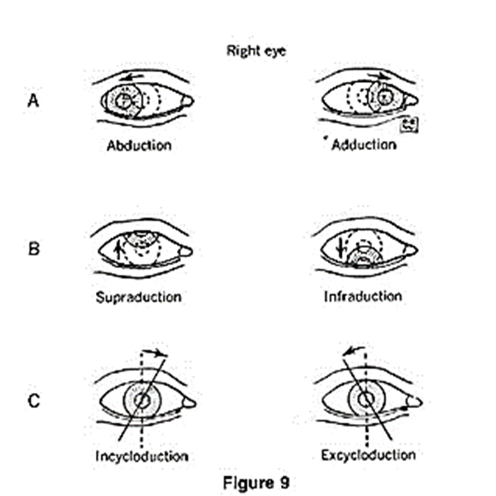
what are ductions and describe the different types of ductions (1:4)
ductions = Rotations of a single eye
–Abduction - turns eye away from the nose (abducting someone is taking them away)
–Adduction - turns eye towards the nose (add - adding)
–Sursumduction (elevation)
–Deosursumduction (depression) d for depression
what are torsions and describe the different types (1:2)
torsions = Rotations about the anterior-posterior axis
–Intorsion (incycloduction) – top of the eye towards the nose - INtorsion so IN towards the nose
–Extorsion (excycloduction) – top of the eye away from the nose - EXtorsion so exiting so AWAY from the nose
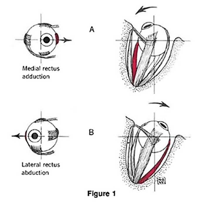
explain the medial rectus (MR) - role, movement and innervation (3)
–Responsible with the lateral rectus for horizontal eye movements.
–Turns the eye towards the nose – adduction.
–Innervated by the IIIrd cranial nerve (oculomotor nerve).
explain the lateral rectus (LR) - role, movement and innervation (3)
–Responsible with the medial rectus for horizontal eye movements.
–Turns the eye away from the nose – abduction.
–Innervated by the VIth (6th) cranial nerve (abducens nerve).
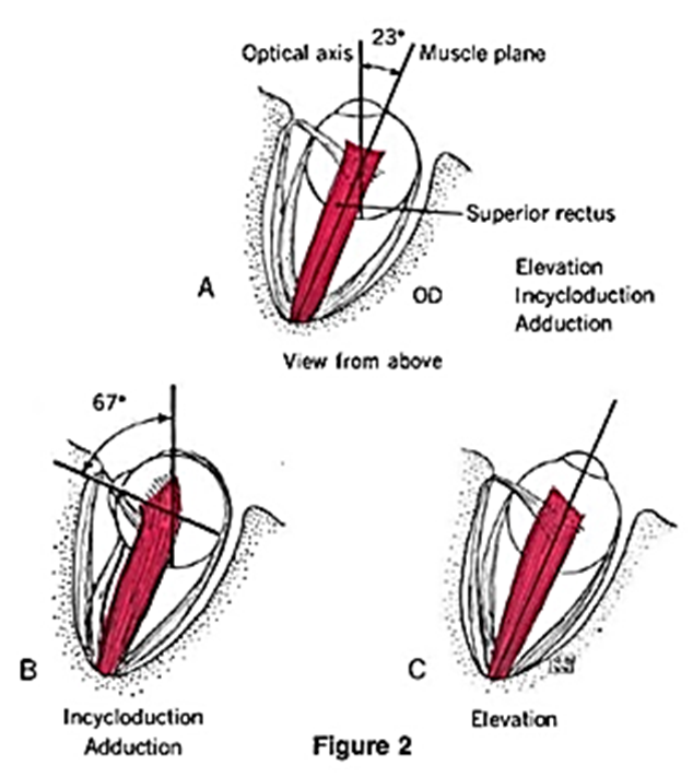
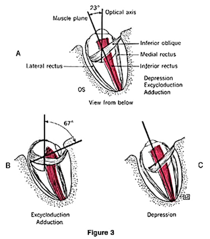
explain the superior rectus (SR) - innervation, eye movements, position and angle (6)
–The superior rectus passes forwards and outwards, at an angle of about 23°, between the optical axis and the muscle plane (Fig A).
–In primary position (straight ahead) action = elevation of the eye (Fig A)
–Secondary actions (Fig B) = adduction (turning the eye towards the nose)
- Tertiary action = intorsion (rolling the top of the eye towards the nose – also called incycloduction).
–When the eye is abducted (~ 23°) (i.e. away from the nose), the superior rectus becomes a pure elevator (Fig C).
–Innervated by the IIIrd cranial nerve (oculomotor nerve).
explain the inferior rectus (IR) - innervation, eye movements, position and angle (6)
–The inferior rectus passes forwards and outwards beneath the globe, at an angle of about 23°, between the optical axis and the muscle plane (Fig A).
–primary position (straight ahead) action = depression of the eye (Fig A)
–Secondary actions (Fig B) = adduction (turning the eye towards the nose)
- Tertiary action = extorsion (rolling the top of the eye away from the nose – also called excycloduction).
–When the eye is abducted (~ 23°) (i.e. away from the nose), the inferior rectus becomes a pure depressor (Fig C).
–Innervated by the IIIrd cranial nerve (oculomotor nerve)
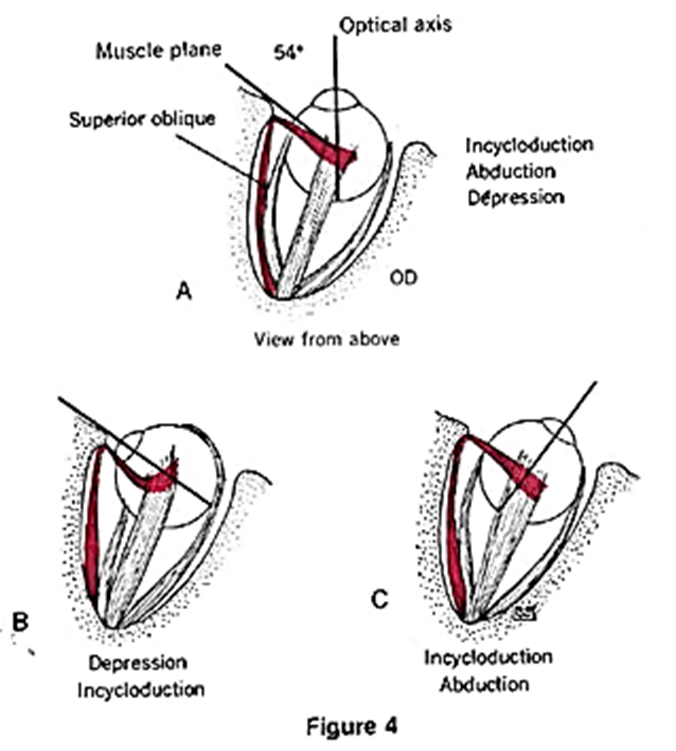
explain superior oblique (SO) - innervation, position and angle, actions (5)
–superior oblique passes near the roof of the orbit, almost to the margin, passing through the trochlear (a small ‘U’ shaped fibro-cartilage structure) and is reflected backwards and outwards at an angle of about 54° between the optical axis and the muscle plane (Fig A).
–In the primary position (straight ahead) action = intorsion (rolling the top of the eye towards the nose – also called incycloduction) of the eye (Fig A).
–Secondary actions (Fig B) = abduction (turning the eye away from the nose) and depression.
–When the eye is abducted (i.e. away from the nose), the superior oblique produces primarily intorsion and secondarily abduction (Fig C).
–Innervated by the IVth cranial nerve (trochlear nerve)
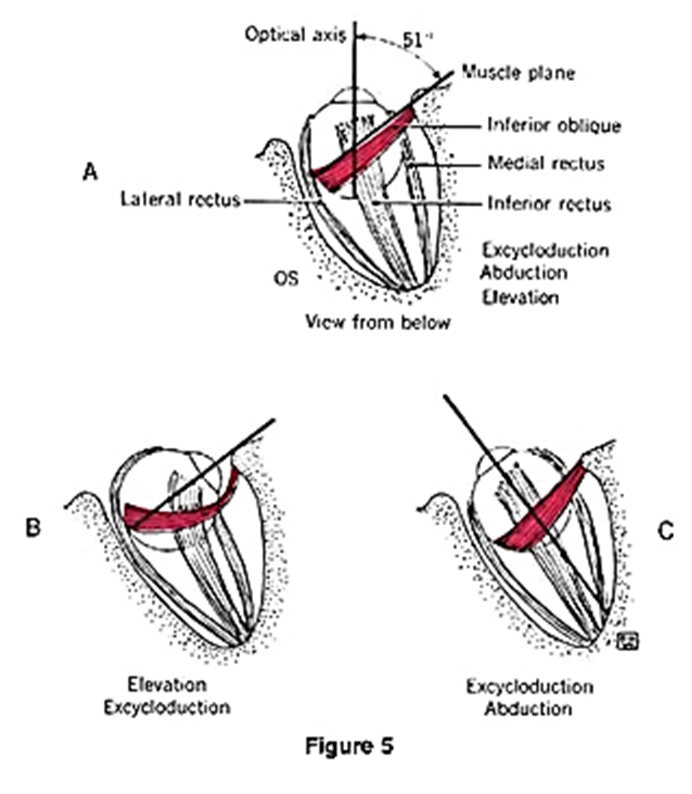
explain inferior oblique (IO) - innervation, position and angle, actions (5)
–inferior oblique is the only muscle that does not arise from the apex of the orbit, rather from the lower orbital margin on the nasal side. The inferior oblique creates an angle of about 51° between the optical axis and the muscle plane (Fig A).
–primary position (straight ahead) action = extorsion (rolling the top of the eye away from the nose – also called excycloduction) of the eye (Fig A)
–Secondary actions (Fig B) = abduction (turning the eye away from the nose) and elevation
–When the eye is adducted (i.e. towards from the nose), the main action of the inferior oblique is extorsion and secondarily abduction (Fig C).
-Innervated by the IIIrd cranial nerve (oculomotor nerve)
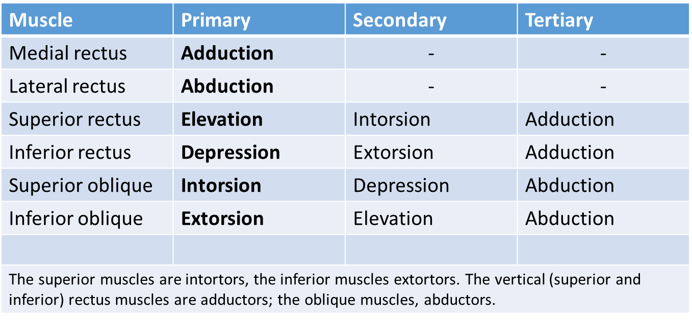
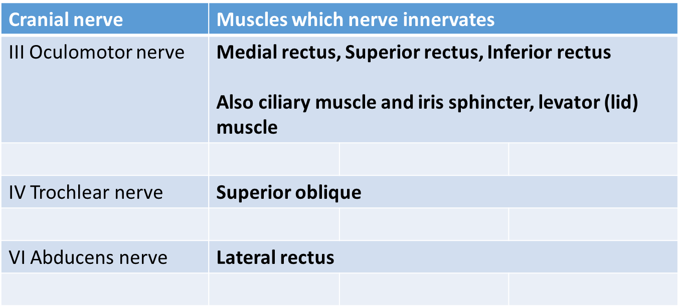
summary table of the muscle actions
 |
summary table of the innervations
 |
define agonist
this is the muscle responsible for the movement.
define antagonist
•produces a movement in the direction opposite to the agonist
give an example of agonist/antagonist muscle pairs in relation to the extraocular muscles
Medial rectus adducts the globe while the lateral rectus abducts: the medial and lateral rectus muscles are antagonists
explain sherrington’s law (2)
Whenever an agonist muscle receives a neural impulse to contract, an equal signal is sent to the antagonist causing it to relax.
This is Sherrington's Law of reciprocal innervation, which suggests that the state of contraction in the agonist regulates the state of contraction in the antagonist - power/strength of one contraction will determine the power of the other contraction in the other muscle
in any one eye movement how many of the extraocular muscles are involved
all 6
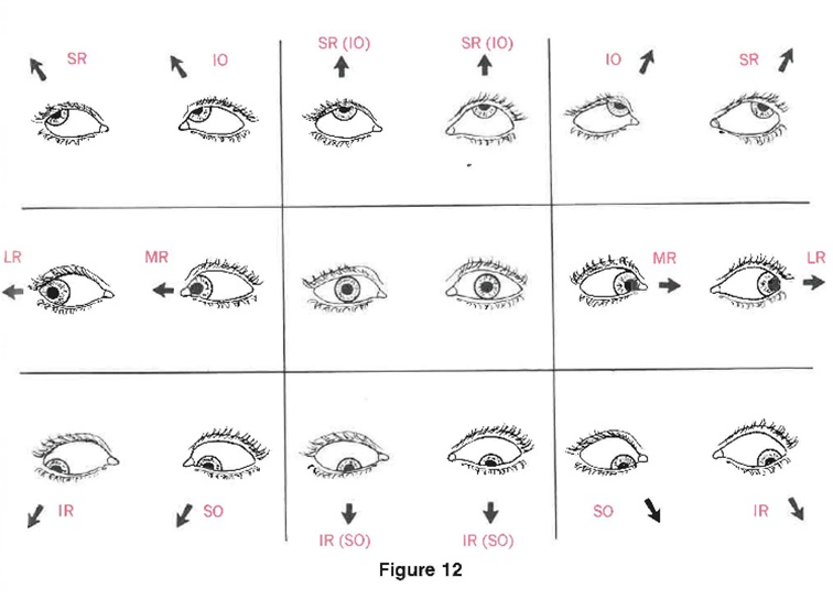
how do we test whether a particular muscle is working properly
•the examiner asks the patient to move into the direction where the muscle has its maximal action.
•For each cardinal position, there is a pair of muscles (agonists) that have the greatest impact.
give an example of when testing ocular mobility forms an essential part of the ocular examination
particularly for patients presenting with an eye turn – strabismus