A&P II - Chapter 19: Circulatory System (Heart) - Smartbook Questions
1/158
There's no tags or description
Looks like no tags are added yet.
Name | Mastery | Learn | Test | Matching | Spaced |
|---|
No study sessions yet.
159 Terms
What is the study of the heart and its disorders called?
Cardiology
The muscular pump that keeps blood flowing through blood vessels is the ______.
Heart
The BLANK circuit carries blood from the right ventricle to the lungs for gas exchange and returns it to the left atrium of the heart.
Pulmonary
As the heart sits in the thoracic cavity, the superior tip end called the ______.
Base
The outer wall of the pericardium is a sac called the ______.
fibrous pericardium
Cardiology involves the study of the ______ and its disorders.
Heart
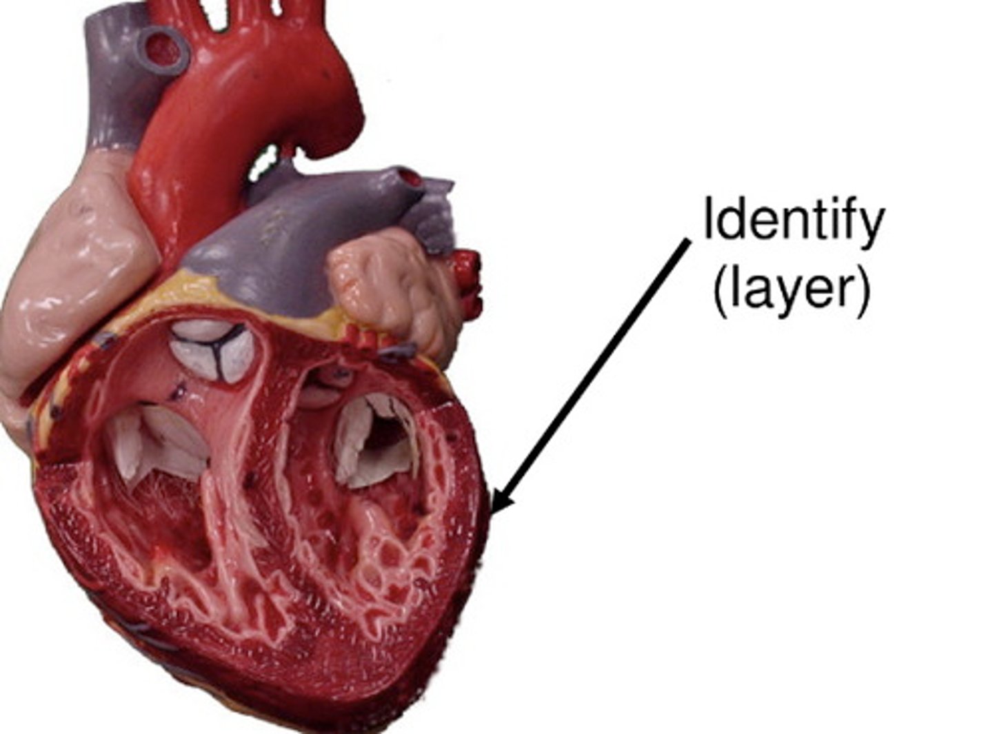
Which are layers of the heart wall?
Epicardium
Endocardium
Myocardium
True or false: The cardiovascular system includes the heart and blood vessels.
True
Which type of epithelium makes up part of the endocardium?
Simple squamous
Which circuit carries blood from the right ventricle to the lungs for gas exchange and returns it to the left atrium of the heart?
Pulmonary
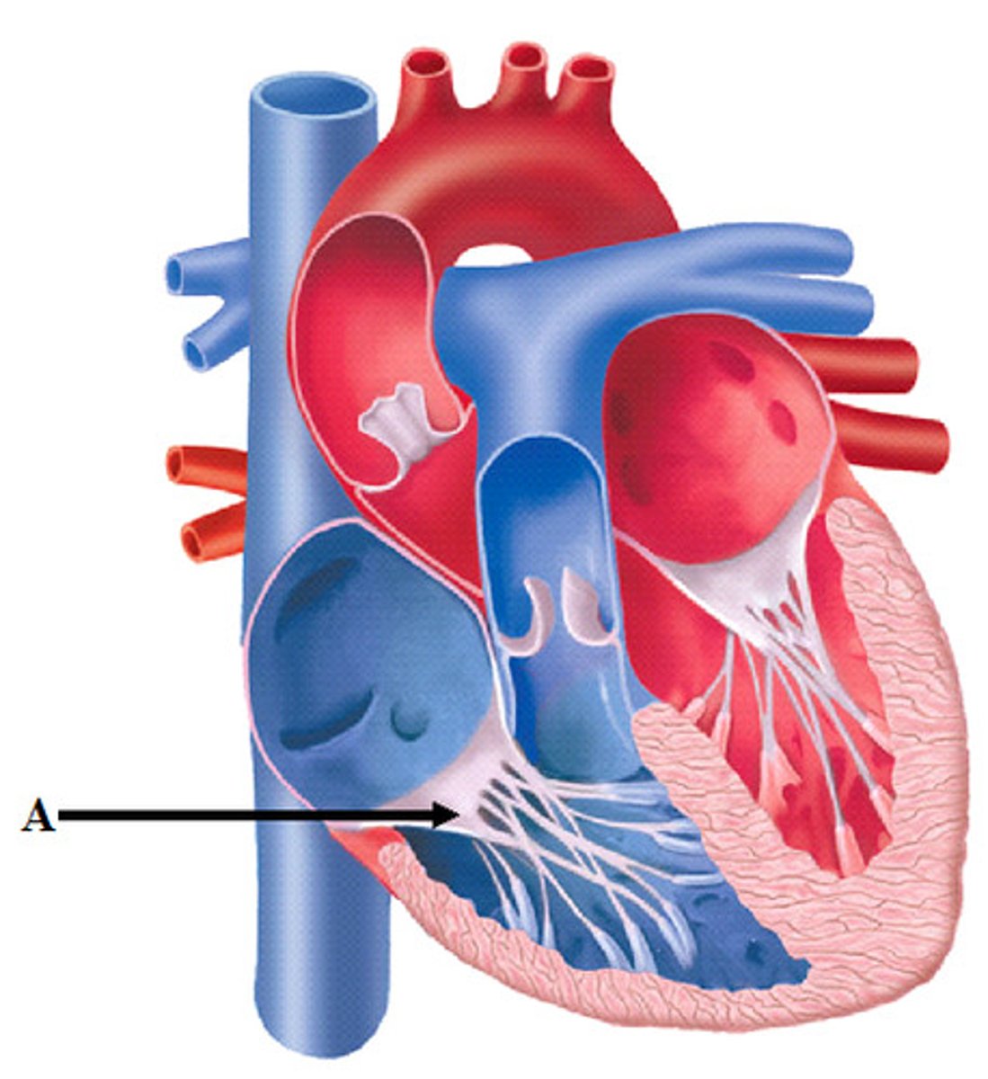
The structure indicated by the letter A in the figure is the BLANK pericardium
Fibrous Pericardium
Place in order the three layers of the heart wall, listing the deepest layer first. (Deepest to surface layer)
1. Endocardium
2. Myocardium
3. Epicardium
Which layer lines the inner chambers of the heart?
endocardium
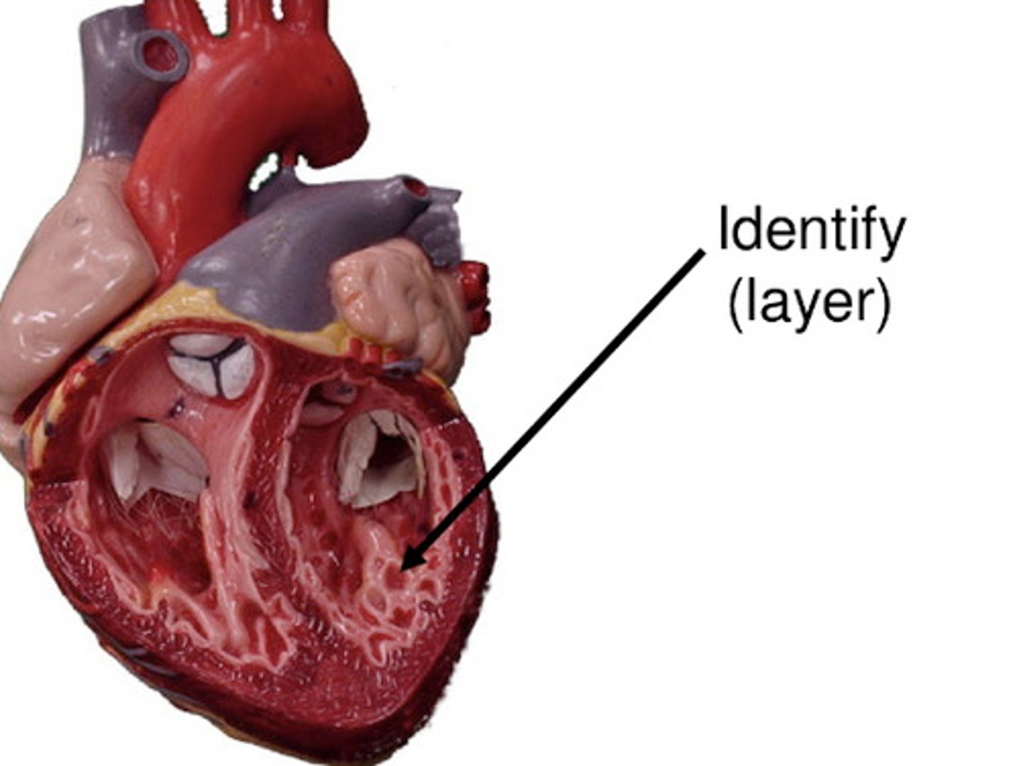
What is the outermost layer of the heart wall called?
Epicardium
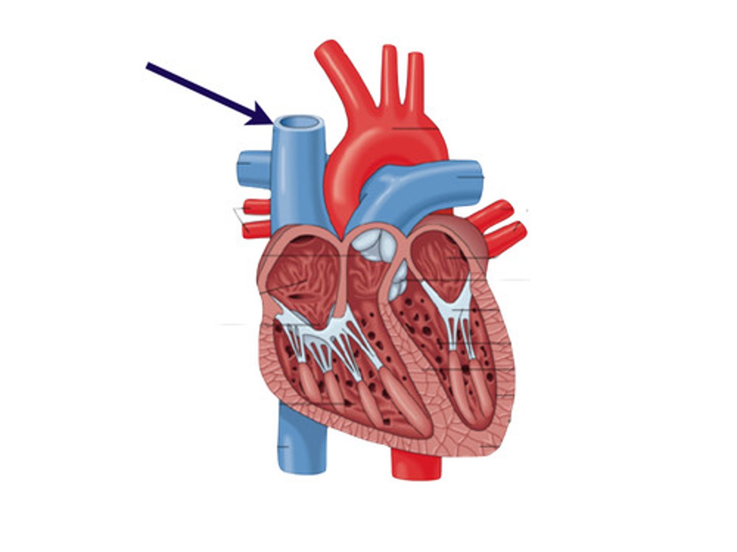
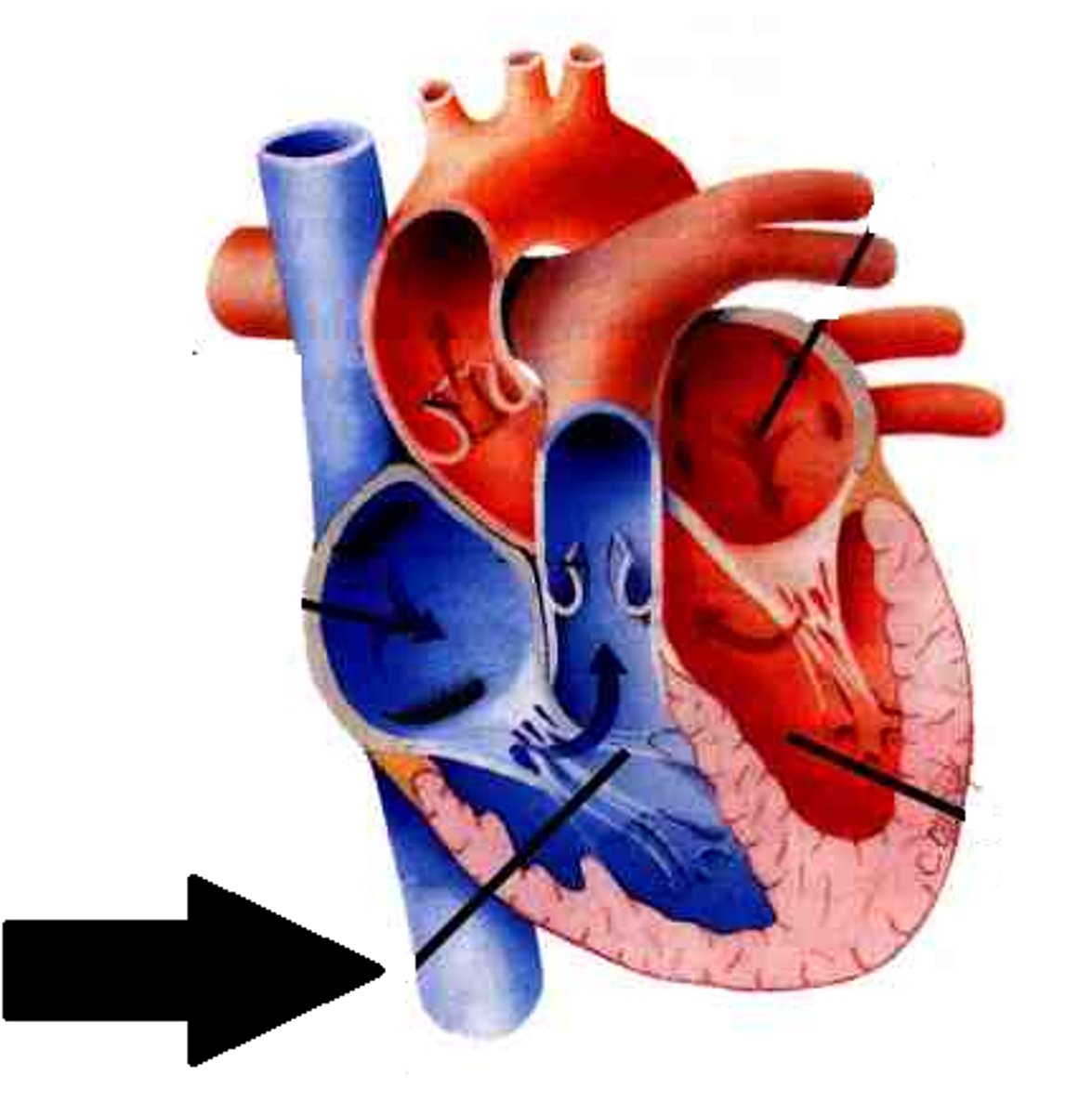
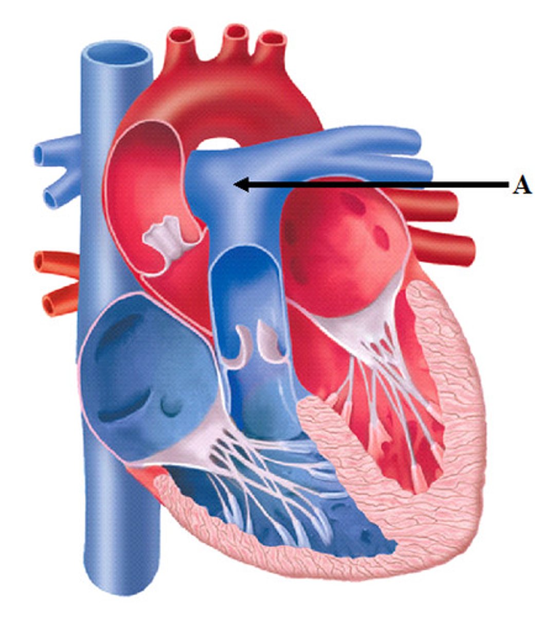
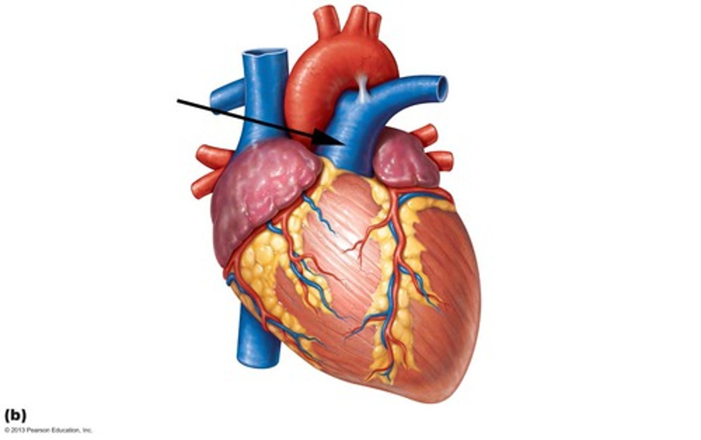
Which blood vessel is indicated by the letter A in the figure?
Superior Vena Cava
The broad superior part of the heart is the BLANK of the heart.
Base
Which blood vessel is indicated by the letter A in the figure?
Inferior vena cava
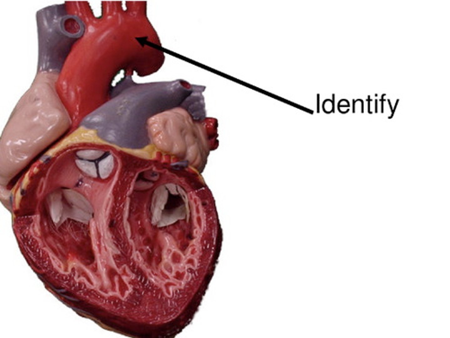
The indicated vessel is a segment of the aorta known as the BLANK arch
Aortic Arch
Another name for the epicardium is the BLANK layer of the serous pericardium.
Visceral
Which is a blood vessel attached to the right atrium?
Superior Vena Cava
As the heart sits in the thoracic cavity, the superior tip end called the ______.
Base
The epicardium is also called what?
Visceral layer of the serous pericardium
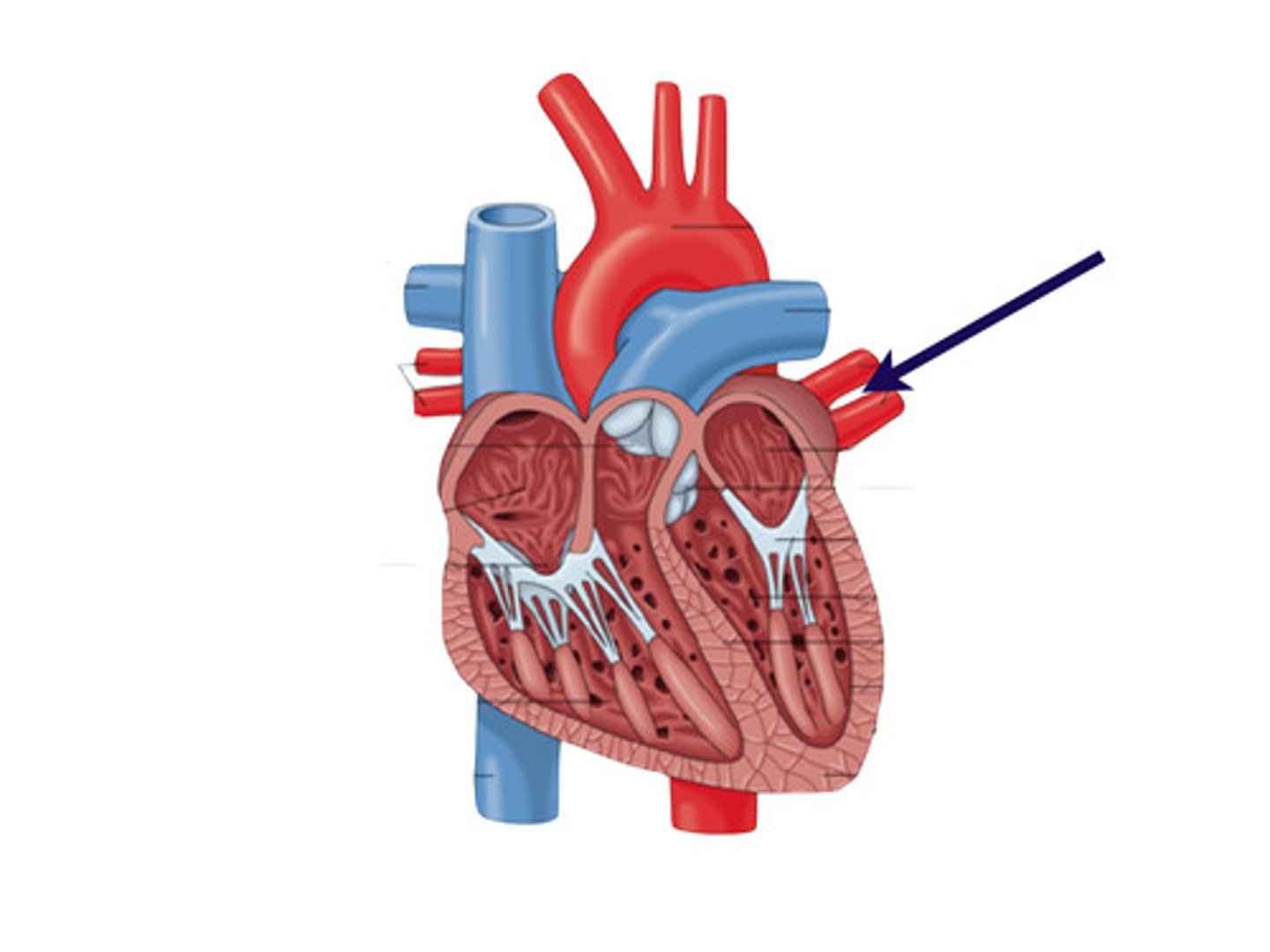
Which blood vessel is a branch off the pulmonary trunk?
Pulmonary artery
The blood vessel that carries blood away from the left ventricle is the ______.
Aorta
an ear-like extension of the heart chamber
auricle
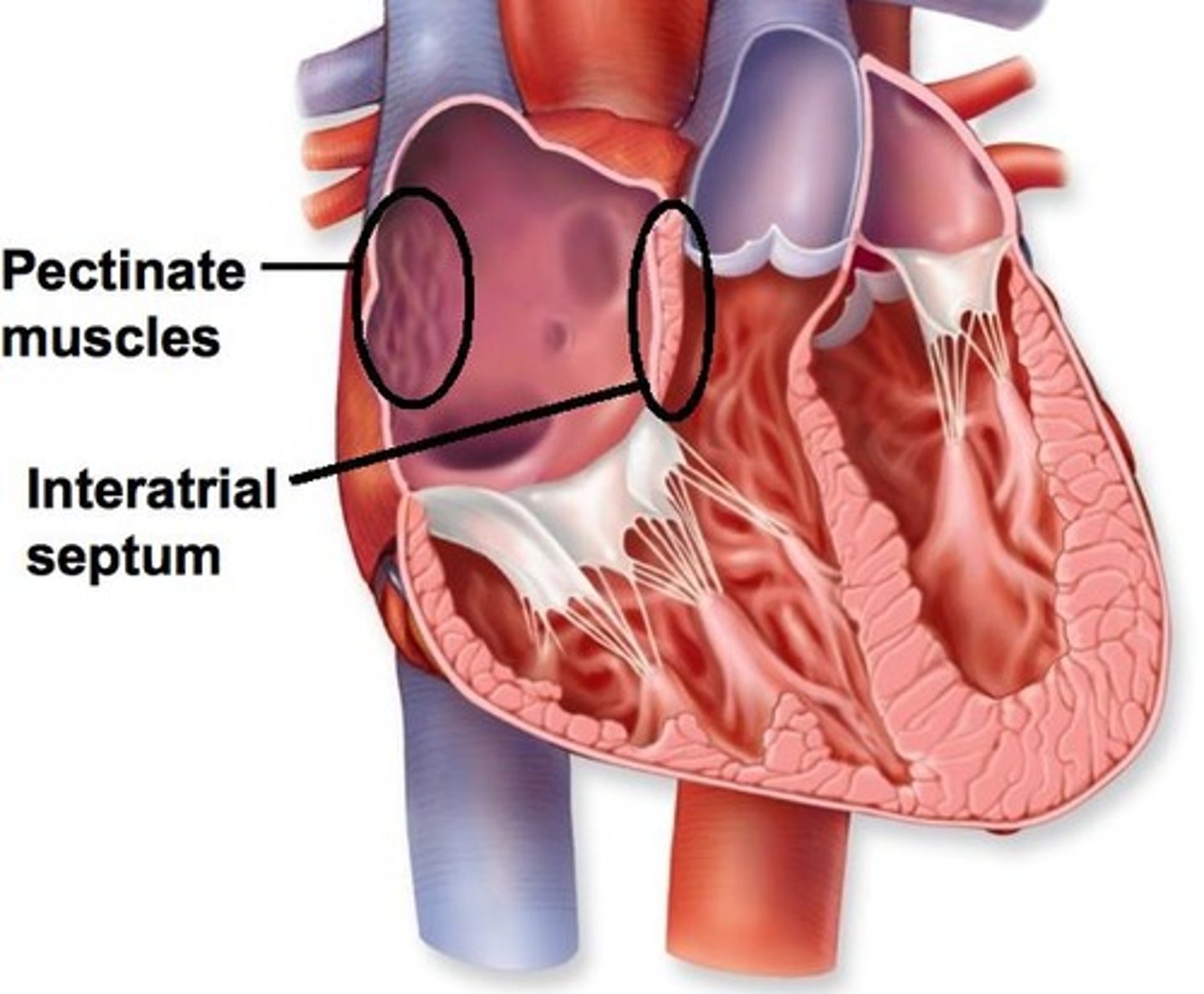
Pectinate muscles
internal ridges of myocardium in right atrium and both auricles
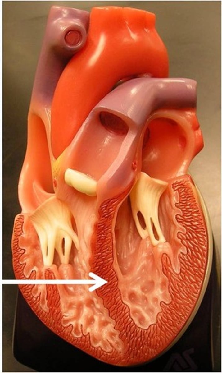
interventricular septum
partition between the right and left ventricles
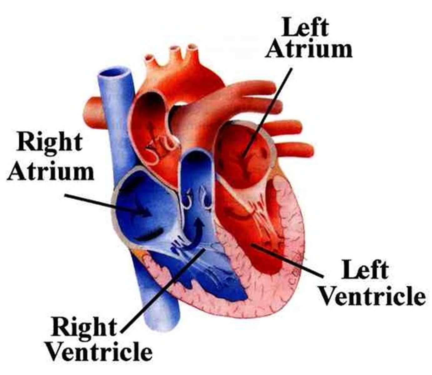
right atrium
receives deoxygenated blood from the body through the vena cava and pumps it into the right ventricle which then sends it to the lungs to be oxygenated.

The blood vessel that carries blood away from the left ventricle is the ______.
Aorta
pulmonary artery
artery carrying oxygen-poor blood from the heart to the lungs
Right Ventricle
pumps oxygen poor blood to the lungs
Pulmonary arteries are branches of the ______.
pulmonary trunk
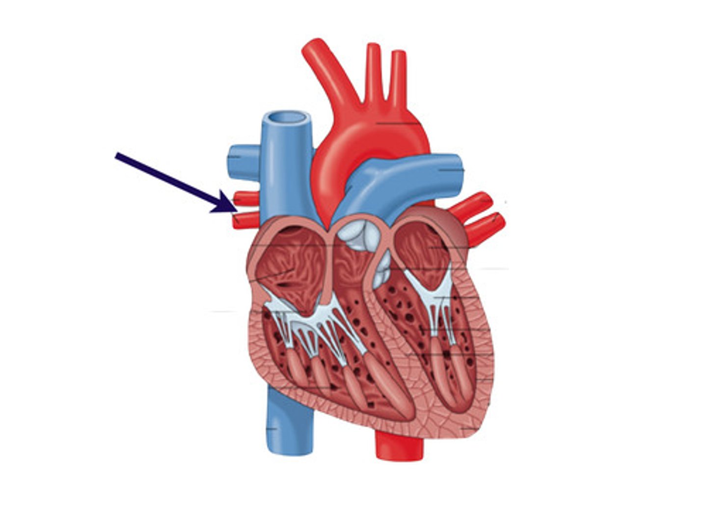
left pulmonary veins
bring oxygen-rich blood from the left lung to the left atrium
right pulmonary veins
Brings oxygen-rich blood from the right lung to the left atrium
left pulmonary artery
carries poor oxygenated blood from the right ventricle to the left lung.
right pulmonary artery
takes oxygen poor blood from the right ventricle to the right lung
The pulmonary trunk carries blood away from the ______.
right ventricle
The BLANK are the lower chambers of the heart that pump blood to the lungs and to the body.
right and left ventricles
right atrioventricular valve
tricuspid valve; blood leaving the right atrium flows into right ventricle through this valve
The right atrioventricular valve has ______ cusps.
Three cusps
The _______ valves regulate the openings between the atria and the ventricles.
atrioventricular or AV
The thick-walled inferior chambers of the heart that pump blood into the arteries are called ______.
Ventricles
The left atrioventricular valve is also called the _______ valve.
Mitral or Bicuspid
Which valve is found between the right atrium and ventricle?
The Tricuspid Valve
Which best represents the correct flow of blood through the heart, immediately after it returns from the venous circulation?
Right atrium, right ventricle, left atrium, left ventricle
semilunar valves
regulate the flow of blood from the ventricles into the great arteries of the heart.
The blood vessels that supply nutrients and oxygen only to the heart muscle make up the specific type of circulation called the ________ circulation.
coronary
The left atrioventricular valve has ______ cusps.
Two cusps
Starting with blood in the superior and inferior vena cavae, place the chambers of the heart in order through which the blood would then flow.
1. Right Atrium
2. Right ventricle
3. Left Atrium
4. Left Ventricle
True or false: The coronary blood vessels are part of the the systemic circulation.
True
The mitral valve is also known as what?
left AV valve
Posterior interventricular vein
Vein that lies in the posterior interventricular sulcus
coronary arteries
blood vessels that branch from the aorta and carry oxygen-rich blood to the heart muscle
Which valves are attached to papillary muscles?
The atrioventricular valves (AV valves)
Which artery travels under the left auricle and then divides into two branches?
Left Coronary artery
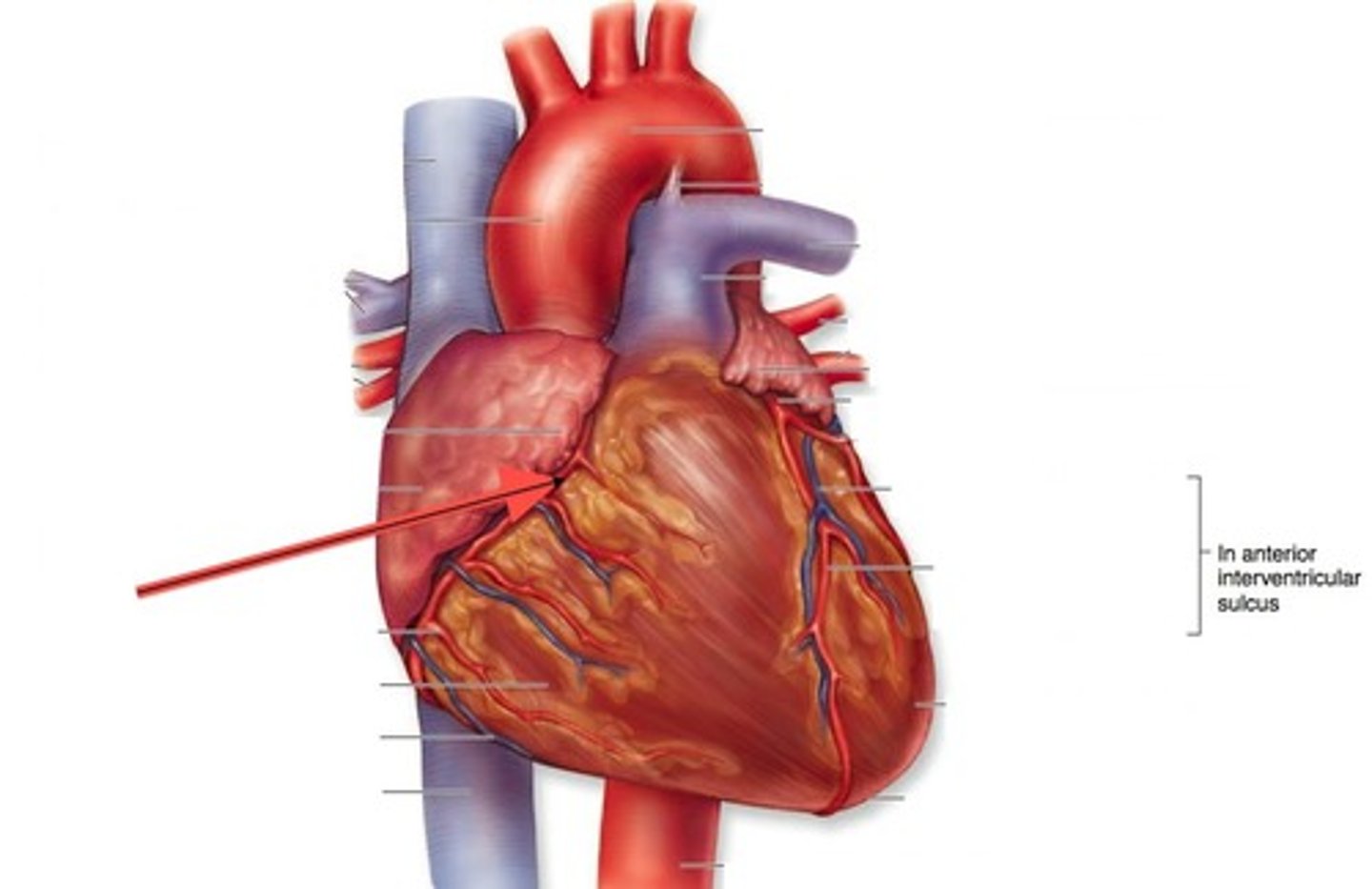
Which arterial branch of the left coronary artery travels down to the apex of the heart?
Anterior interventricular
anterior interventricular artery
branch of the left coronary artery that continues around to the back of the heart in the coronary sulcus.
The left and right coronary arteries arise from which blood vessel?
Aorta
The arterial branch that usually arises from the right coronary artery and supplies the back wall of the heart, both left and right ventricle, is the ________ __________ branch
Posterior Interventricular
The right and left coronary arteries arise from the ascending ________
Aorta
Which artery travels under the left auricle and then divides into two branches?
Left coronary artery
Which branch of the left coronary artery continues around to the posterior of the heart leading to the coronary sulcus?
Circumflex
When two arteries or veins join, this is referred to as a(n) ___________ .
anastomosis
When does blood flow through the coronary circulation increase?
Ventricular diastole
Which artery is a branch of the right coronary artery and supplies the back walls of the ventricles?
Posterior interventricular artery
The left and right coronary arteries arise from which blood vessel?
Aorta
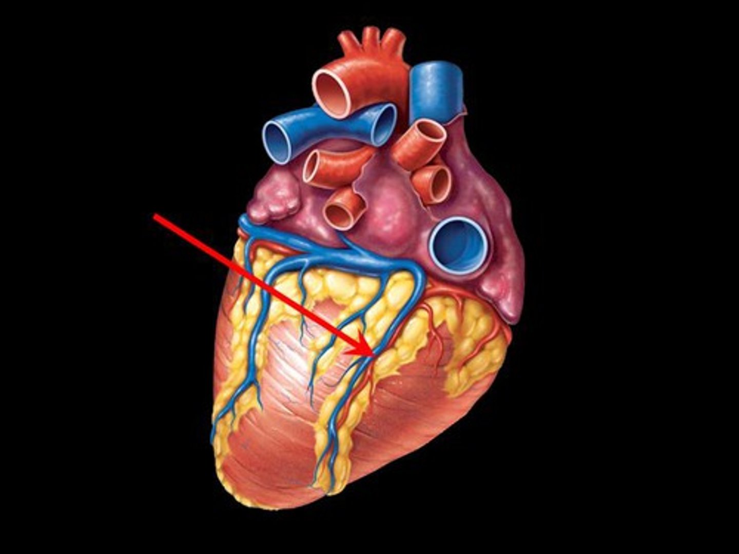
Which vein collects venous drainage from the anterior side of the heart and travels alongside the anterior interventricular artery?
Great cardiac vein
What is a convergence of two arteries called?
Arterial anastomosis
During which phase of the cardiac cycle does blood flow through the coronary circulation?
Ventricular relaxation
What is the ability to rhythmically depolarize without outside stimulation called?
Autorhythmic
Cardiomyocytes are jointed end to end by thick connections called ______.
intercalated discs
What is the benefit of having collateral circulation?
To provide alternative routes of blood flow
When does blood flow through the coronary circulation increase?
Ventricular diastole
Cardiac muscle requires oxygen and therefore relies on ___________ respiration to produce ATP.
Aerobic
The vertebrate heartbeat is said to be ______ because the signal originates within the heart muscle itself.
myogenic
Which vein collects venous drainage from the anterior side of the heart and travels alongside the anterior interventricular artery?
Great cardiac vein
What are the short, thick, branched contractile cells of the heart called?
Cardiomyocytes
Cardiac muscle relies on which process to produce ATP?
Aerobic respiration
The heartbeat is coordinated by the cardiac ___________ system.
conduction
What is the ability to rhythmically depolarize without outside stimulation called?
Autorhythmic
The term ___________ refers to relaxation of the heart.
Diastole
_____________ are short, thick, branched muscle cells of the heart.
Cardiomyocytes
What is the normal heartbeat set by the SA node called?
Sinus rhythm
Nodal rhythm
set by AV node, 40 to 50 bpm (if SA node is damaged)
A spontaneously developing local potential that generates action potentials in the SA node is called what?
Pacemaker potential
Which structures are considered to be part of the cardiac conduction system?
SA node
AV node
Purkinje fibers
Cardiocytes are described as ____________ because individual cells can depolarize on their own without outside stimulation.
Autorhythmic
cardiomyocytes
depolarize quickly but take much longer to repolarize
Muscles cells of the heart, relatively long, branched cells, forms networks throughout each pair of the heart chambers, one network is the artia and the other is the ventricles
Which term refers to relaxation of the heart?
Diastole
Systole
Contraction of the heart
When a normal heart rate is established by the SA node, a _________ rhythm results.
Sinus
The pacemaker potential of SA node cells is due to the influx of which ions?
Sodium
Impulse conduction through the cardiac conduction system is slowest through the _____________ node, allowing a pause between atrial contraction and ventricular contraction.
AV (atrioventricular) node
The heartbeat is coordinated by the cardiac ____________ system.
conduction
What is the ability to rhythmically depolarize without outside stimulation called?
Autorhythmic
Myogenic
heartbeat originates within heart itself
Autorhythmic
The heart is described as such because the heart doesn't depend on the nervous system for its rhythm, it has its own built-in pacemaker and electrical system
Depolarization of a cardiocyte is due to the opening of __________ channels.
sodium
When the AV node acts as pacemaker, the slower heartbeat has what type of rhythm?
Nodal
Which is a recording of all nodal and myocardial action potentials in the heart?
ECG (electrocardiogram)