Integumentary System: Epidermis, Dermis, Hypodermis, etc.
1/51
There's no tags or description
Looks like no tags are added yet.
Name | Mastery | Learn | Test | Matching | Spaced | Call with Kai |
|---|
No analytics yet
Send a link to your students to track their progress
52 Terms
what is the average surface area of the integumentary system?
average weight?
perfect of total body weight?
1.5-2.2 sq m
4-5kg
7%
what is the largest organ in the body?
skin
what are the four functions of skin?
protection, sensation, heat regulation, and water proofing
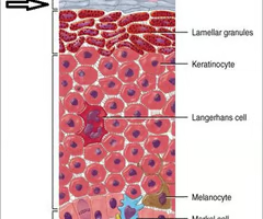
what are the four main cell types?
keratinocytes, melanocytes, langerhans' cells, merkel cells
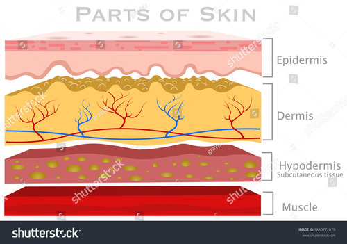
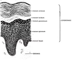
describe basic characteristics of epidermis (thickness, location, composition, vascularity)
- outermost protective shield of body
- 1.5-4.0mm thick
- composed of epithelial tissue (keratinized stratified squamous)
- avascular (needs diffusion)
describe basic traits of the dermis (thickness, composition, vascularity, layers)
- makes up bulk of skin
- mostly dense irregular
- rich supply of nerves, blood vessels, and lymphatic vessels
- majority of hair follicles, oil glands, and sweat glands here
- divided into two layers
what are the two layers of the dermis?
papillary and reticular
describe the papillary layer (thickness, composition, content, noteable traits)
- top 1/3 of dermis
- areolar tissue
- contains capillaries and nerves
- superior surface forms peg-like projections called dermal papillae
how are fingerprints formed?
projections of dermal papillae form friction ridges
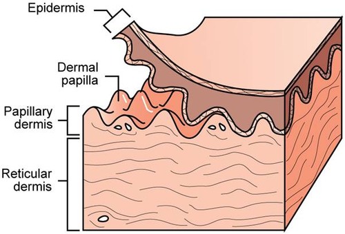
function of papillary layer
keep epidermis binded together
describe the reticular layer (thickness, composition, purpose)
- bottom 2/3 of dermis
- dense irregular
- thick bundles of collagen fibers in different planes give strength, elastin gives some flexibility
cleavage lines
groups of collagen bundles all going the same way in different areas, externally invisible
- determine what will and won't heal up well (does it cut along or across the collagen bundles)
why do we see flexure lines in our palms, but not in areas like our forearms?
palm skin needs to be tightly bound to muscle and collagen for grip, so the skin forms along those instead of being looser like in the forearm
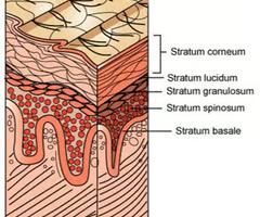
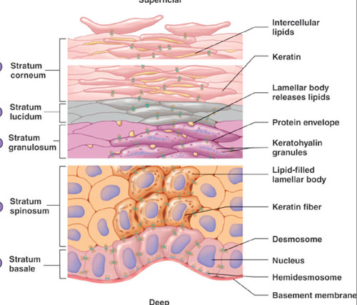
arrange the layers of the epidermis from deep to superficial
basale, spinosum, granulosum, lucidum, corneum
mnemonic device for epidermis layers from superficial to deep
Come, Let's Get Sun Burned
what is another name for hypodermis
subcutaneous layer
describe hypodermis (thickness, purpose, composition)
- adipose tissue, larger blood vessels, nerves
- varies in thickness from person to person
- used mainly for fat storage
- shock absorber
erythema
redness of skin
- may indicate embarrassment, inflamation, hypertension (high BP)
pallor
paleness
- may indicate emotional stress, anemia, hypotension (low BP)
Jaundice
yellowing of skin
- usually indicate a liver disorder
- bile pigment accumulates in skin
bruises
AKA hematomas
- blood has escaped the vessels and clotted below skin
cellulitis
common skin infection
- problem if untreated
cyanosis
bluish tint to skin due to lack of oxygen or blood flow
- may indicate heart failure, cold temp, Raynaud's Syndrome (fingertips go white in cold)
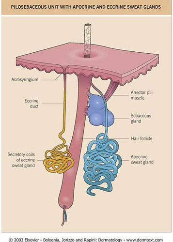
What are the four types of glands?
sudoiferous (two types, eccrine and apocrine), ceruminous, sebaceous
What is in sweat?
Can you actually sweat things "out" that you've eaten?
Sweat is a filtrate of blood containing 99% water with some NaCl and traces of metabolic waste.
So yes.
How many types of sudoiferous glands are there? What are they called?
- two types
- eccrine and apocrine
describe eccrine sweat glands (purpose, composition, location, description within dermis)
- responsible for sweat as we recognize it
- most common gland
- abundant in palms and soles of feet
- excrete sweat for thermoregulation
- coiled, tubular gland that extends directly to surface
describe apocrine sweat glands (location on body, purpose, effects, composition, location within dermis)
- abundant in armpits, wings of nostrils, genital regions
- body odor caused by decomposed waste of skin basteria
- consists of water and fatty substance
- fatty substance causes yellowish appearance (sweat stains on white shirts)
- coils to hair follice
describe ceruminous glands (composition, location within body, function)
- modified apocrine
- lining external ear canal
- secretions mix with sebum to produce cerumen (ear wax)
- Function: prevent debris and dust from entering canal
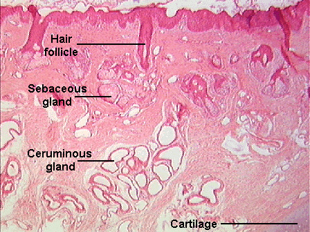
describe sebaceous glands (purpose, composition, location, description within dermis)
- found everywhere on body except palms and soles of feet
- abundant on face, neck, upper chest, and upper back
- secrete an oily substance called sebum
- associated with hair follicle, softens/lubricates hair and skin
- non-coiled to hair follicle
what is the difference between a whitehead and a blackhead?
- both are blocked sebaceous glands
- blackheads are just whiteheads that have been exposed to air and oxidized
define acne
inflammation of sebaceous glands
- usually with pus
what is pus
an indication of infection
- full of leukocytes
define sebborhea
cradle cap, common in infants
- overactive sebaceous glands cause oily scales to form
cradle cap, common in infants
- overactive sebaceous glands cause oily scales to form
muscle attached to hair follicle that pulls hair to stand up
- responsible for goosebumps
describe burns as a whole (threats, process, etc.)
- severity depends on length and intensity of exposure
- immediate threat is dehydration, secondary is infection
- tissue is damaged, which leads to cell death
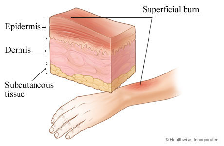
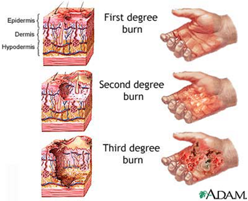
define 1st degree burns
- localized redness, swelling and pain
- 2-3 day healing, quickest overall
- partial thickness burn (only epidermis)
define 2nd degree burns
- partial thickness burn (epidermis and dermis)
- blistering
- splotchy and intense redness
- 3-4 week healing
define 3rd degree burns
- full thickness (epidermis, dermis, hypodermis, into muscle and bone)
- tissue appears gray-ish, charred, cherry red
- initially little to no swelling (edema) or pain (destruction of nerve endings)
- high risk of infection, scarring is common
- takes longest to heal
- usually requires skin grafts
what are the rules of nine?
chart used to quickly assess a patient on how much of their body has been burned
Jane Doe has burns on the posterior side of her left arm and leg, her face, and the upper half of her abdomen. How much of her body has been burned? (chart on back)
27%

John Doe has burns on his face, entire anterior torso, perineal region, and the upper posterior halves of both of his legs. How much of his body has been burned?
32.5%

Jane Doe has burns on her entire right arm, the posterior side of her head, the right side of her back, and the posterior side of her right leg. How much of her body has been burned?
31.5%
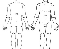
A 1 year old has burns on her entire right hand, the back of her head, back of her neck, and both sides of both of her feet. How much of her body has been burned?
19.5%
What two factors account for the demise of epidermal cells?
limited or nonexistent diffusion and the introduction of keratin
Which layer of the dermis is reponsible for fingerprints?
papillary layer
What glands are activated at puberty?
sebaceous and apocrine
describe the stratum basale
- deepest layer
- single layer of keratinocytes
- the only mitotic layer of the epidermis
- some melanocytes and occasional Merkel
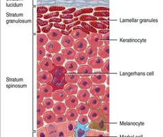
describe the stratum spinosum
- 2nd-deepest layer
- several layers of spiny keratinocytes
- begin to produce keratin here
- layer in which Langerhans' are most abundant
- melanin throughout
describe stratum granulosum
- thin, only 3-5 cell layers
- cells begin to flatten here and accumulate keratin
- extracellular lipids cause waterproofing
- the line between living and dead cells
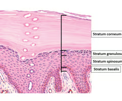
stratum lucidum
- 2nd-most superficial layer
- appear as thin, transparent layer of flat, dead keratinocytes
- only appears in thick skin (i.e. palms and soles of feet)
stratum corneum
- most superficial layer
- shingle-like, dead, keratin-filled cells
- thickness of layer depends on body location (friction)
- average person sheds 40lbs. of these in lifetime