The Heart
 Call Kai
 Learn
Learn Practice Test
 Spaced Repetition
 Match
Match Flashcards
 Knowt Play
1/239

There's no tags or description
Looks like no tags are added yet.
Name | Mastery | Learn | Test | Matching | Spaced | Call with Kai | Chat |
|---|
No analytics yet
Send a link to your students to track their progress
240 Terms
Give the function of:
The heart
Arteries
Veins
Capillaries
Blood
Lymphatic system
Heart: mechanical pump
Arteries: transporting blood away from the heart
Veins: transporting blood to the heart
Capillaries: site of exchange of substances
Blood: transport of cellular components and dissolved substances
Lymphatic system: returning tissue fluid to systemic circulation
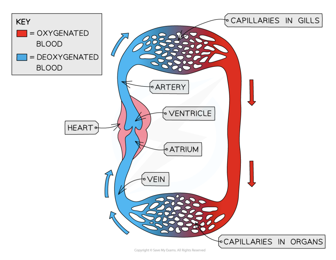
Draw out the circulatory system of fish
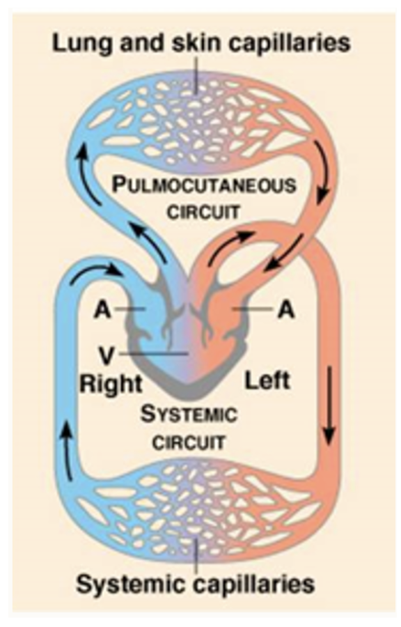
Draw out the circulatory system of amphibians
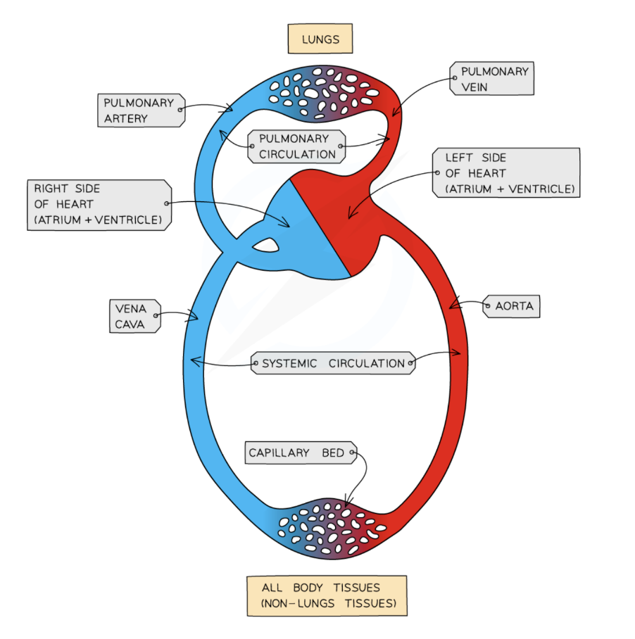
Draw out the circulatory system of mammals
What is the structure/function of:
Muscular arteries
Elastic arteries
Arterioles
Capillaries
Muscular arteries: medium sized artery, changes (vasodilation/constriction) affect blood pressure
Elastic arteries: conducting artery, expands during systole and recoils during diastole
Arterioles: small diameter which changes in response to local conditions
Capillaries: small diameter, endothelial layer only, site of exchange of substances
Describe the structure of:
Tunica interna
Tunica media
Tunica externa
Lumen
Tunica interna: endothelial lining
Tunica media: concentric sheets of smooth muscle
Tunica externa: connective sheath around a vessel
Lumen: cavity of an organ
Draw a labelled diagram of the heart
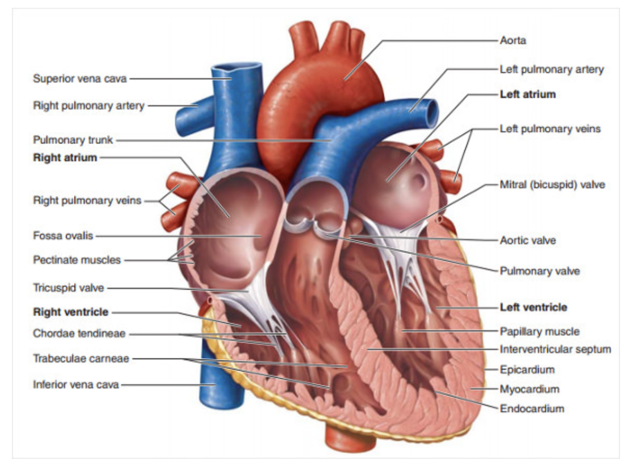
Where are the 4 different heart valves located?
Tricuspid: right atrioventricular
Mitral: left atrioventricular
Pulmonic: right semilunar
Aortic: left semilunar
Describe the different stages of the cardiac cycle
Ventricles fill with blood, atria contract- forcing more blood into ventricles
Ventricles contract, increasing the ventricular pressure
When the ventricular pressure increases above arterial pressure, the atrioventricular valve closes
When the pressure inside the ventricles exceeds the pressure in the outgoing arteries, the semilunar valves open
As a result of ventricular systole and opening of the semilunar valves, blood flows from the ventricles to the arteries
Pressure in the ventricles drops below aortic pressure and the semilunar valves close
When ventricular pressure becomes lower than atrial pressure, the atrioventricular valves open
Draw the pathway of nerve fibres from the central nervous system to:
Skeletal muscle (SMNS)
Smooth muscle, heart muscle, glands (SNS)
Smooth muscle, heart muscle, glands, reticular formation (SNS)
Smooth muscle, heart muscle, glands (PNS)
Include relevant synapses and the neurotransmitters
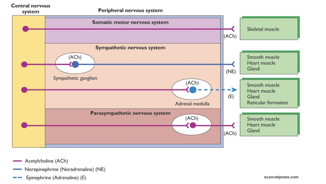
What are the neurotransmitters, designation and receptors associated with the following nerve fibres?
Pre-G SNS
Pre-G PSNS
Post-G SNS
Adrenal medulla
Post-G PSNS
Nerve fibre | Neurotransmitter | Designation | Receptor |
Pre-G SNS | Acetylcholine | Cholinergic | Nicotinic Ach R |
Pre-G PSNS | Acetylcholine | Cholinergic | Nicotinic Ach R |
Post-G SNS | Noradrenaline | Adrenergic | Ɑ and/or β |
Adrenal medulla | Adrenaline | Adrenergic | Ɑ and/or β |
Post-G PSNS | Acetylcholine | Cholinergic | Muscanaric Ach R |
When and why is the SNS activated?
The SNS is activated during times of stress, physical activity or danger to trigger the ‘fight or flight’ response
When and why is the PNS activated?
The PNS is activated when the body is at rest, safe and recovering to trigger the ‘rest and digest’ response
Describe the outflow of the SNS
The SNS has a thoracolumbar outflow. The preganglionic neurons originate from the thoracic (T1) and lumbar (L2/3) segments of the spinal cord
Describe the length of ganglionic fibres in the SNS
Preganglionic fibres are short in length
Postganglionic fibres are long in length
Give the locations of the SNS ganglia
Paravertebral ganglia
Cranial cervical ganglion
Middle cervical ganglion
Caudal cervical ganglion
Thoracic and lumbar ganglia
Sacral and pelvic ganglia
Prevertebral ganglia
Celiac ganglion
Superior and inferior mesenteric ganglia
List the effects of the SNS on various parts of the body
Eye: pupil dilation
Salivary glands: viscous secretion
Heart: increased heart rate, increased force of contraction
Lungs: dilation of bronchioles
Upper digestive tract: motility reduction
Digestive glands: reduced secretion
Liver: increased release of glucose to the blood
Adrenal medulla: increased secretion
Lower digestive tract: reduced motility
Urinary bladder: relaxation of wall musculature, contraction of sphincter muscle
Genitals: ejaculation (male)
Blood vessels/sweat glands: vasoconstriction, increased sweat secretion
Describe the outflow of the PNS
The PNS is activated when the body is at rest, safe and recovering to trigger the ‘rest and digest’ response
The PNS originates in the brainstem, and travels through four cranial nerves:
CN III- Oculomotor
CN VII- Facial
CN IX- Glossopharyngeal
CN X- Vagus
The sacral outflow originates from spinal segments S2-S4 via pelvic splanchnic nerves to supply the lower bowel, bladder and reproductive organs
Describe the length of ganglionic fibres in the PNS
Preganglionic fibres are long in length
Postganglionic fibres are short in length
Give the locations of the PNS ganglia
Sensory ganglia
Dorsal root ganglia
Cranial nerve ganglia
Autonomic ganglia
Sympathetic ganglia
Parasympathetic ganglia
Describe how autonomic reflexes are involved in neural regulation of the ANS, giving examples
These work through feedback mechanisms, which involve the afferent nervous system
Their main function is to maintain homeostasis
Examples
Heart rate (controls speed, manages blood flow)
Digestion (moves food, releases stomach acid and enzymes)
Breathing rate (adjusts air flow, responds to gas levels)
Pupil size (dilation in low light, constriction in bright light)
Give the function and locations of baroreceptors
Baroreceptors detect changes in blood pressure by sensing the stretch of blood vessel walls
Location
Walls of the atria
Walls of pulmonary vessels
Give the function and locations of chemoreceptors
Chemoreceptors monitor chemical levels (oxygen, carbon dioxide, pH) in the blood and brain
Location
Medulla oblongata
Carotid bodies and aortic bodies
How is the SNS involved in innervating the heart and lungs?
Post-G fibres go directly to the heart. The sympathetic cardiac nerves are found within the cardiac plexus
Post-G fibres go to the SA node and AV node (in the walls of the heart)- this controls heart rate and rhythm
Post-G fibres also go to the myocardium- this controls the force of contraction
Lungs
β-2 adrenergic receptors are stimulated to relax the smooth muscle of the airway
How is the PNS involved in innervating the heart and lungs?
Pre-G fibres travel in branches of the recurrent laryngeal and vagal nerves to the cardiac plexus
The post-G fibres end in the atrial walls
Lungs
M3 muscarinic receptors are stimulated to constrict the smooth muscle of the airway
What is the difference between the ‘lub’ and ‘dub’ sound of the heart?
‘Lub’ (S1)
Heard at the beginning of ventricular systole, when the atrioventricular valves shut
‘Dub’ (S2)
Heard at the beginning of ventricular diastole, when the semilunar valves shut
What is the difference between systolic and diastolic murmurs in the heart?
Systolic murmurs
Swishing sounds
Heard between S1 and S2 during ventricular contraction
Diastolic murmurs
Sounds heard after S2 and before S1
How can acquired heart disease cause heart murmurs?
Thickening and weakening of heart valve can cause leakage, causing backflow turbulence (common in older, small-breed dogs)
Diseases that weaken or thicken heart muscle alter chamber size/function, disrupting normal blood flow
Bacterial infections can cause inflammation and growths on the heart valves, preventing proper closure
How can congenital heart defects cause heart murmurs?
Shunts and holes: abnormal openings between heart chambers force blood where it should not go
Stenosis: narrowing of the valves/major arteries forces blood through tight spaces at high speed
What are the functional causes of heart murmurs?
Fever, stress and pregnancy increase metabolic rate which increases blood flow velocity- temporarily producing a functional murmur
Anaemia/low protein can create physical turbulence
Harmless heart murmurs are common in rapidly growing animals, or athletic animals- these disappear with maturity
Describe the structure and function of the annulus fibrosus
The annulus fibrosus consists of 4 interconnected rings of fibrous tissue, situated between the atria and the ventricles
It acts as an electrical insulator, ensuring action potentials from the sinoatrial node only reach the ventricles via the atrioventricular node
This helps ventricular filling to be complete before the initiation of ventricular systole
Give the function of:
The SA node
The AV node
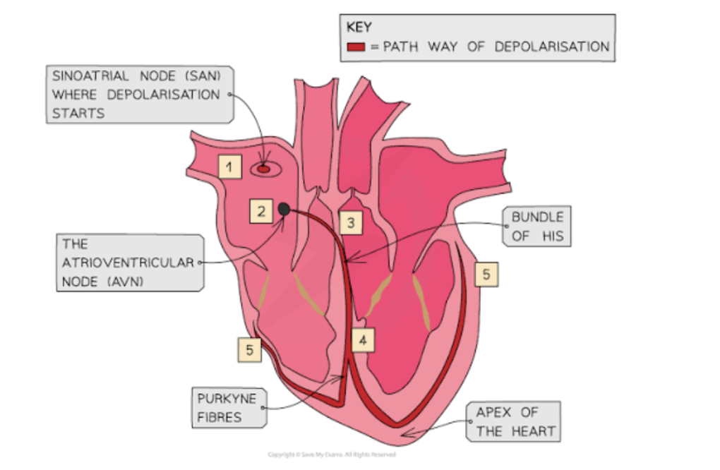
The bundle of His
Purkinje fibres
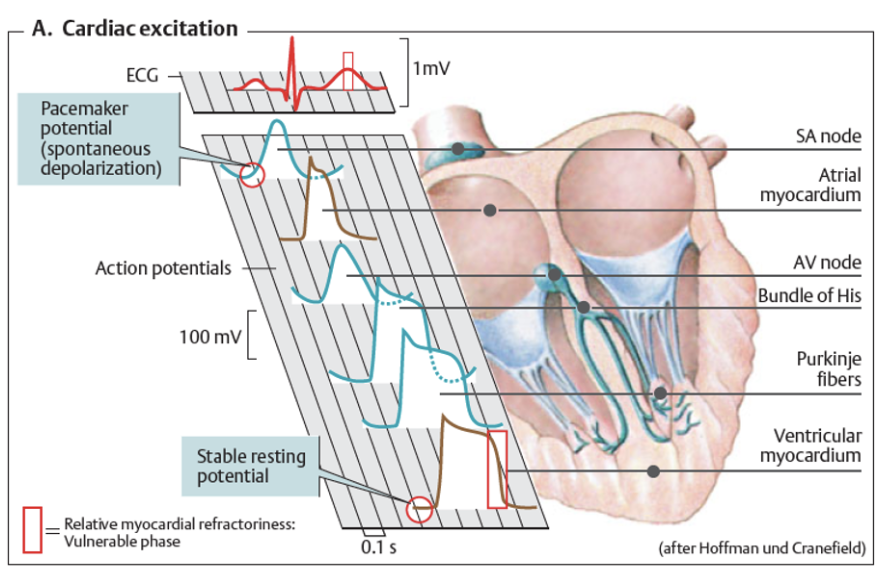
Sinoatrial node: generates electrical impulses that spread through the heart muscle, setting the normal resting heart rhythm, and causing the atria to contract (acts as the natural pacemaker)
Atrioventricular node: delays electrical signals from the SA node, ensuring the atria empty blood into the ventricles before the ventricles contract
Bundle of His: carries electrical signals from the AV node down to the apex
Purkinje fibres: rapidly spread signals from the bundle of His through the ventricular walls, causing the ventricles to contract
Draw a diagram to show the conduction system of the heart
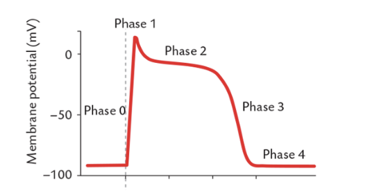
Describe the different phases of conduction in the heart, including a relevant diagram
Phase 0
A stimulus opens voltage gated sodium ion channels
This causes an influx of sodium ions
Membrane voltage sharply increases
Phase 1
Sodium ion channels close
Outward potassium ion channels open briefly, causing a small outflow of potassium ions
This causes a minor decrease in voltage
Phase 2
Calcium ion channels open
Calcium ions enter the cells whilst potassium ions continue to leak out
This holds the voltage steady
Phase 3
Calcium ion channels close
Outward potassium ion channels remain open
This drives the membrane potential back to baseline resting levels
Phase 4
The membrane potential remains stable near -90mV
Inward potassium ion channels stay open
Describe how the action potential is generated in the heart
The SA node, AV node, bundle of His and Purkinje fibres are autorhythmic cells
These spontaneously generate action potentials and undergo slow depolarisation until a threshold potential is reached
These depolarisations occur most rapidly in the SA node- which therefore drives the rhythm of the heart
Action potential at the SA node
Ion channels that are permeable to both sodium and potassium ions open, causing a small influx of sodium ions- this is known as I𝑓 current
Calcium ion channels begin to open
Further calcium ion channels open
Potassium ion channels are activated, allowing an efflux of potassium ions
Potassium ion channels close and I𝑓 channels open
Describe the effects of the SNS on the conduction system of the heart
SA node: speeds up the rate of pacemaker firing (positive chronotropy)
AV node: shortens the delay between atrial and ventricular contraction (positive dromotropy)
Mechanism: noradrenaline binds to beta-1 adrenergic receptors, increasing calcium/sodium entry
Increases heart rate, speeds up electrical conduction, increases strength of contraction and quickens muscle relaxation
Stimulates cardiac myocytes
Describe the effects of the PNS on the conduction system of the heart
SA node: slows down the rate of pacemaker firing (negative chronotropy)
AV node: lengthens the delay between atrial and ventricular contraction (negative dromotropy)
Mechanism: acetylcholine binds to M2 muscarinic receptors, opening potassium channels to hyperpolarise the cells
Has very little direct influence on ventricular muscle cells, because vagal nerve fibres are heavily concentrated in the atria and pacemakers- with very little distribution in the ventricular walls
Draw a diagram to show the conduction of the action potential in both the SA and AV nodes
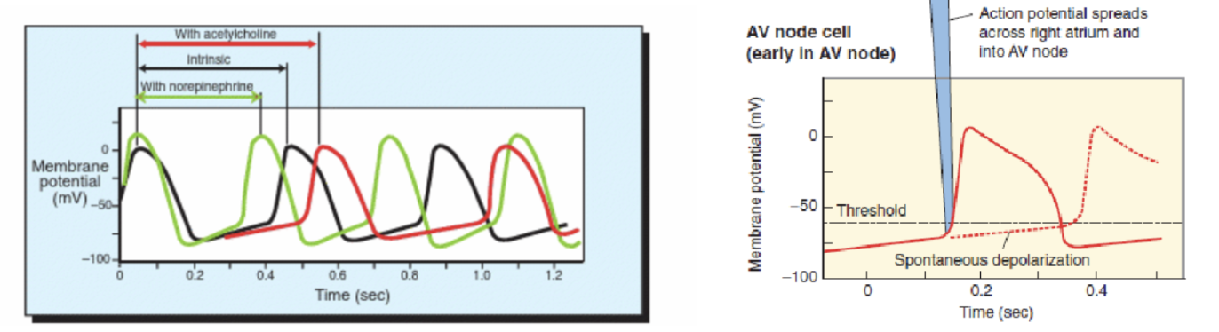
Draw a diagram to show a summary of cardiac excitation throughout the heart and the relevant structures related to each stage of conduction
Describe the structure of the heart, including the locations of the 4 valves
The heart is a simple pump consisting of 4 chambers: 2 atria and 2 ventricles
It has 4 valves- ensuring blood flows unilaterally
AV valves separate the atrium and ventricles
Mitral valve on the left
Tricuspid valve on the right
SL valves separate the ventricles from the aortic and pulmonary arteries
Aortic valve in the left outflow tract
Pulmonic valve in the right outflow tract
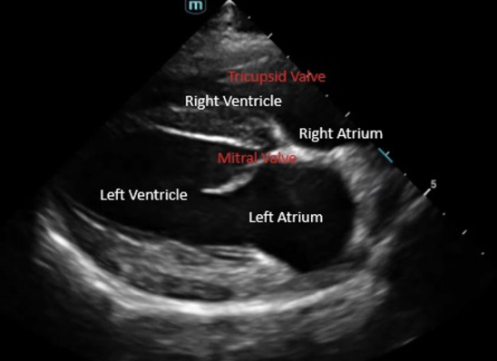
Label an echocardiogram of the heart
Describe the key functions of the cardiorespiratory system
Transport to and from metabolising tissue
Oxygen and carbon dioxide
Nutrients
Waste
Heat
Hormones
Homeostasis
pH, osmolarity, electrolytes
Infection
Other
Generating pressure (e.g renal filtration)
Describe the basics of clinical relevance in relation to the heart
Heart disease is common in both small animal and equine practice
Disease usually involves valve degeneration
Acquired heart disease: valves becoming incompetent, diseases of the heart muscle (resulting in the heart not contracting well, or not filling well)
Disease can also result from issues during the development of the heart (congenital heart defects)
Heart disease does not always result in heart failure
Heart failure: a syndrome in which the heart fails to deliver blood effectively to meet the requirements of metabolising tissues
Describe the structure of the circulatory system of mammals
What is the difference between ventricular systole and ventricular diastole?
Ventricular systole: contraction of ventricles
Results in cardiac output
AV valves close
‘Lub’ sound
Ventricular diastole: relaxation of ventricles
Results in ventricular filling
SL valves close
‘Dub’ sound
Describe the considerations of regulation of cardiac function
Can change heart rate and contractility
Electrical activity (electrophysiology)- can assess using and ECG
Contractile function control
The important role of the ANS
Hormonal mechanisms (local and systemic)
Describe the considerations of regulation of vasculature
Autoregulation: local blood flow regulation, intrinsic ability of an organ to maintain a constant blood flow, despite changes in perfusion pressure
The important role of the ANS
Hormonal mechanisms (local and systemic)
What are the considerations for the heart to act as a pump? Include definitions
Cardiac output: volume delivered into the circulation per minute
Stroke volume: volume delivered by the ventricle per beat
Other pumping mechanisms (venous)
And what control these
What are the considerations for the distribution of blood?
Vascular constriction and dilation (arteries and veins)
The maintenance of unidirectional flow in vital organs
Cardiac valves
Vascular valves
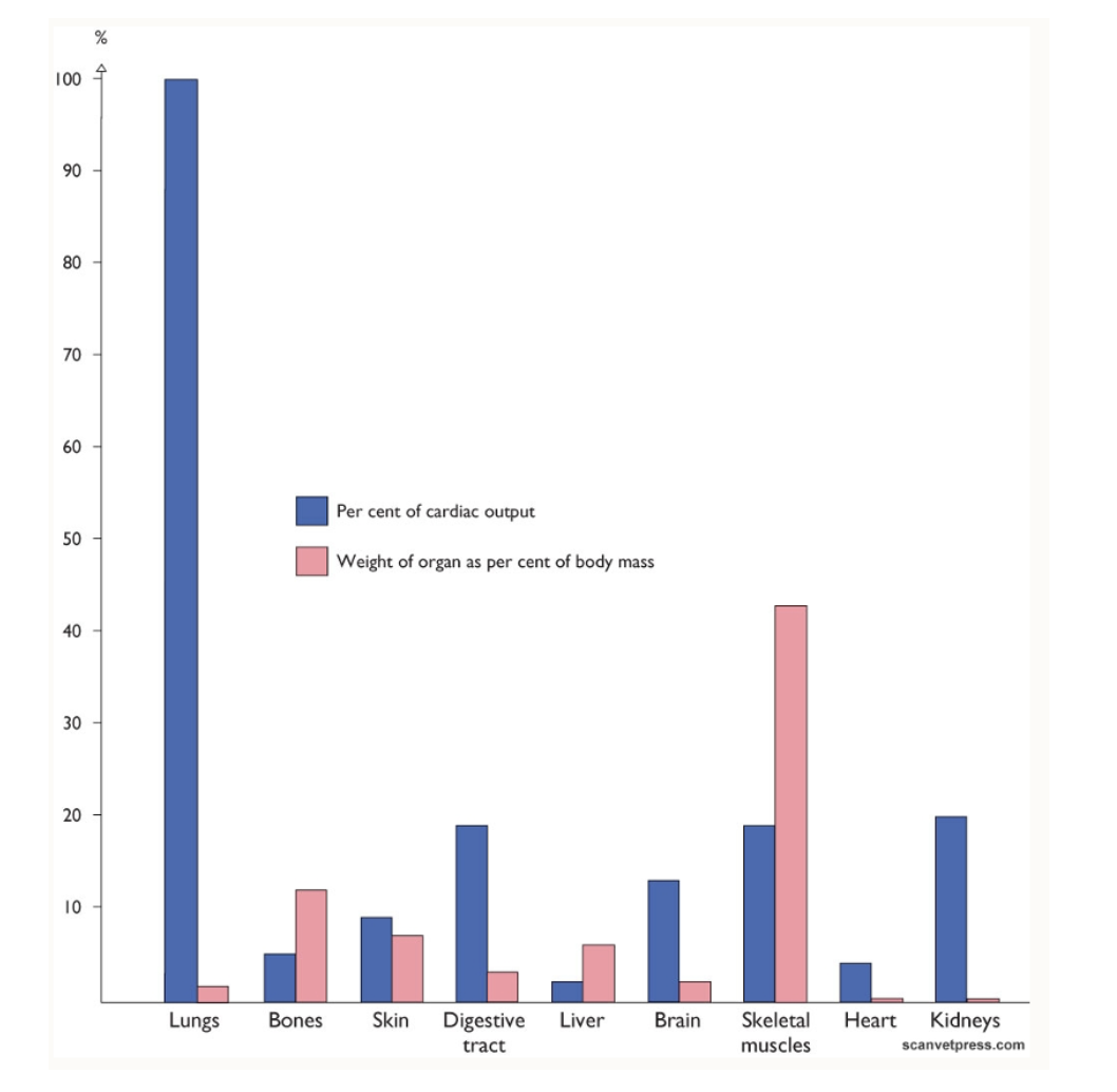
Draw a graph to (roughly) show the percent of cardiac output vs the weight of the following organs:
Lungs
Bones
Skin
Digestive tract
Liver
Brain
Skeletal muscles
Heart
Kidneys
Describe venous return to the heart, including important controlling factors
Deoxygenated blood is returned to the heart
Diastolic cardiac volume is important for cardiac output
Important controlling factors
SNS
Blood volume
Muscle (respiratory) pump
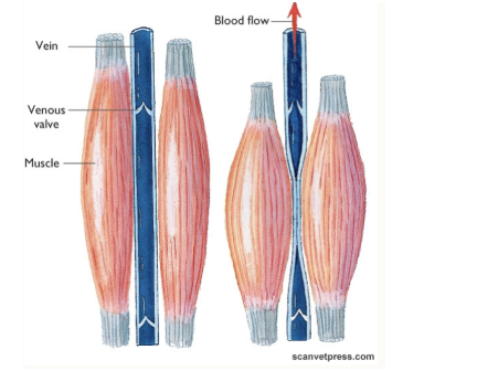
Describe the key components of the vascular system, including a diagram to show various structures
Arteries: from the heart
Capillaries: site of diffusion
Portal veins: between 2 capillary beds
Veins: return to the heart
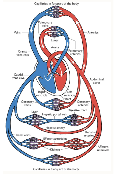
Describe the differences of the cardiac system of birds
Birds have a highly efficient cardiovascular system
To cope with high metabolic demands
Important for oxygen delivery and thermoregulation
Main features of the avian CVS
4-chambered heart, similar to mammals
AV valves are different in structure compared to mammals
The heart is located within the cranial ventral coelom, and is surrounded by air sacs
The term coelom is used because birds do not have a diaphragm
Avian cardiac function
Birds have a relatively high cardiac output (195ml/min)
They can increase cardiac output to over 1 litre/min
During exercise
Mainly by increasing heart rate (115-670bpm)
Heart size fluctuates (increases prior to migration in migratory species)
Ventricles
The left sided ventricular wall is 3x thicker than the right
These empty almost completely on each cardiac cycle
Low end-systolic volume
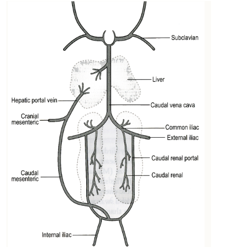
Describe the structure and function of the renal portal system (including a diagram)
Present in reptiles, birds, amphibians and most fish
Receives blood from the caudal body, and returns it to the heart via the kidneys
It functions to supply blood to renal tubules at all times
Portal blood flow is regulated by the renal portal valve
This is important, as it alters how drugs will act
If injected in the caudal half of the body, it may be metabolised before entering general circulation
What is ontology?
Ontology: the development of the embryo from fertilisation to gestation/hatching
Describe the theory of recapitulation
The stages that the mammalian heart goes through during embryological development are evident in the CV systems of fish, amphibians and reptiles
Theory of recapitulation
Embryological parallelism
‘Ontogeny recapitulates phylogeny’
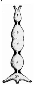
What are the 5 zones of the primitive heart tube? Label a diagram to show these
Arterial trunk/ truncus arteriosus
Ventricle
Primitive ventricle
Atrium
Sinus venosus
Describe the cardiovascular system of fish
Fish have a simple cardiovascular system
2 chambers to the heart
Blood flows in one direction effectively through a single atrium and ventricle
Vascular includes to accommodate oxygenation
The heart is often positioned ventral and caudal to the gills
Describe the cardiovascular system of amphibians, including a labelled diagram
In anurans, we start to see 2 atria (3 chambers in total)
Oxygenation occurs from lungs, skin and buccan cavity- hence vasculature is more complex in comparison to mammals
The heart is mid-cranial coelom
Like birds, they have no diaphragm (hence the herm coelom again)
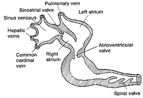
Describe the cardiovascular system of reptiles
Most reptiles have 3 chambers, with 2 atria and 1 ventricle. The ventricle is split into three sections by folds in the muscle wall
Crocodiles are an exception with 4 chambers, but they still have distinct differences from mammals (e.g 2x aortas)
All reptiles can shunt blood away from the respiratory tract when needed
For most species, the heart is mid-cranial coelom, some are very cranial
For snakes, the heart is often found in the cranial third of the body
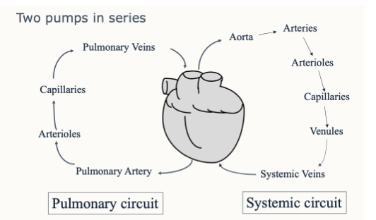
What are the functions and pressures of the left and right side of the heart in mammals? Draw a diagram to show the two pumps in series
The right heart:
Pumps blood without oxygen to the lungs
Low pressure needed
The left heart:
Pumps blood with oxygen to all the organs
High pressure needed
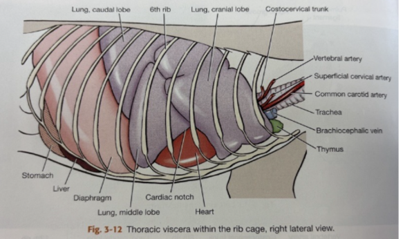
Draw a diagram to show the relevant landmarks close to the heart in dogs
Ventral border of the lungs
Cardiac notch
Lungs (laterally)
Thymus (cranially)
Diaphragm (caudally)
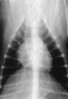
What does this radiograph show?
The heart (dog)
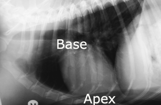
What does this radiograph show?
The heart (dog)
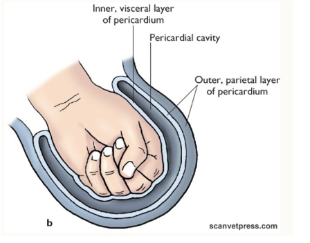
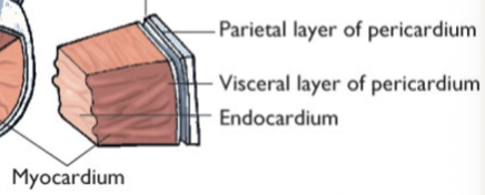
Describe the pericardium, including a diagram
Sac surrounding the heart
Inner visceral layer: on the surface of the heart
Outer parietal layer
No significant lumen
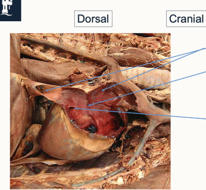
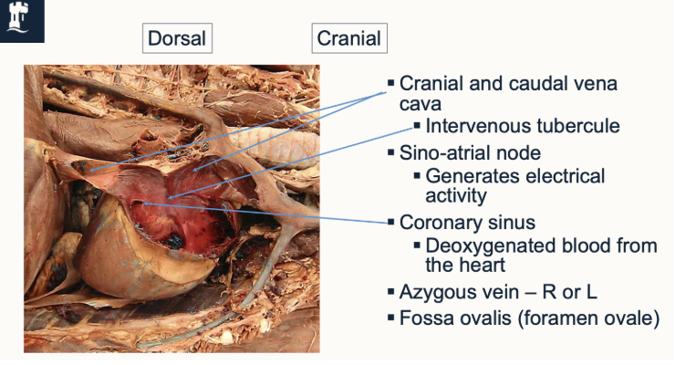
Label this diagram to show the relevant structures of the right atrium of the heart, including their functions
Describe the structure and location of the left atrium
Dorsal and caudal under the tracheal bifurcation
Pulmonary veins enter in groups, into 2 or 3 sites
In the septal wall is the scar of the valve of foramen ovale
Describe the structure and location of the right ventricle
Crescent shaped in section
Wraps around the left ventricle cranial and to the right
Pulmonary artery is cranial and left of the aorta
Trabecular septomarginalis
Septum- outer wall
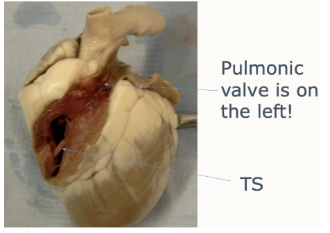
Describe the structure and location of the left ventricle
Circular in section
Occupies all of the apex
Prominent papillary muscles
Aorta is central
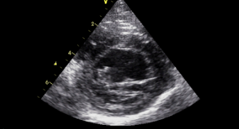
What does this echocardiogram show?
Left ventricle
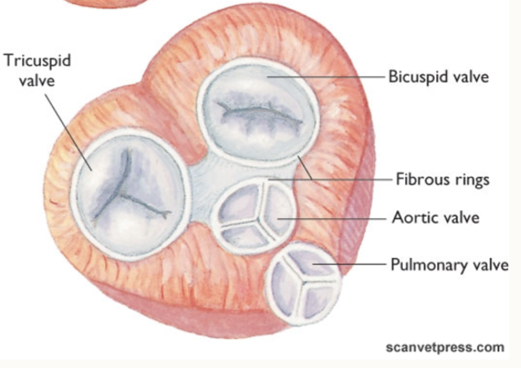
Describe the different types of cardiac valves
AV
Right AV: tricuspid (3 cusps in people, 2 in domestic species)
Left AV: mitral (2 cusps)
Chordae tendinae- papillary muscles
SL
Right SL: pulmonic
Left SL: aortic
Both are 3 cusps
Describe the pressure changes in the heart in relation to the heart valves
As ventricles relax, pressure drops below that in atria (blood moves from atria → ventricle)
Ventricles contract, ventricular pressure rises and AV valves close
When pressure in the ventricles exceeds that of the aorta and pulmonary artery, the SL valves open (ejection phase)
Pressure in the ventricles falls: lower pressure in the aorta and pulmonary artery
All four valves are closed, and the cycle starts again
What is the function of the fibrous cardiac skeleton?
Supports the four valves
Separates atria and ventricles
These are known as ossa cordis
Draw a diagram to show a cross-section of the heart
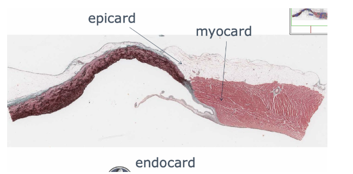
What are the different layers of myocardium?
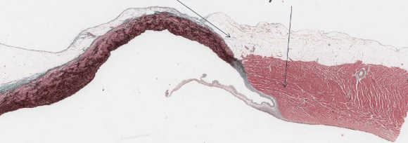
Label the following diagram (there are three labels- one at the bottom)
Describe the structure of cardiomyocytes, including the specialised structures relevant
Large, cylindrical cells
Striated like skeletal muscle
Short, branches fibres
Lots of mitochondria
Intercalated discs: support cell-to-cell communication, required for coordinated muscle contraction
Purkinje fibres: specialised conducting tissue, deliver electrical activity to myocardium
How do cardiomyocytes respond to training and injury?
Training
Mass increase (training/overload) can lead to myocyte hypertrophy
No increase in cell numbers
Injury
Limited capacity to divide/regenerate
Undergo regulated cell death
Release damage signals, which trigger acute inflammation
Prompt neighboring non-myocyte cells to form a structural fibrotic scar
What is the difference between ventrodorsal and dorsoventral radiographs?
Ventrodorsal: animal is on its back
Dorsoventral: animal is on its chest
Why are ventricular sounds louder?
Ventricular sounds are louder, as the ventricles are bigger
Draw a detailed diagram to show pressure and volume changes throughout a cardiac cycle, including a representative phonocardiogram and an electrocardiogram
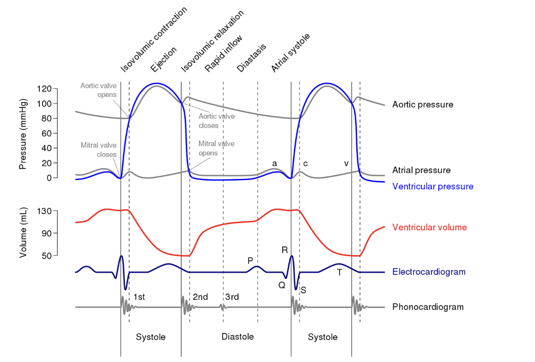
What can affect filling of the ventricles?
Preload
Afterload
If the cardiac cycle is 1 second, how long would systole and diastole be?
If the cardiac cycle is 1 second,
Systole= 0.35 seconds
Diastole= 0.65 seconds
Give a brief outline of the following phases
Filling phase (diastole)
Isovolumetric contraction
Outflow phase (systole)
Isovolumetric relaxation
Filling phase (diastole)
Ventricles fill
Isovolumetric contraction phase
Ventricles contract, but volume remains constant because the heart valves are closed- pressure builds
Outflow phase (systole)
Ventricles contract, valves open and blood flows into the aorta or pulmonary artery
Isovolumetric relaxation
Ventricles relax- ready for refill of blood during the next filling phase
Describe the atrial systole phase of the cardiac cycle
Last phase of diastole (P → R wave)
Depolarisation of the atria leads to atrial contraction
A tiny amount of ‘topping off’ completely fills the ventricle
Describe the isovolumetric contraction phase of the cardiac cycle
First phase of systole
Begins at the peak of the R-wave
No real change in the volume of the ventricles
First heart sound (‘lub’), closing of the AV valves, associated blood turbulence when ventricular pressure exceeds atrial pressure
Describe the rapid ejection phase of the cardiac cycle
During ST segment
When ventricular pressure exceeds that in the aorta or pulmonary artery, semilunar valves open and rapid ejection from the ventricles starts
‘C’ wave in the atrial pressure curve is caused by slight (due to papillary muscles) distension of the AV valves into the atria- normally not measurable
Describe the reduced ejection phase of the cardiac cycle
Final phase of systole
Coincides with the T-wave (ventricular repolarisation)
Blood flow out of the ventricles continues, but more slowly
Eventually pressure in the ventricle falls below that in the artery, and semilunar valves close
Describe the isovolumetric relaxation phase of the cardiac cycle
First phase of diastole
Atria have been filling with blood, and atrial pressure has been gradually rising
Blood flow out of the ventricles stop (ventricles are sufficiently empty)
2nd heart sound ‘dub’ occurs when the semilunar valves close
Describe the rapid ventricular filling phase of the cardiac cycle
When ventricular pressure falls below atrial pressure, the atrioventricular valves open
This allows blood to flow from the atria into the ventricles
Sometimes a sound may be heard (indicative of a congestive heart failure- atrial pressure too high)
Describe the venous pulse, and the structure of the waveform
The venous pulse reflects the dynamic changes in pressure and volume in the heart’s right atrium, and its interaction with venous return
Changes in arterial pressure as a result of the cardiac cycle can be reflected in local veins. Clinicians may therefore be able to feel these waveforms as a venous pulse
The venous pulse waveform typically consists of three positive waves (a, c and v wave) and three descents (x, x’ and y descent)- each linked to specific phases of the cardiac function
A wave: atrial contraction
C wave: tricuspid valve bulging
X descent: atrial relaxation
X’ descent: ventricular systole
V wave: atrial filling
Y descent: ventricular filling
What are the changes when venous return is increased and decreased?
Increased venous return elevates right atrial pressure, and can exaggerate waveforms
Decreased venous return diminishes the magnitude of the pulses
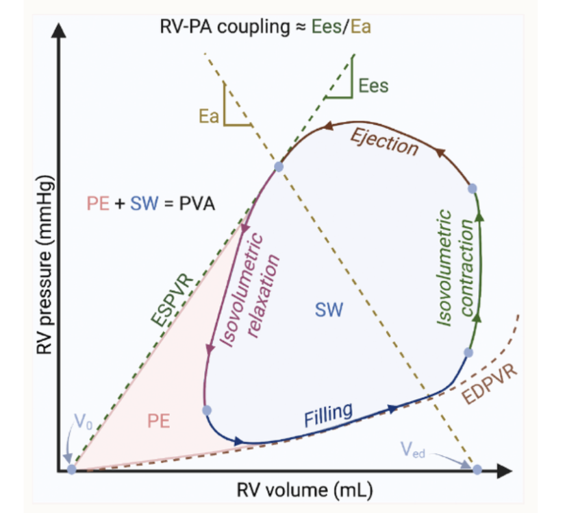
What can you use the ventricular PV loop for? Draw an associated diagram
The ventricular PV loop graphically represents the relationship between pressure and volume in the left ventricle during a single cardiac cycle
This loop is instrumental in understanding cardiac mechanics, as it delineates the phases of ventricular filling, isovolumetric contraction, ejection and isovolumetric relaxation
What are the fundamental units of measurement?
Length: metre (m)
Mass: gram (g)
Volume: litre (L)
How do you convert to:
Milli
Micro
Nano
Pico
Kilo
Milli (m): 10⁻³
Micro (µ): 10⁻⁶
Nano (n): 10⁻⁹
Pico (p): 10⁻¹²
Kilo (k): 10³
What is concentration, and how can it be expressed?
Concentration: the amount of solute dissolved in a given amount of solvent
Concentration can be expressed in a number of ways
Weight in volume (most common)
The number of grams in 100ml of solvent
e.g 5%= 5g per 100ml
Volume in volume
Weight in weight
Molarity
Parts
Mass = concentration x volume
What is meant by ‘dose’?
Dose: the quantity of drug to be delivered to a patient
Why are dilutions used, and what is the dilution factor?
Dilutions are used when the amount of compound required is so small, it cannot be accurately measured
Dilutions often involve combining a volume of a known solution (stock solution) with an appropriate volume of solvent
The dilution factor is the total number of unit volumes, in which your material is dissolved, or how much more concentrated one solution is compared to another
e.g a 1 in 5 dilution combines 1 volume of the solution with four volumes of the solvent
Therefore the dilution factor is 5
Dilution ratio is 1:4
How can you determine the rate of administering fluids, and what is drip rate usually measured in?
The rate of administering intravenous fluids can be determined from:
Volume of fluid needed
Time of administration
Number by the drops per ml delivered by the giving set
Drip rate: usually how many seconds per drop
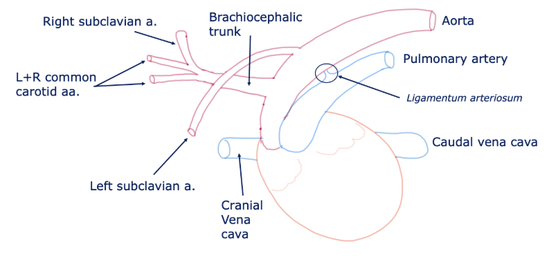
Draw out the basic structure of the cat/dog heart