Family Medicine: EENT
 Call Kai
 Learn
Learn Practice Test
 Spaced Repetition
 Match
Match Flashcards
 Knowt Play
1/91

There's no tags or description
Looks like no tags are added yet.
Name | Mastery | Learn | Test | Matching | Spaced | Call with Kai | Chat |
|---|
No analytics yet
Send a link to your students to track their progress
92 Terms
Barotrauma
Injury to the middle/inner ear from failure to equalize pressure across the tympanic membrane during rapid changes in ambient pressure
-Presentation: ear pain, fullness, hearing loss, vertigo, tinnitus, occasionally bleeding. Can have an effusion, hemotympanum, or TM perforation
-Dx: clinical
-Tx: equalization maneuvers, oral/nasal decongestants, keep ear dry if perfed, no diving/flying and ENT referral
BPPV
Most common cause of peripheral vertigo and is caused by displaced otoconia in the semicircular canals, which is most common in the posterior canal
-Presentation: brief episodes of spinning vertigo triggered by head position changes, no hearing loss, no tinnitus, no aural fullness, nausea is common
-Dx: Dix-Hallpike maneuver reproduces vertigo with upbeat-torsional nystagmus
-Tx: Epley maneuver
Cerumen Impaction
Accumulation of earwax that occludes the external auditory canal, which is often caused by cotton swab use pushing wax inward, hearing aids, narrow canals, or elderly patients
-Presentation: conductive hearing loss, ear fullness, itching, tinnitus, occasional vertigo or cough
-Dx: otoscopy
-Tx: irrigation (CI in TM perforation), manual removal, or cerumenolytics

Conductive
What type of hearing loss is being described?
-Rinne BC > AC
-Weber lateralizes to affected ear
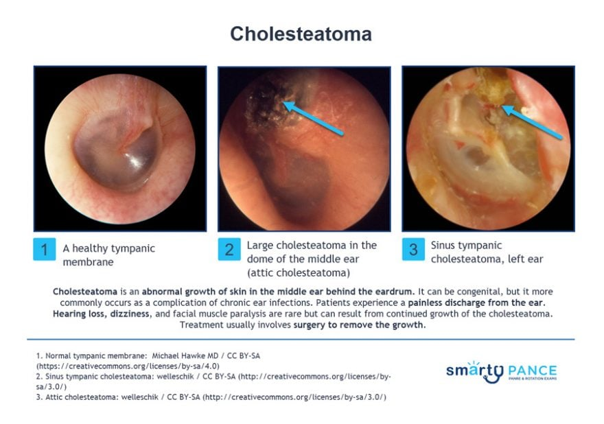
Cholesteatoma
Painless otorrhea, brown/yellow discharge with a strong odor due to chronic eustachian tube dysfunction, leading to chronic negative pressure that causes the formation of granulation tissue
-Presentation: granulation tissue and conductive hearing loss
-Dx: otoscopic visualization, CT and audiogram to evaluate hearing loss
Eustachian Tube Dysfunction
Impaired ventilation/drainage of the middle ear caused by edema, allergies, URIs, smoking, GERD, or anatomic obstruction
-Presentation: ear fullness, popping, crackling, muffled hearing, mild discomfort with pressure changes, autophony
-Dx: clinical, otoscopy shows retracted TM and decreased mobility, negative middle-ear pressure on tympanometry
-Tx: treat underlying cause, autoinsufflation, tympanostomy tubes
Nasopharyngeal carcinoma
In adults with unilateral persistent ETD or effusion, you should perform nasopharyngoscopy to rule out what?
Ear Foreign Body
Most common in children due to playing with beads, beans, and small toys
-Presentation: ear pain, decreased hearing, drainage
-Dx: otoscopy
-Tx: direct removal with alligator forceps, suction, curette, or irrigation. Insects require lidocaine/mineral oil. Button batteries are an emergency
Sensorineural
What type of hearing loss is being described?
-Weber lateralizes to the unaffected ear
-Rinne shows AC > BC
Presbycusis
Most common cause of sensorineural hearing loss
-Presentation: symmetric bilateral high-frequency loss in older adults
ENT referral, steroids
What are the two things that should be done in a patient with sudden sensorineural hearing loss?
Labyrinthitis
Acute onset of continuous vertigo + hearing loss + tinnitus that lasts several days to a week, which is associated with a viral URI and absence of neurologic deficits
-Associated with nausea and vomiting
-Dx: clinical dx, absence of neuro deficits, + Romberg, MRI
-Tx: Meclizine + Zofran + Prednisone
Acute Mastoiditis
Suppurative infection of the mastoid air cells, which is almost always a complication of acute otitis media
-Pathogens: Strep Pneumo (MC)
-Presentation: postauricular pain, erythema, swelling, and fluctuance with a forward-displaced auricle
-Dx: clinical + CT temporal bone with contrast
-Tx: admit + IV ceftriaxone + ENT consult

Meniere Disease
Excessive endolymph fluid in cochlea overstimulates hairs causing vertigo and sudden hearing loss with aural fullness, where the vertigo can last hours
-Presentation: low-frequency hearing loss, tinnitus with aural fullness, vertigo
-Dx: clinical diagnosis
-Tx: low salt diet + diuretics
Otitis Externa
Infection of the external auditory canal, most often due to Pseudomonas aeruginosa
-Presentation: ear pain and pruritus with pain on tragal manipulation, edematous canal with white/purulent debris, conductive hearing loss
-Dx: clinical, ESR, CT/MRI of temporal bone
-Tx: fluoroquinolone drops
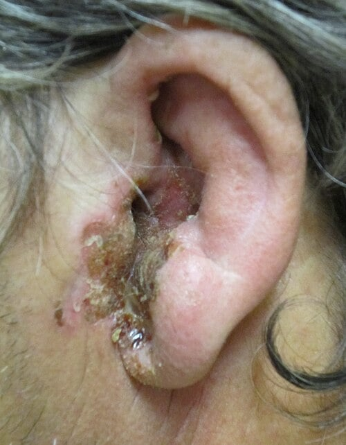
Malignant Otitis Externa
Ear pain out of proportion to exam findings and granulation tissue on the canal floor at the bony-cartilaginous junction, which is seen in elderly or immunocompromised patients
-Tx: admit + IV cipro or Zosyn
Aminoglycoside
What class of ear drops should never be used if the TM is perforated, tubes are present, or TM cannot be visualized?
Otomycosis
Fungal otitis externa, which typically follows prolonged topical antibiotic use or in humid climates
-Presentation: pruritus, fullness, weeping, and hearing loss with a swollen, moist canal
-Dx: clinical
-Tx: canal cleaning + topical antifungal
Acute Otitis Media
Bacterial infection of middle ear fluid that typically follows a viral URI, peaking at 6-24 months
-Presentation: middle ear effusion, bulging TM, otorrhea, mild bulging with acute otalgia, conductive hearing loss
-Pathogens: H. influenzae or S. Pneumo
-Dx: otoscopy
-Tx: high dose amoxicillin

Cefdinir
What agent should be given for AOM if patient has a mild PCN allergy?
Amox-Clav
What antibiotic should be given if a beta-lactam was given in the past 30 days, concurrent conjunctivitis, or amoxicillin failure for AOM?
Otosclerosis
Autosomal-dominant disorder causing abnormal bony remodeling at the stapes footplate, fixing the ossicles and producing a progressive conductive hearing loss
-Dx: audiometry (Carhart notch at 2000 Hz)
-Tx: hearing aids, stapedectomy

Tinnitus
Sensation of sound in the absence of an external acoustic stimulus is often described as ringing, hissing, buzzing, or whooshing
-Dx: refer to audiology, rule out acoustic neuroma with MRI
-Tx: identify and treat the cause
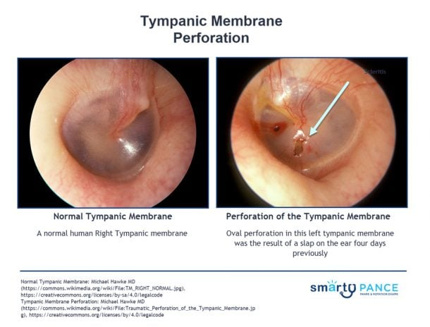
TM Perforation
Pain, otorrhea, and hearing loss/reduction
-Dx: clinical
-Tx: Floxin drops, surgery
Viral Conjunctivitis
A red eye with copious watery discharge, scant mucoid discharge
-MC due to adenovirus
-Tx: supportive care

Bacterial Conjunctivitis
A red eye with purulent discharge and crusting that is usually worse in the morning
-Can be due to S. pneumo, S. aureus, N. gonorrhea
-Tx: gentamicin/tobramycin drops, trimethoprim/polymyxin B drops

Fluoroquinolone drops
What is the treatment of choice for bacterial conjunctivitis in someone who wears contacts?
Allergic Conjunctivitis
Red eyes, itching and tearing that is usually bilateral with cobblestone mucosa on the inner/upper eyelid
-Tx: systemic antihistamines and topical antihistamine like azelastine

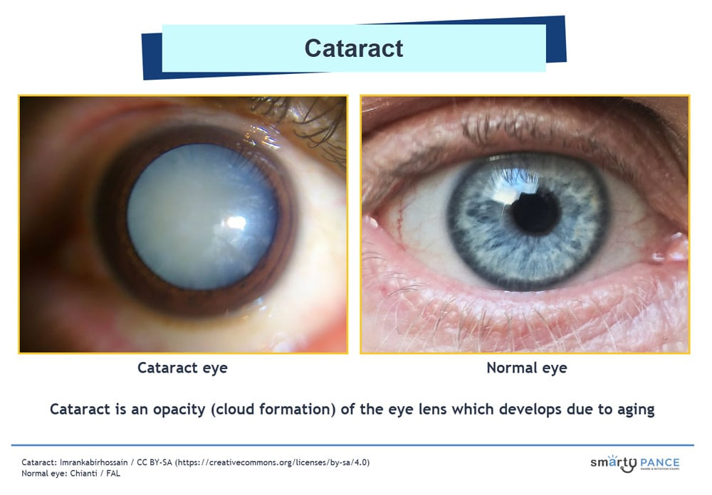
Cataract
Opacification of the crystalline lens producing gradual painless vision loss, which is the MCC of reversible visual impairment worldwide
-RF: aging, diabetes, smoking, chronic steroid use, UV exposure, trauma
-Presentation: gradual painless decreased visual acuity, glare, halos around lights, faded color vision, and second sight. Diminished red reflex, cloudy lens on slit lamp
-Dx: clinical
-Tx: early can be managed with glasses, definitive is IOL implantation
Corneal Abrasion
Sudden onset of pain, photophobia, tearing, foreign body sensation, blurring of vision, and/or conjunctival injection
-Dx: fluorescein staining shows increased absorption in the devoid area
-Tx: antibiotic eye ointment, no patching
Corneal Ulcer
White spot on the surface of the cornea that stains with fluorescein, which is seen in contact lens wearers due to a deep corneal infection
-Dx: fluorescein stain, cultures before starting abx
-Tx: immediate referral to ophthalmology, topical abx
Keratitis
Inflammation of the cornea, which can be bacterial, viral, fungal, or UV light related
-Dx: slit lamp exam with fluorescein staining, corneal scrapings for culture and gram stain
-Tx: depends on etiology
HSV Keratitis
Recurrent unilateral eye pain + photophobia + dendritic ulcer with fluorescein
-Dx: slit lamp exam with fluorescein staining
-Tx: topical trifluridine or ganciclovir gel + oral antiviral, avoid topical steroids

Pterygium
Elevated, superficial, fleshy, triangular-shaped “growing” fibrovascular mass that is most common in the inner corner of the eye
-Associated with increased sun exposure and climates where the wind, sand, and dust is high
-Presentation: extends onto the cornea and can affect vision
-Dx: clinical
-Tx: artificial tears, NSAID drops for inflammation, UV protection

Pinguecula
Yellowish, raised conjunctival nodule on the bulbar conjunctiva that does not cross the limbus onto the cornea
-Dx: clinical
-Tx: artificial tears, no surgery unless cosmetic, UV protection
Open-Angle Glaucoma
Most common type of glaucoma that affects the outflow of aqueous humor
-Presentation: peripheral to central gradual visual loss in an older African American individual
-Dx: tonometry shows increased IOP, peripheral field testing, optic disc monitoring
-Tx: prostaglandin analog drops (latanoprost), topical timolol, trabeculoplasty to open meshwork
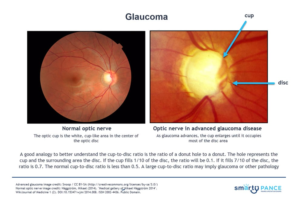
Glaucoma
All patients at age 40 should be screened for what eye condition?
-Routine fundoscopy with cup to disc ratio
Acute Angle-Closure Glaucoma
Type of glaucoma where the iris is against the lens
-Presentation: injected conjunctiva, steamy cornea, and fixed dilated pupil after acute loss of vision, nausea, and vomiting
-Dx: tonometry, gonioscopy is gold standard
-Tx: acetazolamide IV, topical timolol, pilocarpine, peripheral iridotomy
Iritis
Inflammation of the iris, often associated with HLA-B27 spondyloarthropathies (ankylosing spondylitis, IBD, psoriatic arthritis, sarcoidosis)
-Presentation: unilateral red painful eye, photophobia, blurred vision, tearing. Ciliary flush, miotic and irregular pupil, cells and flare in the anterior chamber, consensual photophobia is classic
-Dx: slit lamp exam
-Tx: topical corticosteroids + cycloplegic, ophthalmology referral

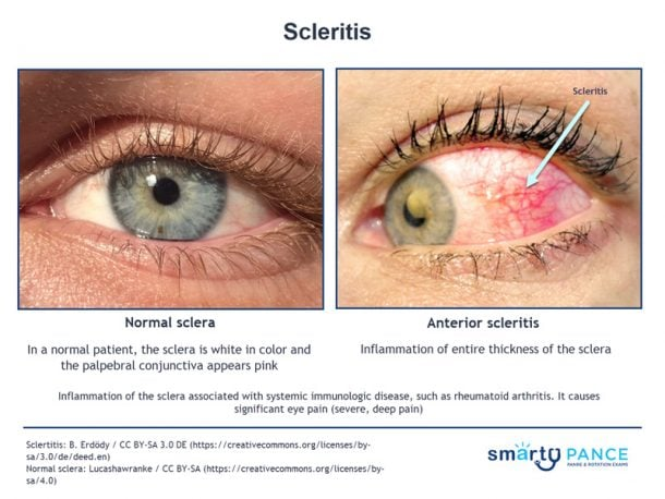
Scleritis
Inflammation of the sclera, which is usually severe, vision-threatening, and associated with systemic autoimmune disease (SLE, RA)
-Presentation: severe deep boring eye pain that radiates to the face/jaw which is worse at night and wakes patient up from sleep, violaceous scleral hue, tearing, photophobia, redness does not blanch with topical phenylephrine
-Dx: slit lamp exam
-Tx: oral NSAIDs, systemic corticosteroids + immunosuppressants
Uveitis
Inflammation of the uveal tract, which is classified by location
-Types: anterior (MC), intermediate, posterior, and panuveitis
-Presentation: pain, photophobia, floaters, blurred vision
-Dx: slit lamp + dilated fundus exam
-Tx: topical corticosteroids + cycloplegics, steroids

Dacryoadenitis
Inflammation of the lacrimal glands, usually caused by bacteria or a virus that initiates the inflammation
-Dx: clinical, CT orbits if chronic
-Tx: rest and warm compress if viral
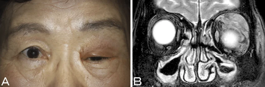
Dacryocystitis
Infectious obstruction of the nasolacrimal duct
-Dx: tear duct massage to express material for culture and gram stain
-Tx: systemic antibiotics if acute, surgery if chronic

Blepharitis
Chronic inflammation of eyelid margins caused by seborrhea, staph, or strep and is a sign of Meibomian gland dysfunction
-Presentation: crusting, scaling, red-rimming of the eyelid and eyelash flaking, adherent eyelashes, hyperemic lid margins, dandruff-like deposits and fibrous scales, slightly injected conjunctiva
-Dx: slit-lamp exam
-Tx: warm compresses, daily wash with baby shampoo, topical antibiotics

Hordeolum
Painful, warm, swollen red lump on the eyelid with S. aureus being the most common etiology
-Dx: clinical
-Tx: warm compress and topical antibiotics

Chalazion
Painless, chronic granulomatous inflammation of a Meibomian gland due to obstruction
-Presentation: firm, rubbery, non-tender nodule on the eyelid that is painless and cold to the touch
-Tx: warm compress + gentle massage (most resolve in weeks), referral to ophthalmology if > 6 weeks

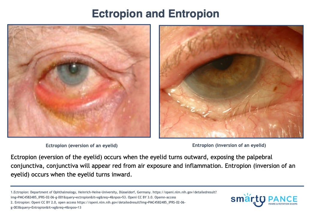
Ectropion
Eversion of the eyelid that occurs when the eyelid turns outward, exposing the palpebral conjunctiva
-Presentation: outward turned eyelid with red conjunctiva
-Dx: clinical
-Tx: tear supplements, ocular lubricants at night, surgery is definitive

Entropion
Inversion of the eyelid that occurs when it turns inward, most commonly due to age-related tissue relaxation
-Dx: clinical
-Tx: tear supplements and ocular lubricants at night, surgery is definitive
Ocular Foreign Body
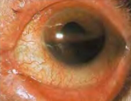
Superficial conjunctival debris to intraocular foreign body in someone with a history of high-velocity activity, blast injury, or eye protection failure
-Presentation: pain, photophobia, foreign-body sensation, tearing, blurred vision
-Dx: slit-lamp, fluorescein stain, Seidel test positive indicates emergency, CT orbits
-Tx: irrigation if superficial, shield the eye if suspected globe penetration, irrigation if chemical burn

Hyphema
Blood in the anterior chamber of the eye, which may cover part or all of the iris
-Dx: orbital CT if indicated, ophthalmology consult
-Tx: elevate head at night at 30 degrees, Tylenol for pain, NO NSAIDs
Optic Neuritis
Inflammatory/demyelinating disease of the optic nerve, which is most common in young women with multiple sclerosis
-Presentation: unilateral subacute vision loss + pain with eye movement + color desaturation, Marcus Gunn pupil on swinging-flashlight test, normal exam otherwise
-Dx: MRI orbits with contrast
-Tx: IV methylprednisolone then oral taper

Preseptal Cellulitis
Infection of eyelid and soft tissues anterior to orbital septum
-Presentation: eyelid swelling/erythema, no proptosis, no ophthalmoplegia, no vision loss
-Dx: clinical, CT orbits with contrast
-Tx: outpatient oral abx (clinda, amox-clav, bactrim)
Orbital Cellulitis
Infection posterior to orbital septum, which is vision-threatening and can spread intracranially
-MC from ethmoid sinusitis
-Presentation: proptosis, pain with eye movement, ophthalmoplegia, decreased visual acuity, RAPD, fever
-Dx: CT with contrast
-Tx: admit for IV abx (vancomycin + ceftriaxone)

Papilledema
Optic disc swelling that is caused by increased intracranial pressure, which is usually bilateral and can occur over a period of hours to weeks
-Caused by malignant hypertension, brain tumor/abscess, meningitis, cerebral hemorrhage, encephalitis
-Presentation: bilateral, develops slowly, disc appears swollen, margins blurred, obliteration of the vessels
-Tx: tx underlying cause

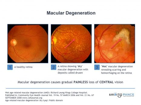
Macular Degeneration
Gradual painless loss of central vision
-Presentation: metamorphopsia on Amsler grid
-Dx: fundoscopic findings are diagnostic
-Tx: VEGF (wt), zinc + antioxidant vitamins (dry)

Dry
What type of macular degeneration is being described?
-Most common
-Atrophic changes with age, which is a slow gradual breakdown of the macula
-Drusen → yellow retinal deposits
Wet
What type of macular degeneration is being described?
-Hemorrhage, neovascularization
-New abnormal vessels grow under the retina, which leaks and bleeds, causing retinal scarring
-Tx: VEGF inhibitors, photodynamic therapy
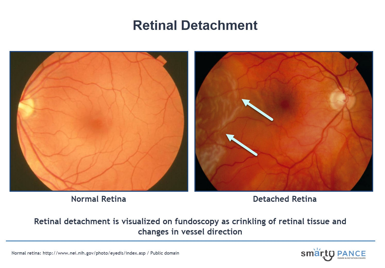
Retinal Detachment
Separation of the retina from the pigmented epithelial layer, causing the detached tissue to appear as a flap in the vitreous humor which can occur spontaneously or secondary to trauma / extreme myopia
-Presentation: vertical curtain coming down across the field of vision may sense floaters or flashes at the onset, loss of vision over several hours, myopia, peripheral visual field defects
-Dx: fundoscopy
-Tx: stay supine with head turned towards the side of the detached retina, pneumatic retinopexy
Central Retinal Artery Occlusion
Occluded flow through the CRA
-Presentation: sudden, painless, unilateral, and usually severe vision loss
-Dx: fundoscopy shows cherry red spot and pale opaque fundus with red fovea and arterial attenuation, rule out carotid artery stenosis with carotid US
-Tx: emergent ophthalmology consult, reduction of IOP with ocular hypotensive drops, intermittent digital massage

Central Retinal Vein Occlusion
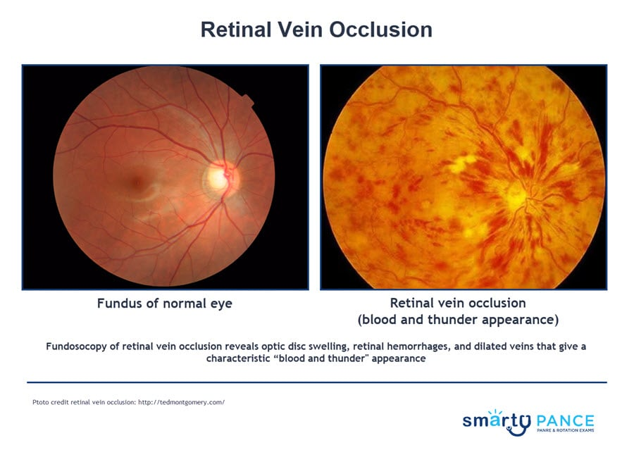
“Blood and thunder fundus”, which usually occurs secondary to a thrombotic event that is most common in ages 50+, associated with hypertension, glaucoma, diabetes, and hyperviscosity states
-Dx: fundoscopy shows retinal hemorrhages, optic disc swelling
-Tx: vision resolves with time, workup for thrombosis
Diabetic retinopathy
What is the leading cause of blindness in adults?
Hypertensive Retinopathy
Retinal damage caused by high blood pressure
-Presentation: blurred vision, floaters, and difficulty seeing in low light in a patient with hypertension
-Can lead to blindness
Nonproliferative
Early stage of diabetic retinopathy in which the symptoms are mild or nonexistent
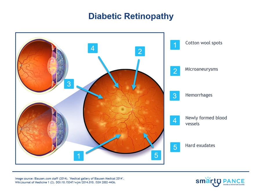
-Fundoscopy shows → microaneurysms, hard exudates, cotton wool spots, blot and dot hemorrhages, and venous dilation
-Tx: manage blood sugar, get yearly dilated eye exam
Proliferative
Advanced form of diabetic retinopathy characterized by by neovascularization on fundoscopic exam
-Tx: blood glucose control, retinal laser photocoagulation, yearly eye exam

Myopia
Nearsightedness, where the eyeball is too long
-Distant objects are blurry but close objects are okay
-Treat with concave lenses or LASIK
Hyperopia
Farsightedness, where the eyeball is too short
-Near objects are blurry
-Treat with convex lenses
Presbyopia
Age-related lens stiffening (> 40 years), need reading glasses
Amblyopia
Decreased vision in an anatomically normal eye from disuse during visual development, which can be caused by strabismus, refractive error, and deprivation
-Tx: patching the good eye
Strabismus
Misaligned eyes, which is diagnosed with corneal light reflex and cover-uncover test
Amaurosis Fugax
Transient monocular vision loss lasting minutes, typically due to a carotid embolus from ipsilateral atherosclerotic plaque
Vestibular Schwannoma
Benign tumor of cranial nerve VIII
-Presentation: unilateral sensorineural hearing loss, tinnitus, disequilibrium
-Dx: MRI with contrast
-Tx: surgery or stereotactic surgery
Sinusitis
Sinus pain/pressure that is worse with bending down/leaning forward
-Usually viral but can progress to bacterial
-Dx: plainview x-ray but CT is gold standard
-Tx: Amox-Clav or Doxy
Allergic Rhinitis
Clear nasal drainage and pruritis in someone with a history of allergies
-Presentation: drainage, pale bluish boggy mucosa, allergic shiners
-Dx: history
-Tx: avoid allergens, antihistamines, Flonase (intranasal ICS) are first line
Epistaxis
Nose bleeds due to nasal trauma, dryness, hypertension, cocaine, or alcohol
-Occur most often at Kisselbach’s or Wodruff’s plexus (anterior vs posterior)
-Tx: direct pressure while seated and leaning forward, anterior nasal packing + antibiotics to prevent TSS, Ayr gel for dryness, and posterior balloon packing

Nasal Polyps
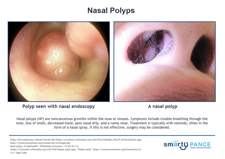
Teardrop-shaped growths that form int he nose or sinuses, which is part of Samter’s triad with asthma and aspirin sensitivity
-Presentation: growths in the nose + chronic congestion + decreased sense of smell
-Dx: clinical
-Tx: topical nasal corticosteroid is initial treatment of choice, oral steroids can help reduce the size, surgery if polyps are large
Aphthous Ulcers (canker sore)
Single or multiple small, shallow ulcers with a yellow-gray fibrinoid center with red halos
-Dx: history and physical, biopsy if ulcers lasting more than 3 weeks
-Tx: viscous lidocaine 2-5% applied to ulcer QID after meals until healed

Dental Abscess
Localized pus collection from untreated dental caries, fractured teeth, or periodontal disease
-Presentation: severe throbbing tooth/jaw pain, swelling, foul breath, fever, tenderness on percussion, fluctuant swelling
-Complication: can lead to Ludwig angina (emergency)
-Dx: clinical
-Tx: I&D by dentist, clindamycin or amox-clav

Dental Caries
Tooth decay caused by acid-producing bacteria fermenting dietary sugars and demineralizing enamel, which is the most common preventable chronic disease in childhood
-Presentation: asymptomatic chalky white spots → brown discoloration on teeth → sensitivity to hot/cold/sweets, cavitation, toothache. Can lead to pulpitis and pulp necrosis
-Dx: clinical, dental x-rays
-Tx: restoration, prevention with fluoride every 3-6 months from 6 mo to 5 years

Fluoride
What should be applied in the primary care office for children aged 6 months to 5 years to help prevent dental caries?
Acute Epiglottitis
Life-threatening cellulitis of the epiglottitis and supraglottic structures with impending airway obstruction
-Presentation: drooling, dysphagia, dysphonia, distress, tripod position. Abrupt onset of symptoms over hours, high fever, sore throat, and toxic appearance
-Dx: do not examine the throat, lateral neck x-ray shows thumbprint sign, definitive dx is direct laryngoscopy in the OR
-Tx: secure the airway first, IV ceftriaxone

HSV-1
What is the most common cause of viral stomatitis?
Herpetic Gingivostomatitis
HSV-1 infection in young children (6 mo to 5 years)
-Presentation: fever, irritability, refusal to feed, painful vesicles and ulcers throughout the oral cavity + gingivitis + cervical adenopathy
-Dx: clinical, PCR is gold standard
-Tx: oral acyclovir, watch for dehydration
Cold Sores
Blisters appearing at the vermilion border of the lips, associated with a tingling prodrome
-Presentation: prodrome → grouped vesicles on an erythematous base → crusting that is triggered by sun, stress, illness, fever, or menses
-Dx: clinical, HSV PCR
-Tx: valacyclovir 2 g PO BID x 1 day, suppressive therapy if > 6 outbreaks per year

Herpetic Whitlow
HSV vesicles on fingers
Laryngitis
Acute hoarseness following a URI, which is almost always viral (M. catarrhalis and H. influenzae)
-Dx: clinical diagnosis, laryngoscopy required if > 3 weeks of symptoms
-Tx: vocal rest, supportive therapy, PO/IM steroids may help recovery in performers
Leukoplakia
Premalignant white patch on oral mucosa that cannot be wiped or scraped off and cannot be attributed to another disease
-RF: tobacco, alcohol, HPV-16, chronic irritation
-Presentation: white patch on oral mucosa, red patch, or speckled on lateral tongue / floor of mouth / ventral tongue
-Dx: excisional / incisional biopsy of any persistent oral white patch > 2 weeks
-Tx: eliminate risk factors, surgery if cancer

Thrush
Opportunistic infection by Candida albicans of oral mucosa
-RF: infants, elderly, corticosteroid use, antibiotics, diabetes, immunocompromised
-Types: pseudomembranous, erythematous, angular cheilitis, denture stomatitis
-Dx: clinical, KOH prep
-Tx: nystatin swish-and-swallow, systemic for moderate to severe with fluconazole

Peritonsillar Abscess
Penetration of infection through the tonsillar capsule and the involvement of neighboring tissue
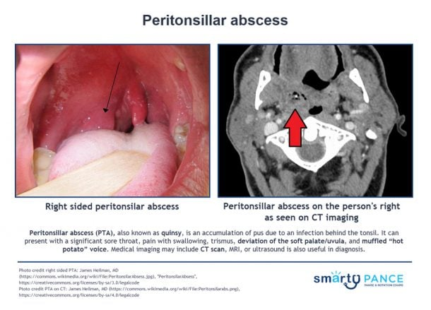
-Presentation: severe sore throat, lateral uvula displacement, and a bulging tonsillar pillar. Associated with a muffled “hot potato” voice
-Dx: x-ray, CT, or US of neck. Needle aspiration of the mass and cultures
-Tx: aspiration, I&D, parenteral amoxicillin/clindamycin or PO if less severe, tonsillectomy
Retropharyngeal Abscess
Deep neck space infection in the potential space between the posterior pharyngeal wall and the prevertebral fascia, which is MC in children 2-4 years
-Presentation: fever, severe sore throat, dysphagia, drooling, neck stiffness, torticollis, muffled voice, and stridor
-Dx: lateral neck x-ray shows widened prevertebral soft tissue, contrast CT neck is imaging of choice
-Tx: admit + airway monitoring + IV zosyn, clindamycin, or unasyn, surgical drainage by ENT
Pharyngitis
Sore throat, which is usually due to a viral infection
-Dx: Centor Score for strep throat
Parotitis
Inflammation of one or both parotid glands
-Presentation: fever and chills, periauricular, mandibular pain, and swelling. May also have trismus, dysphagia, and purulent drainage
-Causes: S. aureus, parainfluenza, mumps
-Dx: clinical, US, CT scan
-Tx: self-limiting

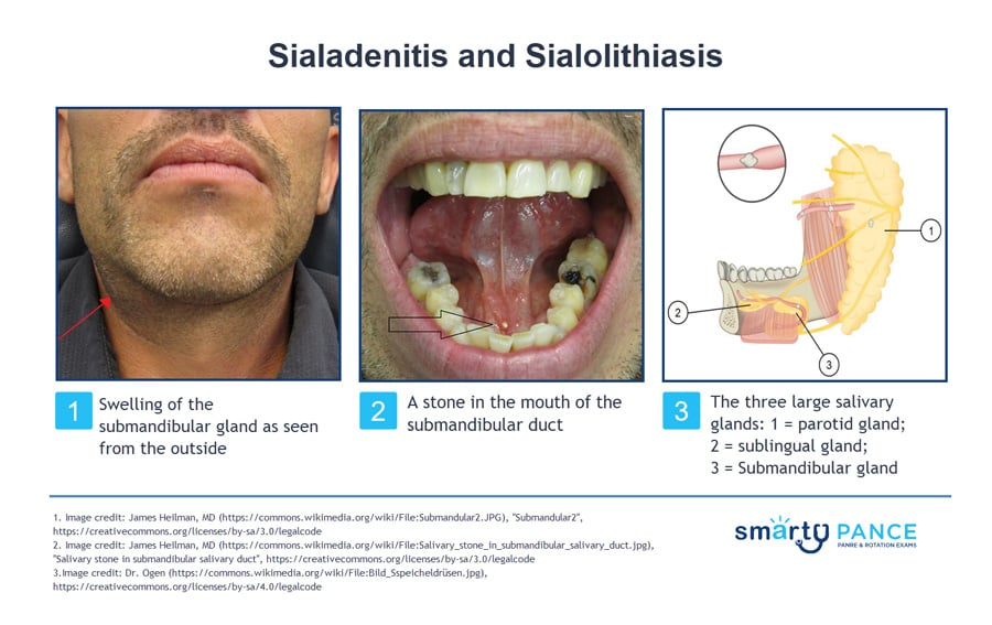
Sialadenitis
Bacterial infection of a salivary gland usually caused by salivary stones
-Presentation: acute swelling of the cheek, which worsens with meals
-Dx: CT, US, MRI
-Tx: IV abx, hydration, warm compresses, gland massage