HN220 - Unit 3 The Nervous System
1/168
There's no tags or description
Looks like no tags are added yet.
Name | Mastery | Learn | Test | Matching | Spaced |
|---|
No study sessions yet.
169 Terms
What are the Four Functional Regions that all neurons have?
1. Input
2. Trigger = axon hillock
3. Conducting = connection of input to output region
4. Output
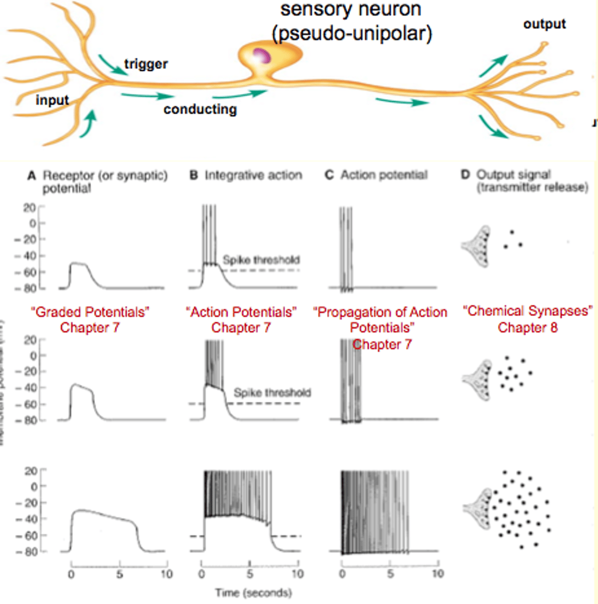
Sensory = pseudounipolar neuron, cell body is in the middle of axon
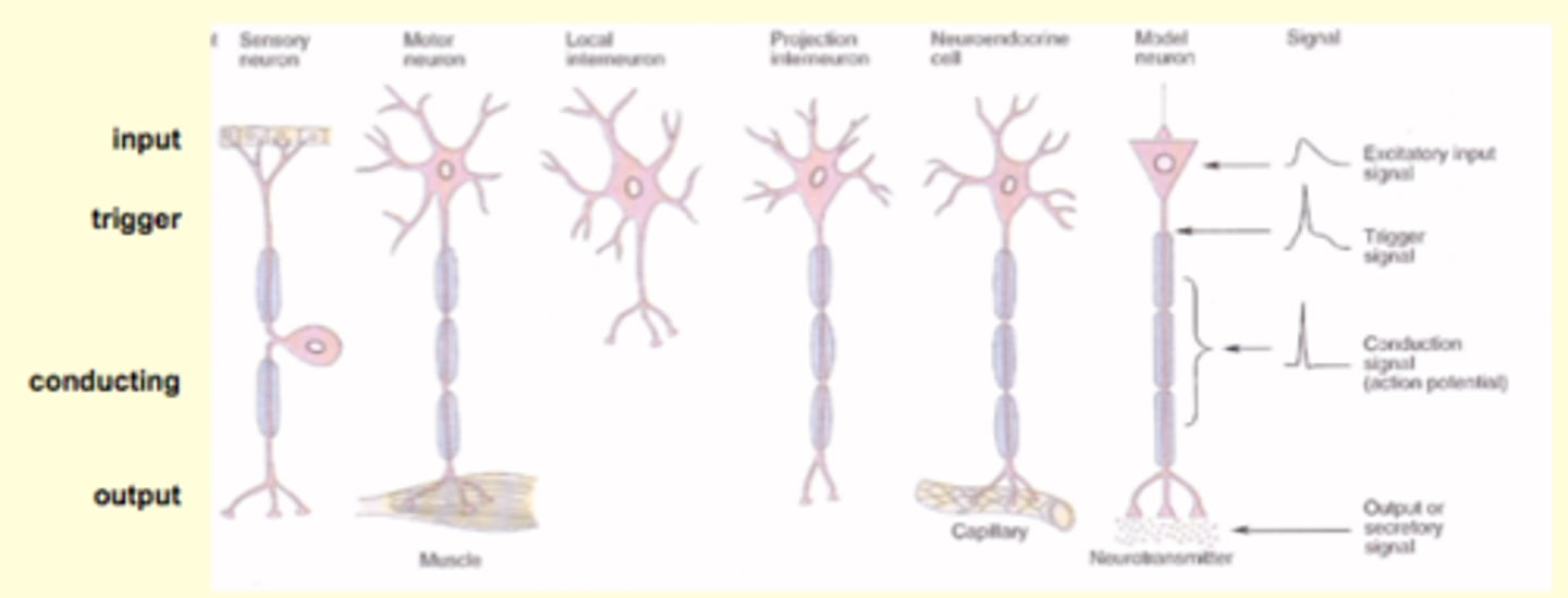
Describe the Sensory
- nucleus in the middle is irrevelant to function(?)\

Input = Receptor = synaptic potential
Triggor region = axon hillockk = action potential
Conductial region = no potential
Output signal =
Why is the resting membrane potential -70 mV?
potential = voltage across the membrane
Why is resting -70?
1. Ions are present in different concentrations in the fluid compartments on either side of the cell membrane
2. The cell membrane is selectively permeable
3. There are two different forces that determine the direction that an ion travels across a cell membrane
If the membrane was only permeable to K+ then the resting potential would the equilibrium force of K+ (E = -90 mV)
However the cell is a little permeable to Na+ (E = +60 mV)
Resting is more similar to E = K+, because it is more permeable to K+
What are Neuronal Ion Channels?
leak channels - open all the time (a lot for K+) which means that the membrane is fairly permeable for K+
Voltage-gated channels: changes from rest (-70 mV) to something, the channels will open; what opens them is the change in membrane potential
Ligand-gated channels: opens when something binds to them
What is the GHK Equation?
- similar to Nernst Equation (x 3)
- have to know permeability of each ion
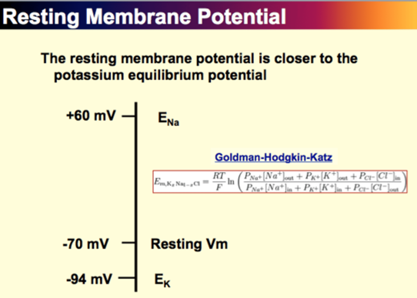
Resting Membrane Potential
- K+ channels >> Na+ channels
- membrane is more permeable to K+ (~ 20x)
- even with driving forces, bc of the dispartity in channel numbers, the outward movement (K+) is more than the inward movement
- membrane becomes more negative (+ charges leaving)
- Resting is -70 mV - much closer to the equilibrium potential of K+ than Na+
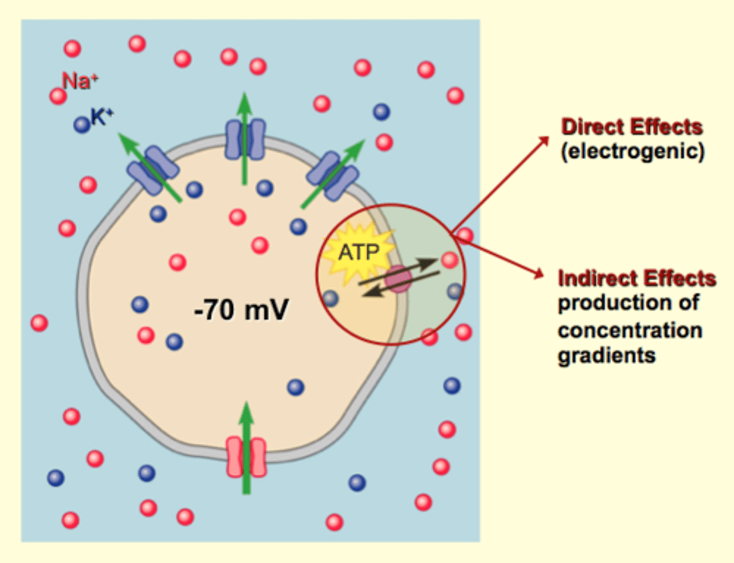
- neither K+ nor Na+ are at equilibrium - therefore forces are acting on them, causing Na+ to leak out which creates a membrane potential
- pumps avert the possibly of a membrane potential of zero, by establishing and maintaining concentration gradients
Vocabulary
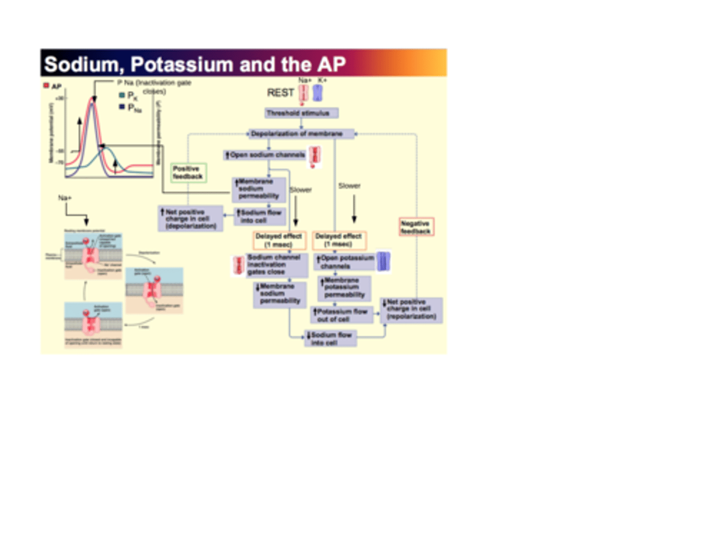
What is the Action Potential?
The self-propagating nerve signal from axon hillock to axon terminals
- once initiated it does not change the amphitude and frequency
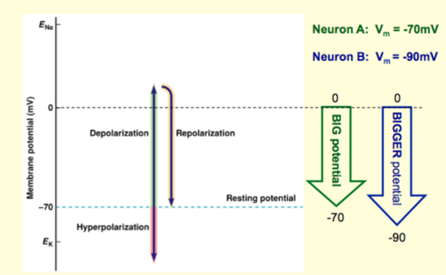
1. Rapid depolarization (-70 mV to +30 mV) - dramatic increase in Na+ permeability
2. Repolarization (+30 mV to -70 mV) - back to rest with open K+ channels
3. After hyperpolarization (approaches -94 mV) - K+ remains elevated and membrane potential is even more negative
What are Graded Potentials?
Small changes in membrane potential that occur when ion channels open or close in response to a stimulus acting on the cell
- varies or is "graded"
- can be EXCITATORY or INHIBITORY
- gets smaller as it travels - decremental - bc of leaks
- if it's big enough (threshold) when it hits the trigger region, it will create an action potential
Depolarization phrase
- increase in sodium permeability (ie. channel opens)
- potassium permeability increases
- membrane permeability is changed during action potential
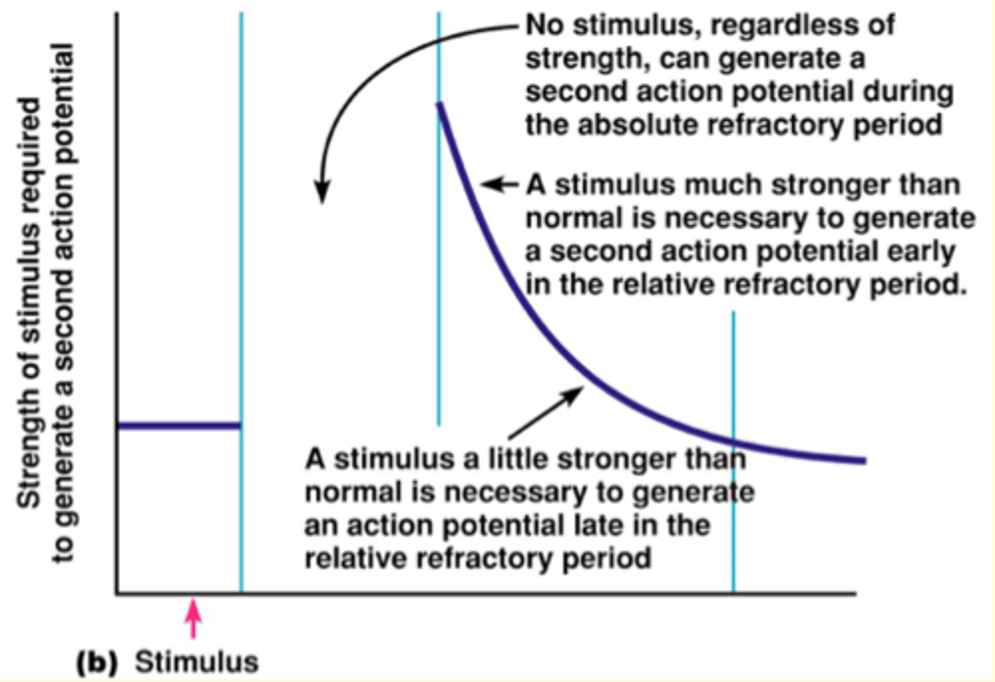
Refractory Periods
- establish action potential's All-or-None property, frequency at which an action can be generated by a single neuron, and unidirectional propagation along an axon
- important for coding of information that arrives in the form of action potentials
**prevent action potentials from travelling in the reverse direction
Back to the Big Picture
Bigger input (larger stim.) leads to higher firing rates
*Because bigger inputs overcome the relative refractory period
The bigger the depolarization: (sodium channels are open) the earlier in Relative Refractory Period you can overcome potassium current (which wants -90 mV)
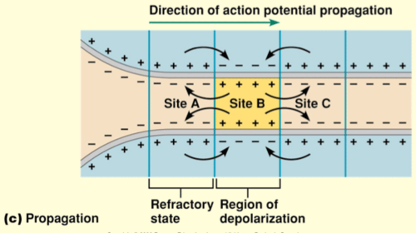
Initiation of Conduction
When an action potential occurs at site A on the membrane, separation of charge occurs in the intracellular fluid and extracellular fluid; the charge separation serves as a force for current to move; the local currents produced by positive ions moving toward the negative regions of the intracellular fluid
Electric conduction - current travels to adjacent areas of the cell membrane causing voltage changes in these areas
No action potential at original site @ initiation
Why doesn't it go backwards: Can't get an action potential if you don't have a voltage-gated sodium channels
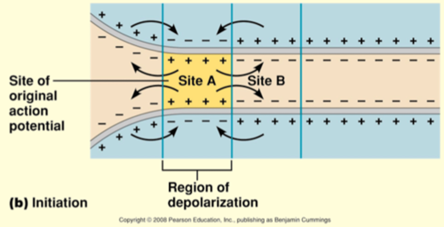
Propagation of Conduction
Not moving backwards bc these voltage gated are closed
The current depolarizes the adajcent region of the membrane (site B) to threshold, eliciting an action potential there
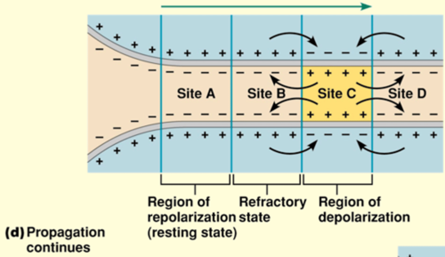
Propagation continues
Not moving backwards bc these voltage gated are closed - refractory
Depolarization of adjacent regions continues until the action potential has been propagated all the way to the axon terminal
What is Myelin? And what is Saltory Conduction?
- fatty
- produced by Shawann cells
- ions cant pass through
When you gave an action potential (bc we have Na gated voltage channels):
- wave of depolar wil ltravel via electricial conudction move along myleinated region not
- prevents the loss of charge as it travels the length of the axon
- until it reaches un-myelinated where neurons will cluster Na gated voltage channels
- wave (starts at +30 mV) lowers (ie. +10 mV) as long it is above threshold
- NO JUMPING
- just movement of positive of charge and reinitiation of potential
Why isn't every axon Myleinated?
Maintainence is energy consuming and damage is extensive
The Length Constant
- current travels down both directions (getting more dispersed as you go along)
- membrane is a bad insulator aka charge leaks OUT
- bc of this, membrane potential drops as u travel length of neuron
distance that current travels, before the neuron drops down to 37% of value - The Length Constant
further/long length constant = current is faster
essentially a measure of speed
What are the determinents of The Length Constant?
1. resistance across the membrane or r m (little a) (flow, solution = mylein, which has more resistance)
2. axial resistence r a (little a) - resistence to current flow; large neurons = lower axial resistence
Why do large neurons have lower axial resistence?
ie. bigger the diameter, the less resistence (traffic/lanes/resistence to traffic flow)
How do nerves generate signals?
[ image]
if sum = -55 = action potential
![<p>[ image]</p><p>if sum = -55 = action potential</p>](https://knowt-user-attachments.s3.amazonaws.com/afba5474-4849-4a9d-8489-39d39af1833b.png)
Neural signaling and synaptic function
depolarization = excitatory post synaptic potentials (membrane becomes less polarized (negative)
hyperpolarization: inhibitatory post synaptic potentials - membrane becomes more polarized (negative)
post synpatic = bc membrane
Neural signalling
action potential (-55 mV) that goes all the way to the other side of cell
Ca+ acts as a signal, thru a series of proteins Ca+ will trigger synaptic vesicles to travel to active zones (adjacent to post synaptic membrane) of synaptic terminal
proteins help diffuse - so we get exocytosis
synaptic cleft -
bind to receptor and illicit response to target cell
how to get rid of neurotransmitter after? - sometimes just as important/more than reaching target cell:
6. neurotransmitter can bind to enzyme and will break down more quickly
7. re-uptake metabolic byproducts of neurotransmitter
8. (least efficient) let it diffuse away
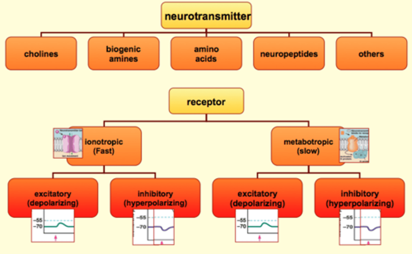
Post synaptic effects of neurotransmitters
neuropeptide: ADH or vassopressin
amino acid: GABA glycine
CH: Ach
biogenic amines: Epi Ne
Some receptors are build for speed/fast not as long lasting responses
Others are more complex and slow longer lasting effects
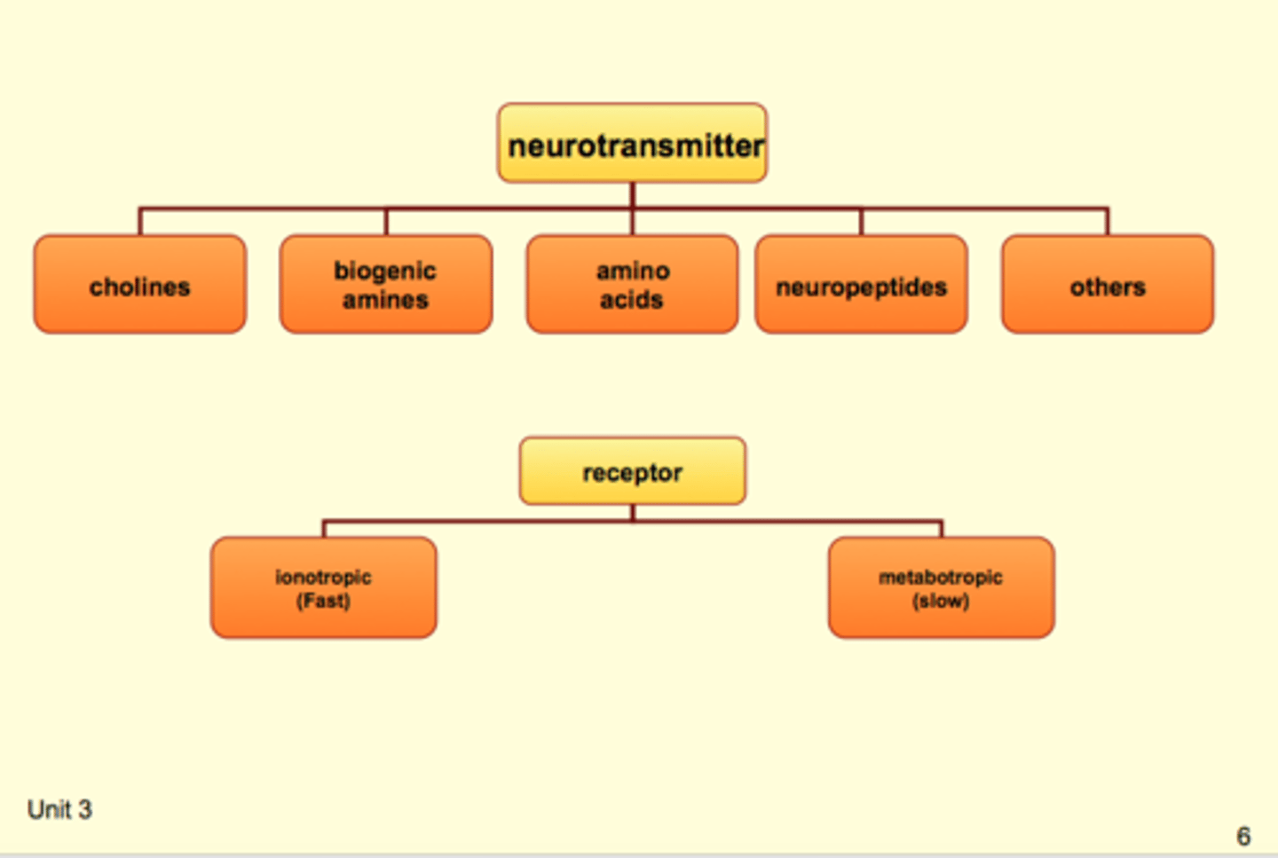
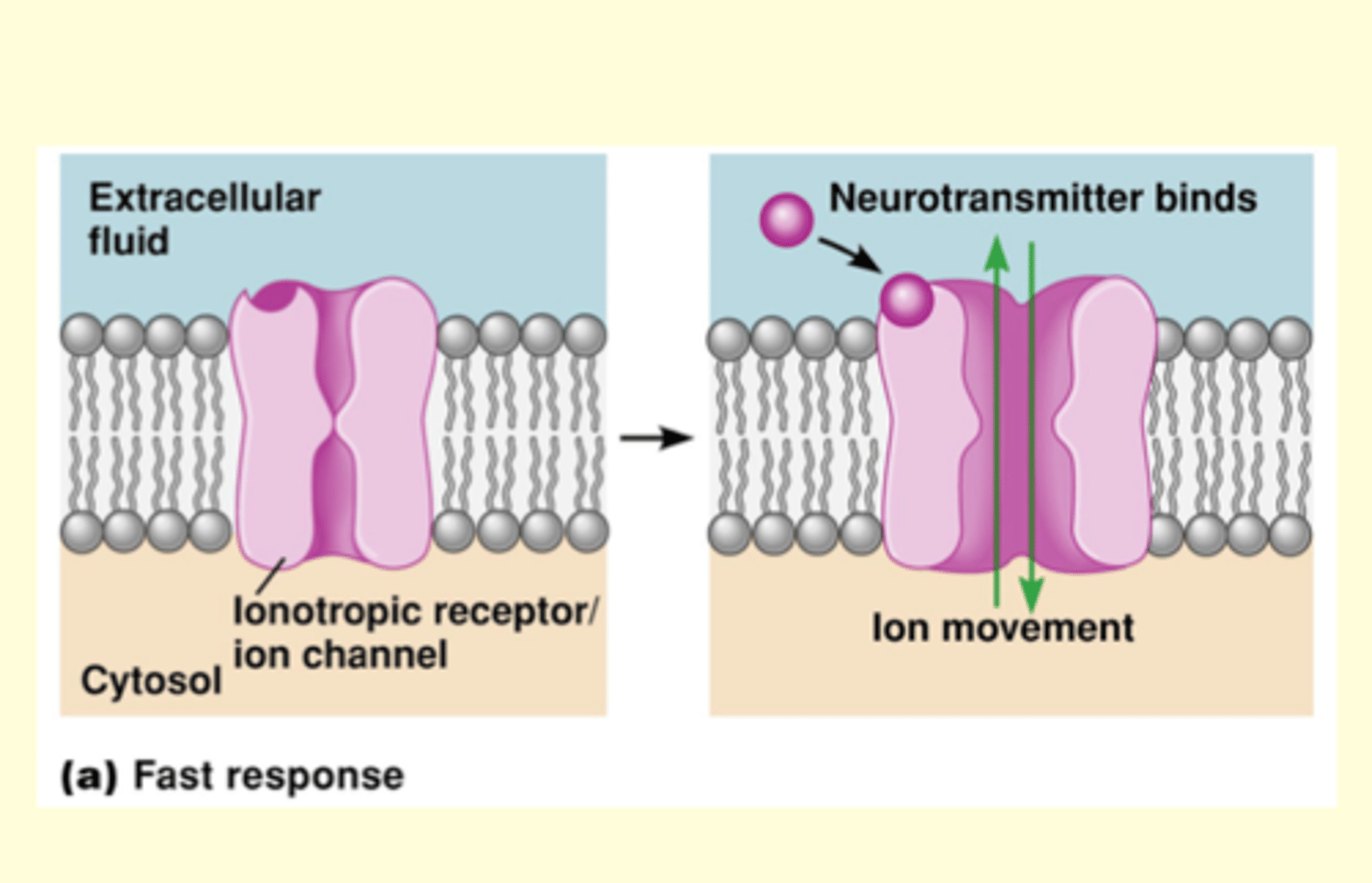
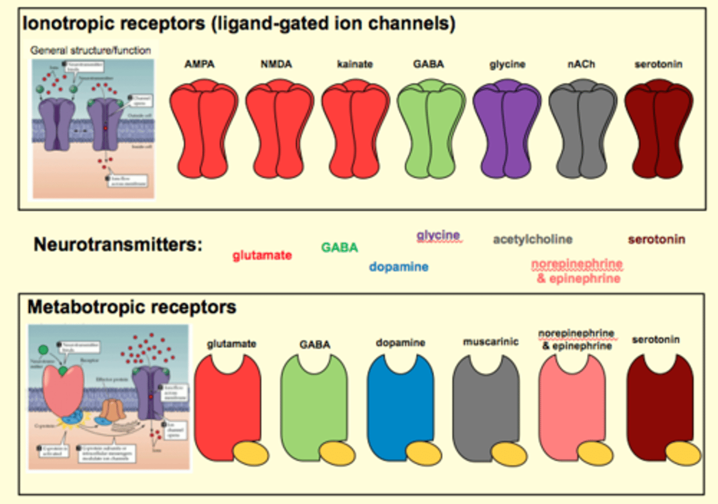
Ionotropic
iontropic receptor is a ligand gated channel
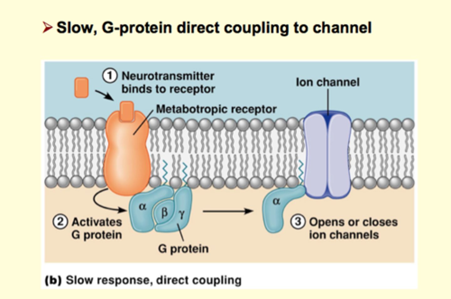
Metabotropic: slow response
1. by direct communication with ion channel - neurot. binds to receptor (not a channel), receptor is G-protein which has alpha subunit, when neurot binds to receptor it triggers activation of alpha subunit which is free to translocate along intercellular membrane until it reaches it target, which is an ion channel (opens/closes it)- takes longer
2. slpha subunit in this case activates second messenger system (uses a lot of protein), subunit activates enzyme that is binded to membrane and inititates a casscade of events and the messages could be to simply to open/close channel OR second messenger can activate a variety of other response, for example gene regulation (gene transcription, can make another channel) **receptors are very powerful
SOme Neurotransmitters have both iontropic and metatropic receptors
Neurotransmitters can bind to more than one receptor
Ionotropic and Metabotropic can be broken down further
Fast Response EPSP
-pic: non-specific cation channel (Na/K+)
membrane at rest: Na+ moves the most (Na+ would like cell to be +60 and not -70 mV) ompared to k+, theres a bigger driving force for Na+
gets: EPSP
preventing Na+ from leaving: causes excitatory potential
can also get EPSP that's slow??
ie. +30 mV: K+ would move the most
Inhibitory Synapse: K+ Channels
opens K+ that were previously closed
hyperpolarization
fast IPSP
Inhibitory Synapse: Cl- Channels
ionotropic channel
increases permeability to Cl-, makes more negative IPSP
opens Cl- channel, no driving force for C-: co-localized around other channels
neut: opens channel, postive charge going into cell, creates electrochemical driving force for Cl- (neg.), channels act as membrane stabilizers
want EPSP: not activate Cl- channels/dont do anything
not all cells have Cl- transporters
Nicotinic Cholinergic Receptor
nAchR
Muscarinic Cholinergic Receptor
- slow receptor, second messenger system
- big player in heart rate
hint
convergence and divergence on quiz?
Temporal summation**
Input (graded potential) of one source does not disappiate before another one from the same source occurs and potentials SUM
Neuron A is firing, if they fire in close enough in time, for EPSPs to ADD UP
Spatial summation
Input from different sources
have to be close together in space but also in time to add EPSP
Presynaptic Modulation
axo-axonic synapse
presynaptic neuron: functions to decrease or increase neurot release and it only does it at this particular synapse
advantage: modulate actvitiy of single synapse and not the whole cell
presynaptic is DIVERGENT
Presynaptic Facilitation
axo-axonic will have net effect of increasing neurot release - will be bigger
ie. EPSP acting on post synaptic membrane to produce EPSP more neut, will get bigger depolarixation and get action potential - bigger efefct on post synaptic membrane
Presynaptic Inhibitation
decrease
causes short circuit
What is the Peripheral Nervous System?
Neurons that provide communication between the CNS and organs throughout the body
- divided into Afferent and Efferent
What do neurons in the Afferent Division do?
- transmit sensory and visceral info from the organs to the CNS
- this includes somatic senses (skin, muscles, joints), special senses (vision, hearing, equilibrium, smell and taste), and visceral info for the internal environment
What do neurons in the Efferent Division do?
- transmit info from the CNS to organs in the periphery - effector organs - that preform functions to commands from neurons
- further subdivided into :
(1) somatic nervous system - motor neurons for skeletal contractions
(2) autonomic nervous system - neurons that regulate the function of internal organs and other structures that are not under voluntary control
Autonomic nervous system (opposite actions)
(1) parasympathetic nervous system
(2) sympathetic nervous system
What are "Excitable Cells"?
Neurons, which are capable of producing large, rapid electrical signals (action potentials)
What are "Glial Cells"?
- 90% of the cells in the nervous system
- provide various types of support to the neurons (structural integrity and metabolic support) in the CNS
What is the Axon Hillock?
The site where the axon originates from the cell body
- specialized in most neurons for the initiation of action potentials
Explain the
- for axon terminal to carry out function it must have enzymes for synthesizing neurotransmitters, molecules that transport neurotransmitters/substrates across membranes and vesicles to store neurotransmitters until an action potential triggers exocytosis
- simple diffusion would take days/months to complete the process of cell body to axon terminal
Name and explain the two basic mechanisms neurons have for moving products.
(1) anterograde transport - from the cell body to the axon terminal
(2) retrograde transport - from terminal to cell body
Distinguish between slow and fast axonal transport.
- both slow and fast axonal transport involve proteins (microtubules and neurofilaments)
Slow (0.5-40 mm/day) - is associated with movement of small soluble molecules in the cytosol
Fast (100-400 mm/day) - is associated with movement of vesicles (ie. synaptic vesicles)
- kinesins "walk" down track (microtubules) carrying vesicles with them; requires ATP
What are Leak Channels?
- found in the plasma membrane throughout a neuron
- are always open
- responsible for the resting membrane potential
What are Ligand-Gated Channels?
- open or close in response to the binding of a chemical (ligand) to a specific receptor in the plasma membrane
- in neurons: most densely located in dendrites/cell body (communication from neurontransmitters)
What are Voltage-Gated Channels?
- open or close in response to changes in the membrane potential
- necessary for the initiation and propagation of actions potentials
- Na+/K+ channels are most densely in the axon/hillock but are located throughout neuron
- Ca+ most densely located in axon terminals
What are Bipolar Neurons?
- sensory neurons with 2 projections; (1) an acon and (2) a dendrite, coming off the cell body
- typical ones functions in the senses of olfactory and vision
- most are pseudo-unipolar (subclass), axon and dendrite appear as a single process and dendrite is a functional continuation of the axon - called the peripheral axon - and transmits action potentials. axon is called the central
What are Multipolar Neurons?
- the most common neuron
- have multiple projections from the cell body
- one projection is the axon, the rest are dendrites
Name the three Functional Classifications of Neurons?
1. Efferent Neurons
2. Afferent Neurons
3. Interneurons
What are Interneurons?
- account for 99% of neurons in the body
- located entirely in the CNS
- preform all the functions of the CNS
examples include: processing sensory info from from afferent neurons, creating/sending commands to effectors through efferent neurons, complex brain functions (thought/memory/emotions)
How do the terms Pathways, Tracts and Commissure relate to the Central Nervous System?
They are bundles in which cell bodies of neurons travel together when they are in the nuclei form
How does the term "Nerves" relate to the Peripheral Nervous System?
The name for how cell bodies of neurons - ganglia - travel together in bundles
What are the Four Types of Glial Cells?
1. Astrocytes (CNS)
2. Microglia (CNS)
3. Oligodendrocytes (CNS)
4. Schwann Cells (only one in peripheral nervous system)
What is the primary function of Oliodendrocytes and Schwann Cells?
The primary function is to form an insulating layer of myelin around the axons of neuron, for more rapid and efficient transmission of action potentials
Oliodendroctyes - form myelin around axons in CNS, 1 oliodendrocyte sends projections for many axons
Schwann cells - form myelin around axons in PNS and only provides projections for one axon
What is Na+ electrically balanced by?
Cl- ions
What is K+ electrically balanced by?
A- (primarily proteins)
What are Mechanical-Gated Channels?
- open or close in response to a mechanical force on the membrane
- these channels are associated with sensory or visceral receptors located at the ends of afferent neurons
What are Activation Gates?
- responsible for the opening of Na+ channels during the Depolarization phase of an action potential
- Open during depolatization
- Closed and incapable of opening once inactivation gate opens after repolarization
- regenerative mechanism
What are Inactivation Gates?
- responsible for the closing of Na+ channels during the Repolarization phase of an action potential
- Closed but capable of opening at rest
- Closed and incapable of opening after activation gate is opened
What is the All-or-None Principle?
Whether a membrane is depolarized to threshold or greater; the amplitude of the resulting action potential is the same; if the membrane is not depolarized to threshold, no action potential occurs
What does Subthreshold mean?
A depolarization that is produced by a stimulus that is less than threshold that opens some Na+ channels, however not enough of them to produce an inward flow of Na+ large enough to over the outward flow of K+ through leak channels.
What does Suprathreshold mean?
When a stimulus is greater than threshold, which elicits an action potential, however the action potential does not increase in size nor are they graded
What are the 2 reasons for an Absolute Refractory Period?
1. During rapid depolarization phrase of action potentials - regenerative opening of Na+ channels goes to completion and is not affected by a second stimulus
2. Beginning of repolarization phase, most of the Na+ channels are closed and incapable of opening
- all of depolar and most of repolar
Relative refractory period
- occurs after absolute refractory period
- can produce action potential if stimulus stronger than threshold
- due to increased permeability to K+ that continues beyond repolarization phase
- determinant: potassium current
- the further you get into relative refractory, the less of a problem potassium current is
What is an Axodendritic Synapse?
Synapses between the presynaptic neuron's axon terminal and the postsynaptic neuron's dendrite
- same function as axosomatic synapses
What is an Axosomatic Synapse?
Synapses between the axon terminal and the soma of the postsynaptic cell
- same function as axodendritic synapses
What is an Axoaxonic Synapse?
Synapses between presynaptic neuron's terminal axon and the post synaptic neuron's terminal axon
- modulating communication at axodentric and axosomatic synapses
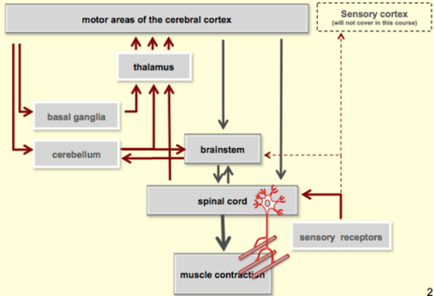
What is the motor unit?
The smallest force-producing unit of the intact neuromuscular system (organism)
- comprised of a motor neuron and all of the muscle fibres that it innervates (has muscle and motor unit)
part of motor unit is in peripheral nervous system, ends in muscle and activates muscle cells
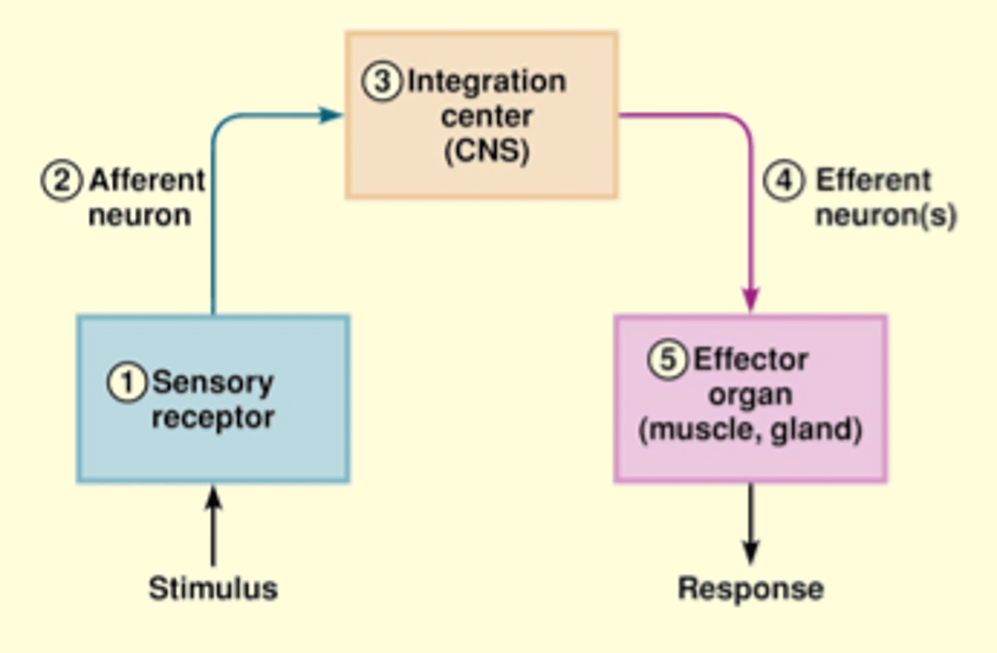
Image: know the functions of all the "boxes"
can only activate alpha motor neurons, each motor neuron (alpha)
motor units can innervate 10s or 100s
Why are muscle fibres spread out (instead of clumped)?
innervated by one nerve; flow of tension??
What is the reflex activation of the motor unit?
- muscle spindles (1a afferents)
- Golgi tendon organs (1b afferents)
- pain afferents (group III, IV afferents)
- cutaneous receptors
- joint receptors
What does Di or Polysynaptic mean?
- more than one synapse
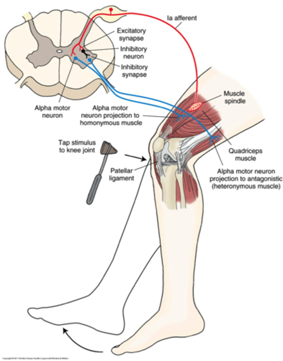
The Stretch Reflex
- also called "tendon tap"or "knee jerk"
- monosynaptic reflex (no interneuron)
"big wings" = ventral side
ventral = motor side
dorsal side = sensory
muscle spindle = coil like, bc its wrapped around something
sensory neuron = 1a afferent =
tap tendon = stretches nerve = sensory neuron fires = singal travels up peripheral nerve = enters dorsal = through dorsal horn = monosynaptic synapses with alpha neuron = releases gluatamate (excitation - depolarixation or motor neuron) = excited motor neuron = travels back down = contraction
***monosynaptic is like a negative feedback
pseudounipolar neuron (cell body in the middle)
neuron root: visible structure that projects from the spinal cord (dorsal root)
- it has swelling - which is called a dorsal root ganglion (ganglion = cluster of cell bodies)
ventral root and dorsal root connect together just outside spinal cord - called a spinal nerve root (sensory and motor component coming together), you'd have a right spinal nerve and a left from each vertebrae (ie. L4 for quadracieps) come together as "mixed nerves" or "peripheral nerves" (ie. femoral nerve)
reciprocial inhibition:
in order to contract the muscle, you bave to relax the opposite muscle :
branch -> inhibitory neuron; hyperpolarizes its target (also alpha motor neuron), this motor neuron travels through different mixed nerve, goes to antagonist muscle (hamstrings) = inhibits nerve of hamstring (sciatic) = relax
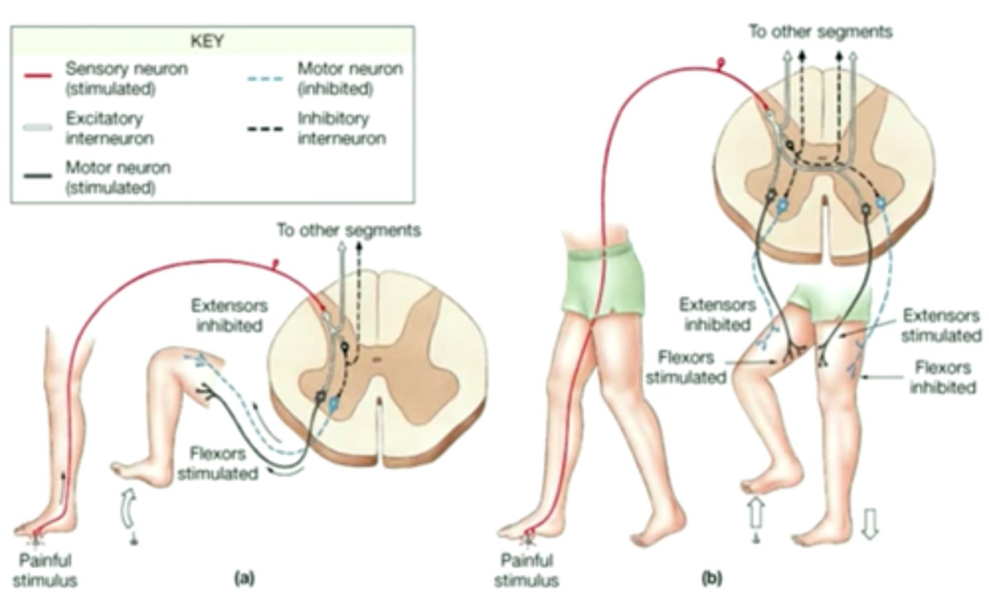
Activation of the Motor Unit
Crossed Extensor Withdrawal
stimulus = noxious stimulus (has the potential to cause pain/damage)
2 cross to opposite side of spinal cord, 2 will stay on the same side (not shown on diagram, but it happens around (3.)
- reflax quadriceps to flex knee (inhibitory)
- constract flexor on painful side (excitatory - releases gultamate) and it depolarizes next neuron, which activates the nerve thast goes down to the hamstring so that you can contract the flexor (lifts up foot)
- example of divergence: interneurons with their diff. neurot. that allow for different responses in different targets
What is Innervation Ratio?
The number of muscle fibres that one neuron innervates
What is Slow Oxidative?
Why are neurons with smaller cell bodies innervated/activated first?
Smaller cell bodies are easier to get to threshold.
smaller cell bodies and dendrites - more resistance to current
- any little bit of current, is gonna cause a large membrane potential (ie. -70 mV to a big +++)
**as to keep going/can't quit even after you've activated larger neurons
What is Henneman's Size Principle?
States that under load, motor units are recruited from smallest to largest.
In practice, this means that slow-twitch, low-force, fatigue-resistant muscle fibers are activated before fast-twitch, high-force, less fatigue-resistant muscle fibers
Which nerve emerges from the top of the coccyx?
A single coccygeal nerve.
The Gray Matter
The "butterfly" region of the the interior spinal cord
- contains interneurons, cell bodies and dendrites of the efferent neurons and the axon terminals of the afferent neurons
What is the Dorsal Horn?
The area that encompasses the dorsal half of the gray matter on either side
- afferent fibers originate in the periphery as sensory receptors and terminate the dorsal horn, where they synapse on interneurons or efferent neurons
What is the Ventral Horn?
The area that encompasses the ventral half of the gray matter on either side
- efferent neurons originate here and travel to the periphery, where they form synapses with skeletal muscles.
Where are the cell bodies that the afferent fibres (originated in the periphery as sensory receptors) are located?
Not located in the spinal cord itself, but rather outside of the spinal cord in clusters called dorsal root ganglia.
Where are the cell bodies of efferent neurons located?
Located in the spinal cord.
Where are the Lateral Horn and the Intermediolateral cell column located?
In the thoracic and upper lumbar regions of the spinal cord - between the dorsal and ventral horns on either side
Where are the origins of the efferent neurons of the autonomic nervous system?
The Lateral Horns
What is the name of the bundles containing Afferent Neurons?
Dorsal Roots.
What is the name of the bundles containing Efferent Neurons?
Ventral Roots.
Why are Spinal Nerves called "Mixed Neves"?
Because they contain both afferent and efferent axons
What are Ascending Tracts in white matter?
They transmit information from the spinal cord to the brain.
What are Descending Tracts in white matter?
They transmit information from the brain to the spinal cord.
Name the Ascending Tracts.
1. Dorsal columns - transmit sensory infromaion from the periphery to the brain
2. Lateral spinothalamic tract
3. Spinotectal tract
4. Anterior spinothalamic tract