VSP 3 Lecture 3 Color Vision Part 2
 Call Kai
 Learn
Learn Practice Test
 Spaced Repetition
 Match
Match Flashcards
 Knowt Play
1/89

There's no tags or description
Looks like no tags are added yet.
Name | Mastery | Learn | Test | Matching | Spaced | Call with Kai | Chat |
|---|
No analytics yet
Send a link to your students to track their progress
90 Terms
Trichromatic Theory of Color Vision
3 types of photoreceptors
3 different photopigments
Each sensitive to a different wavelength
Yong-Helmholtz Trichromatic Theory
states that within your eye are tiny cells that can receive waves of light and translate them into one of three colors: (S) blue, (M) green, and (L) red. These three colors can then be combined to create the entire visible spectrum of light as we see it.
cyanolabe
426 nm
Short
SWS
S cones
Chlorolabe
530 nm
medium
MWS
M cones
Erythrolabe
552 or 557 nm
long
LWS
L cones
Univariance
photoreceptors cannot register the wavelength of the photoms they catch
output depends on quantum catch
have higher likilihood of respondong to light that correspond to its peak wavelength than of a different wavelength
LWS absorb maximally at
yellow
so its a misnomer to call them red cones!!
The MWS and LWS cones respond to light
over almost nthe entire spectrum
both have the same reduced sensitivity to shorter wavelengths
SWS cones make up ____% of the cone populaiton
6-7%
L and M cones make up _____ of cone populatino
90-95%
ratio varies from person to person
Range of L:M cones in normal trichromatic vision
1:1 to 16:1
why we may view colors different!
LWS and MWS are desnsest in
the fovea
SWS are absent in
central .3 to .4 degrees of foveal pit
Normal color vision with very small, centrally fixated stimuli is
tritanopic bc SWS are absent
tritanopic
lack of blue cones!
THEREFORE SWS are less sensitive overall
Trichromat: Changing the wavelength
changes the response of each type of cone
each wavelength has
a unique ratio of the 3 cone types
Grassman's LAw
- additive property
- scalar porperty
-associative propery
Additive Proptery
if the same radiation L(x) is added to 2 metamers, they remain metamers
if: L(a) + L(b) = L(c) + L(d)
then: L(a) + L(b) + L(x) = L(c) + L(d) + L(x)
Scalar Property
if the intensities of 2 metamers is increaed or decreaed by the smae amount, they remain metamers
Associative Property
a match will be maintained if one metamer is subsituted for another metamer
if: L(a) + L(b) = L(c) + L(d)
and: L(f) + L(g) + L(h) = L9b)
then: (a) + L(f) + L(g) + L(h) = L(c) + L(d)
Color labes are based on 3 perceptual atrtibutes
hue
saturation
brightness
hue
perception of color based on wavelength
510 appears
green
400 appears
violet
hue is lay persons term for color
but color is mcuh broader
Saturation (and desaturation)
colorimetric purity or fullness
how much white light is in the sample
based on wavelength and purity
Saturation P =
p = Ly/(Ly +Lw)
(luminance of the test wavelength)/(luminance of the test wavelength + luminance of the white light that is combined with the test wavelength)
colorimetric purity
the proportional amount of the spectral light in a mixture of the spectral light and a white light
When there is no light added the colorimetric purity is
1
p = Ly/(Ly +Lw)
the closer to 1
the more its made up of only the spectral light
a sample of 100% white light would have a colormetric purity of
0
a 540 nm stimulus with a colormetric purity of .4 is a combination of
60% white light and 40% 540 nm light
ON EXAM
Presit and Brickwedde and Kaiser found that monochromatic light at 570 nm
appears less saturated than a monochromatic wavelength at any other wavelength (for trichromats)
This is not true for other ppl w color def
Brightness
the subjective correlate of photometric luminance or luminous intensity
follows the luminance function which is the addition of M and L cones (dominate cone poopulation)
S cones less becuase frewer and lower luminance profile
therfore 555 will appear
brighter than other monochromatic stimuli of equal energy in photopic conditinos
Wavelength discrimination
How much does a stimulus of wavelength need to be changed to appear different
W curve
W curve of color discrimination: We need the least amount of change to distinguish between two colors
495 nm (cyan blue) and 590nm (yellowish orange)
our best discrimination!
less than 2 nm
we can see the difference between 495 and 498nm!

Which two hues will appear the most similar to an observer
445 and 450
500 and 505
595 and 600
445 and 450
wavelength discrimination is poor
need to be within 6 nm to disciminate at this point on the W curve
Bezold Brucke Phenomenon
the change in hue of most spectral colors that accompanies a change in their intensity or luminance
for most wavelength hue changes slightly as intensity is adjusted
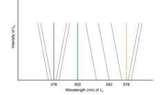
Bezold Brucke Effect example
as increase luminance the light appears more yellow therefore must decrease wavelength to keep the color the same as intensity increases
Bezold Brucke: Hue contour line
all stimulu that fall on this line (have the same hue)

Bezold Brucke: Invariant wavelengths or invariant points
most lines are tilted - as intensity increases hue changes and adjustments are made to keep the color the same
some are not called Invariant Wavelengths
Invariant Wavelengths
478nm (475)
503 nm (510)
578 nm (570)
Unique hues
hues associated with these invariant wavelengths
appear pure
not mixing with other wavelengths
--- are the neural elements that begin the processes of color vision, asife from sepctral composition
Cone
Other factors affect how we see colors
in the retina, after PR
though the visual cortex an on to
higher cortical processing
color constancy
is the stability of our perception of the color of objects even though there may be changes in the composition of incident light
Land's Mondrian Experiment
we tend to perceptive colors as stable regardless of changes in illumination
Retinex theory
the brain and eye work together through comparison
color depends on context!
Opponent Color Theory
Interpreted the results of color mixing
- certain pairs of colors that are never seen together in the same place at the same time
Opponent Color Theory Consisting bipolar hue channels
Red Green
Blue Yellow
black White (codes brighntess)
no such thing as reddish green
when mixed together = white
Herring Primary Colors
Yellow
Blue
red
Responses to one color of an opponents channel are ----- to those to the other color
antagonists
opponenent process stats that the human visual system inteprests informaiton about color by processing signals in an antagonsiic manner
Hering After Images
after images with chromatic stimuli of complement color
Red --> Green (Cyan) after image
Blue -> Yellow after image
Young Helmholtz Theory was correct
at the level of the PR
Hering Opponent Theory was correct for
subsequent levels of visual pathwayT
Today
Three cones and three oponent pathways
Leo Hurvich and Dorothea jameson
provided the first quantitative color opponent model based on psychophsyical experimentation
Hue Cancellation
Hue Cancellation
If a light appeared red, the redness could be canceled by adding green
the amount of green required to neutralize would reflect the strength of the red
'chromatic valence functions'
perception of color can be explained by relative activity of these red-green, blue-yellow, and brightess channesl
Crossover points
only one of the two channels os active at these points resulting in the perception of a true hue
unique blue
478nm
unique green
503 nm
Unique yellow wavelength
578 nm
Unique red?
falls outside the spectrum
(contains yellow in spectrum)
Bezold-Brucke Effect Unique Hues
appear pure and are not mixed
478 nm
503 nm
578 nm
The opponents processing curves and Hue cancellation predict the correct location of the
unique hues from the Bezold-brucke effect
Gunnar Svaetichin founded the field of intracellular recording of light responsed from retinal neurons in fish
Gunnar Svaetichin discovered
S potentials or Retinal Horizontal ell responses of two types
Color opponents
Non color opponent
discovered objective neural color opponency !!
Non color opponent celsl
- no inhibition
- excitation only at all wavelengths
- monochromatic
- most peak at 550nm
- Input from M and L cones
- based on spectral sensitivity
Trichromatic vision
expains color mathcing
begins at the pothotrecpots
Color opponency
helps code color inforamtion
ebgins at bipolar cells
Diffse bipolar cells are
non color opponent
midget bipolar cells are
color opponent
bipolar cells receive input from
more than one cone
directly from PR
or indirectly from horizontal cells
____ begin the process of color opponency
Bipolar cells
for Midget Bipolar cells
single M or L cone connects to a Midget Bipolar Cell centrally and in mid periphery
forming the receptive field
H1 Horizontal contribute to the bipolar cell surround
for Midget Bipolar cells M or L cones
oppose each other to produce L-M opponent cells
For diffuse bipolar cells
center has input from 5-10 cones
more than 1 cone type may contribute to center
H1 contribute to surround (also input from M and L)
spectrak sensitivity in the central and surround may be similar
NO color opponency
spectral similar to non color opponent cells
S cones Bipolar Cells
synapse on bistratified GC
the center respond to only 1 cones in the fovea
absent on central .3 to .4 degrees fovea
Yellow on, Blue off
Blue on, yellow off
S cones are opposed by
Addition of L and M cones
Yellow on, blue off bp cell
What cells contribute to the center
M cones and L cones
On center Midget BP synpase with
on center midget ganglion cells (retinal parvo) cells
On center diffuse BP cells cynapse with
on center parasol (retinal magno) ganglion cells
Off center midget BP cells synpase with
off center midget ganglion cells (retinal parvo) cells
OFF center diffuse BP cells synapse with
off center parasol (retinal magno) ganglion cellsM
Midget Ganglion Cells (retinal parvo cells) allow for
- high spatial resolution (poor temporal resolution)
- Are color sensitive (color opponency)
- Project to the parvocellular layers of the LGN
Parasol ganglion cells (retinal magno cells) allow for
- good temporal resolution
(sensitive to motion, poor spatial resolution)
- no color sensitive (non color opponent)
- project to the magnocellular layers of the LGN
Smalll Bistratided Ganglion Cells
S cones synpase with these
color sensitive
prokect to konio layers of LGN