💀LAB: MSK Anatomy
1/134
There's no tags or description
Looks like no tags are added yet.
Name | Mastery | Learn | Test | Matching | Spaced | Call with Kai |
|---|
No analytics yet
Send a link to your students to track their progress
135 Terms
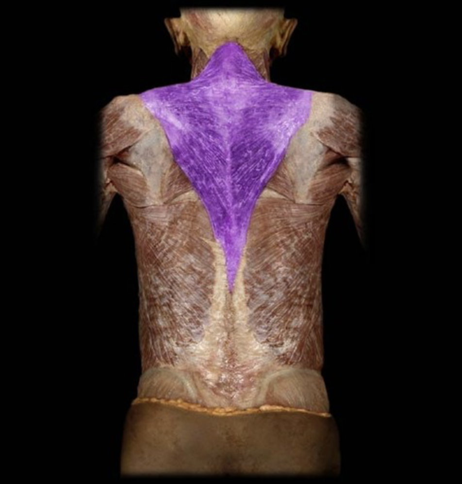
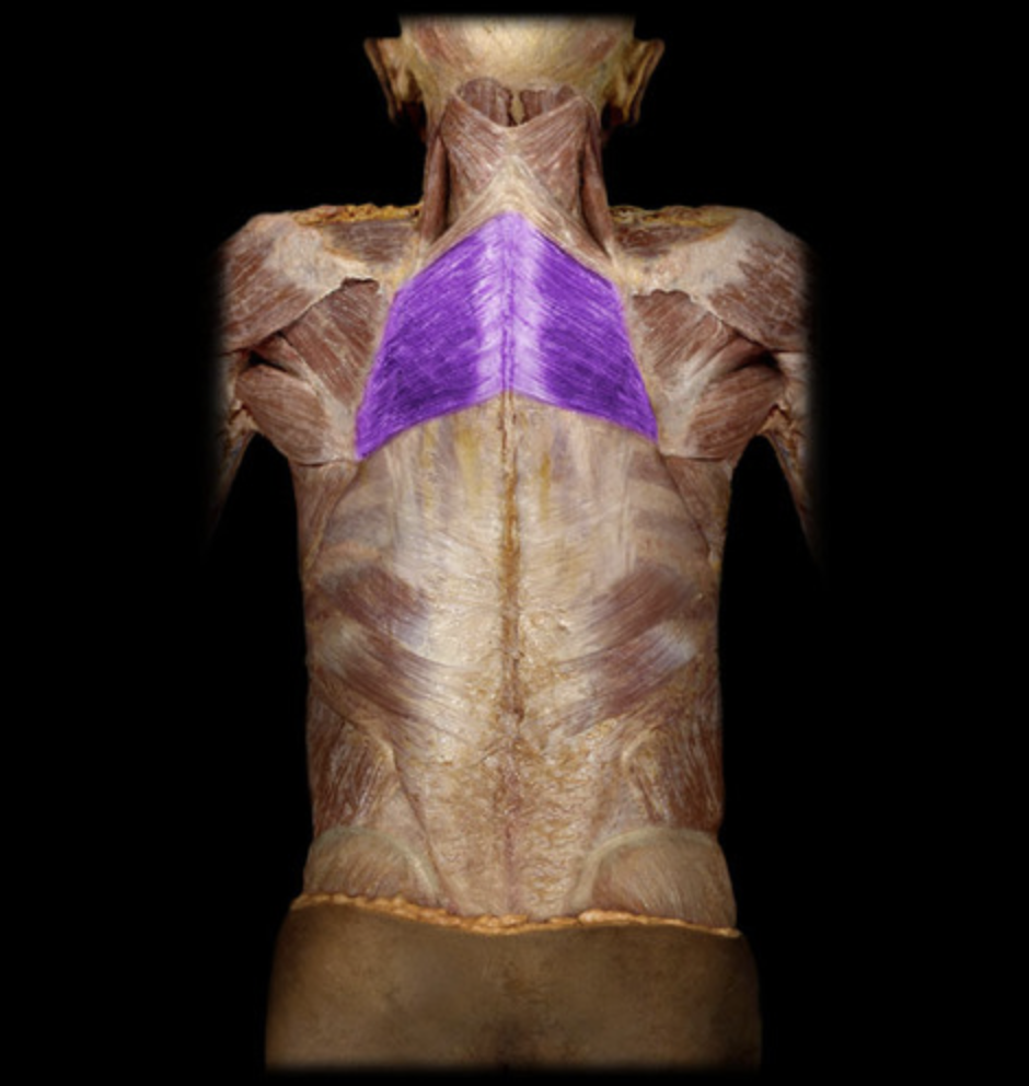
Trapezius
table 1, 3
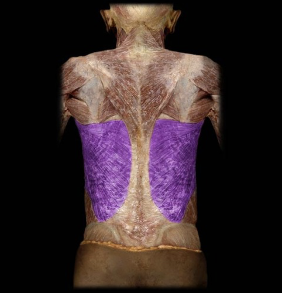
Latissimus dorsi
table 1, 3
I: intertubercular (bicipital) groove of humerus
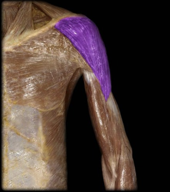
Deltoid
table 1, 3
Rhomboid major
table 3
Rhomboid minor
table 3
Levator scapulae
table 3
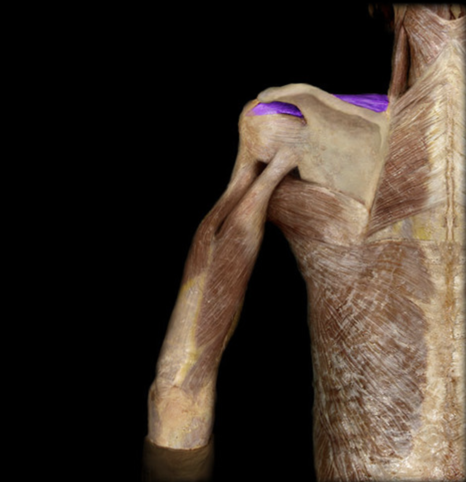
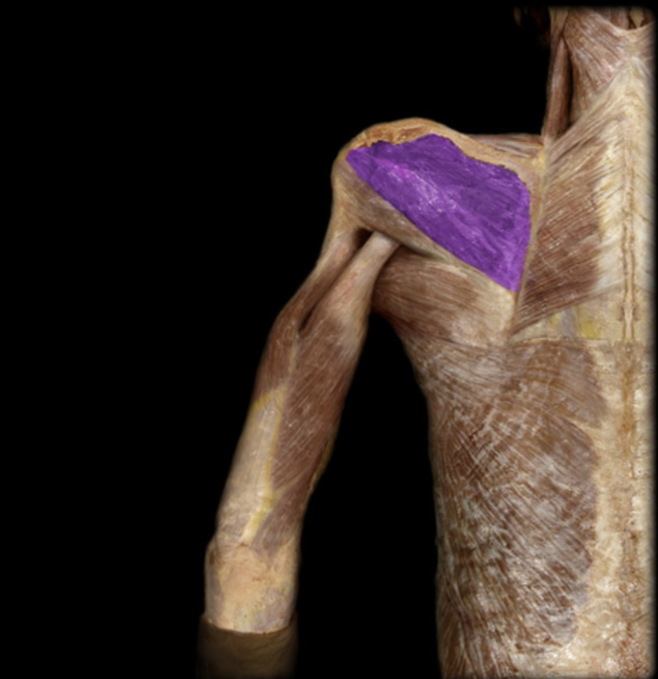
Supraspinatus
table 1
O: supraspinous fossa of scapula
I: greater tubercle of humerus
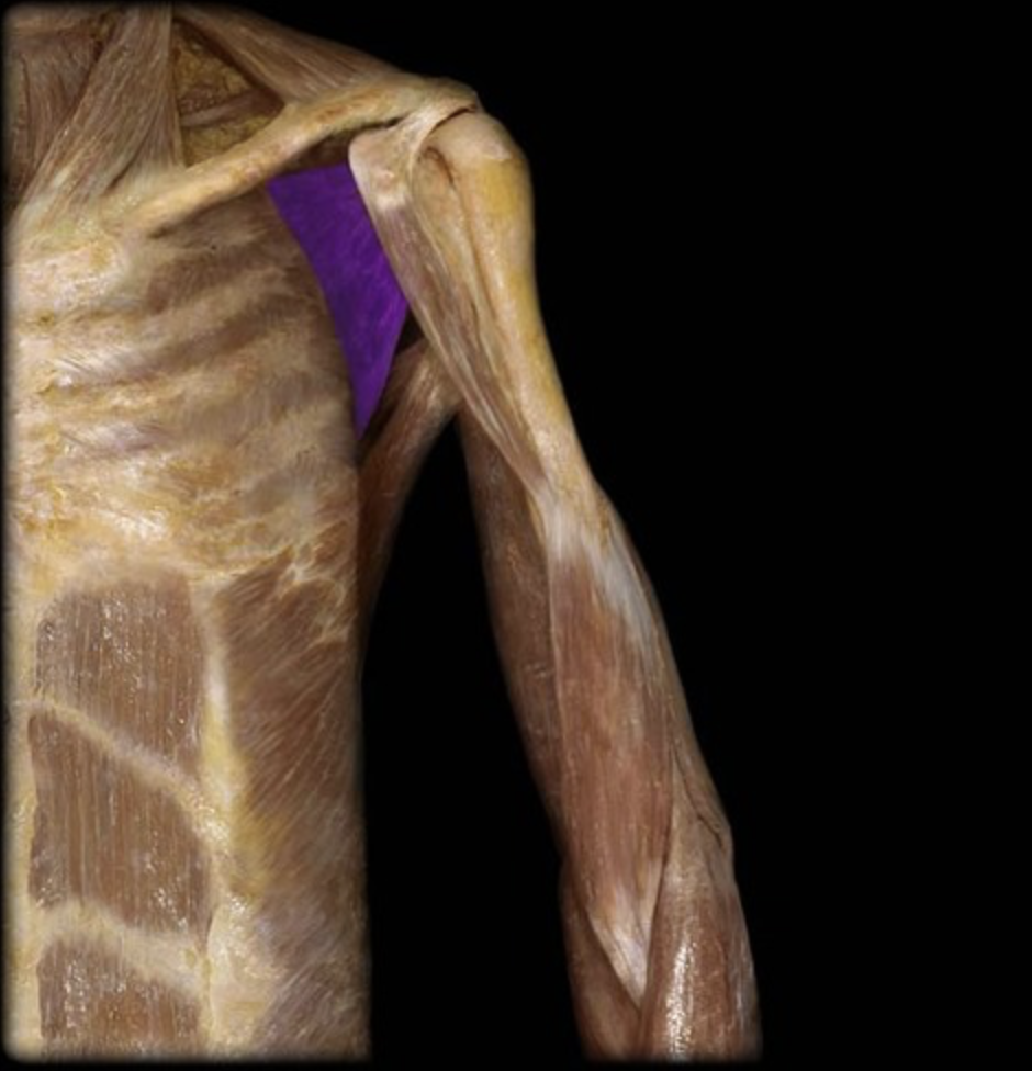
Infraspinatus
table 1
O: infraspinous fossa of scapula
I: greater tubercle of humerus
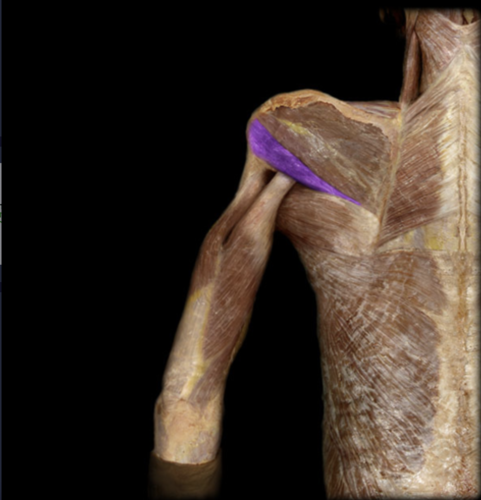
Teres minor
table 1
O: mid-lateral border of scapula
I: greater tubercle of humerus
Subscapularis
table 1
O: subscapular fossa
I: lesser tubercle of humerus
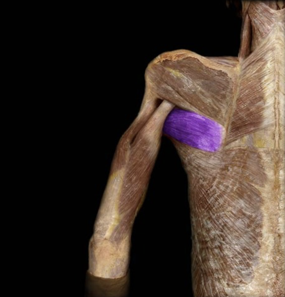
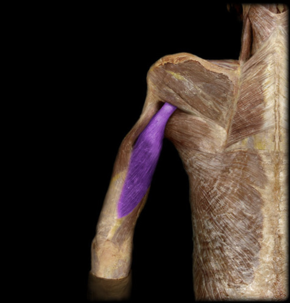
Teres major
table 1
I: intertubercular (bicipital) groove of humerus
Serratus anterior
table 3
Serratus posterior superior
table 3, deep to rhomboid major
Serratus posterior inferior
table 3, deep to latissimus dorsi
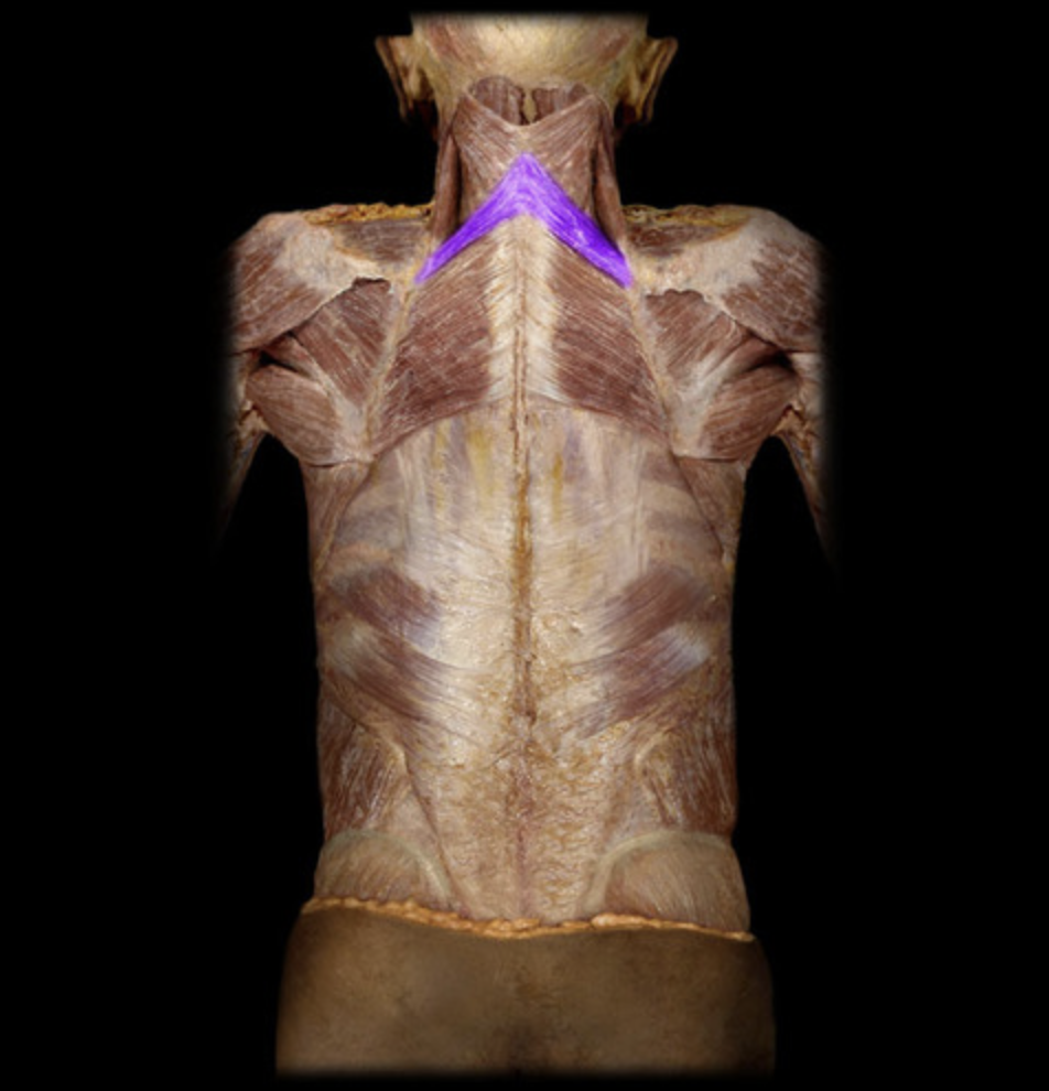
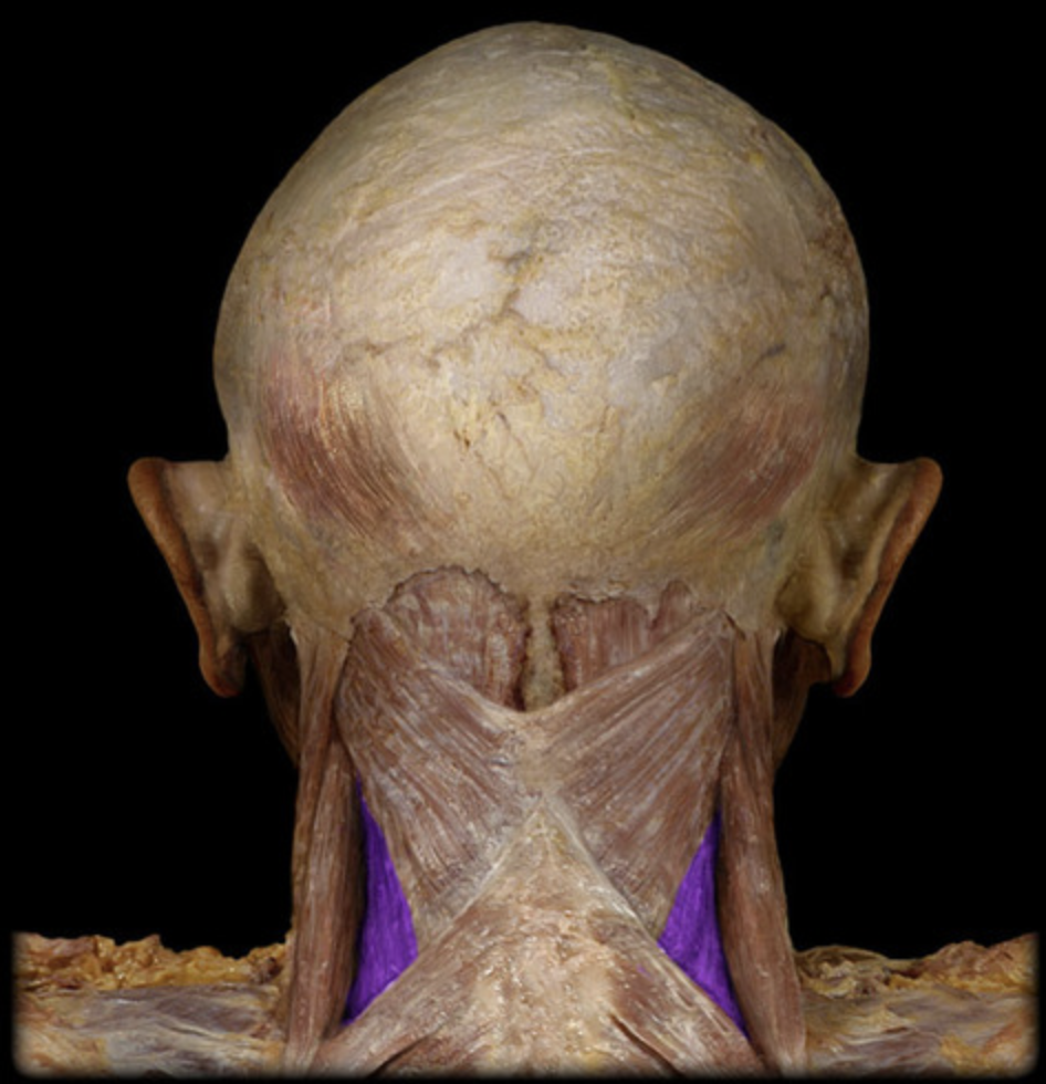
Splenius capitis
table 3, connects to scalp
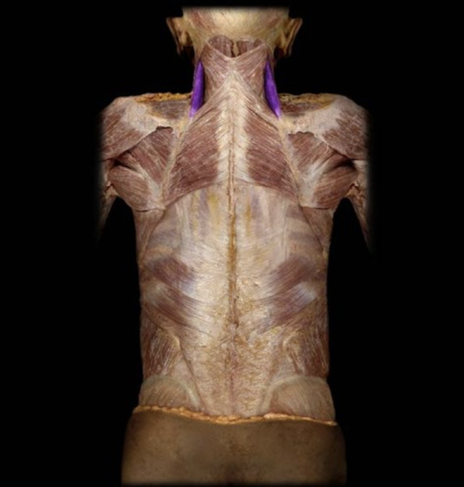
Splenius cervicis
table 3, connects to neck
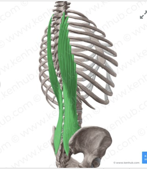
Iliocostalis thoracis
table 3, pink color, most lateral of erector spinae
Iliocostalis cervicis
table 3, pink color, most lateral of erector spinae on neck

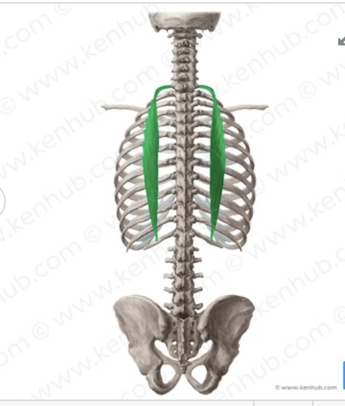
Longissimus thoracis
table 3, white color, intermediate erector spinae
Longissimus cervicis
table 3, white color, intermediate erector spinae at neck
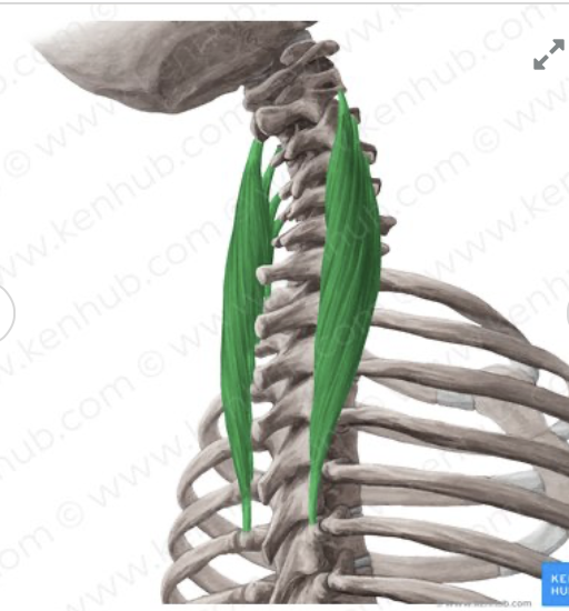
Spinalis thoracis
table 3, most medial of erector spinal superior to spine

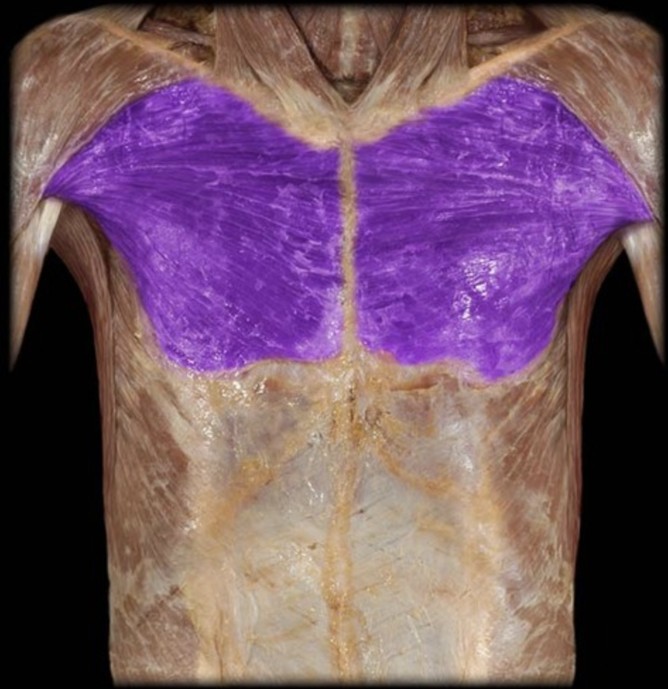
Pectoralis major
table 3
I: intertubercular (bicipital) groove of humerus
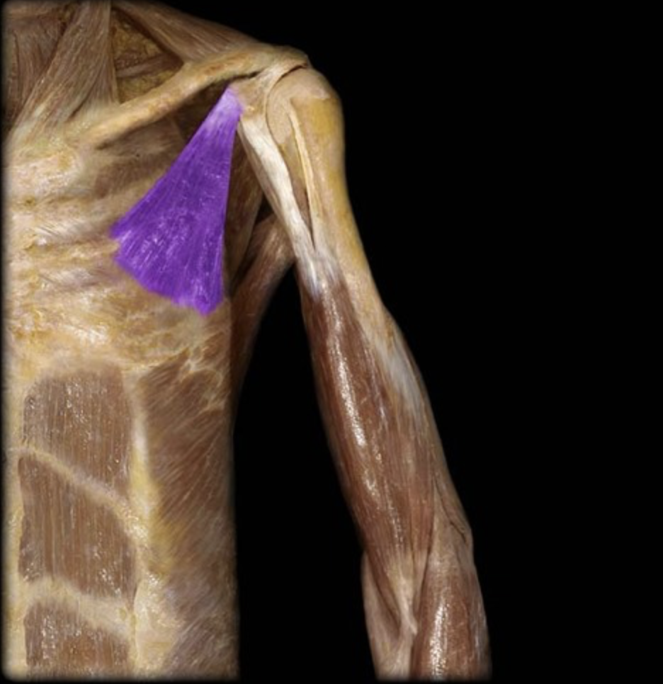
Pectoralis minor
table 3
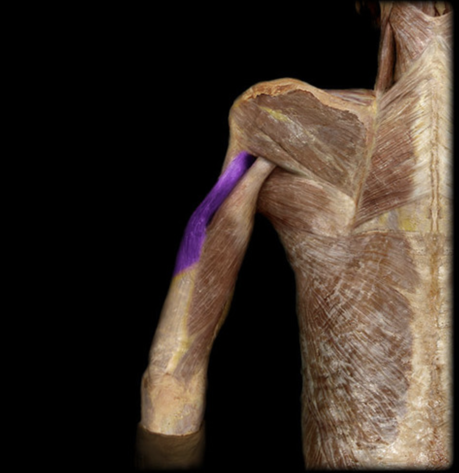
Triceps brachii long head
table 1
Triceps brachii
table 1, radial n (C6-C8), elbow extension
I: olecranon process of elbow
Triceps brachii medial head
table 1
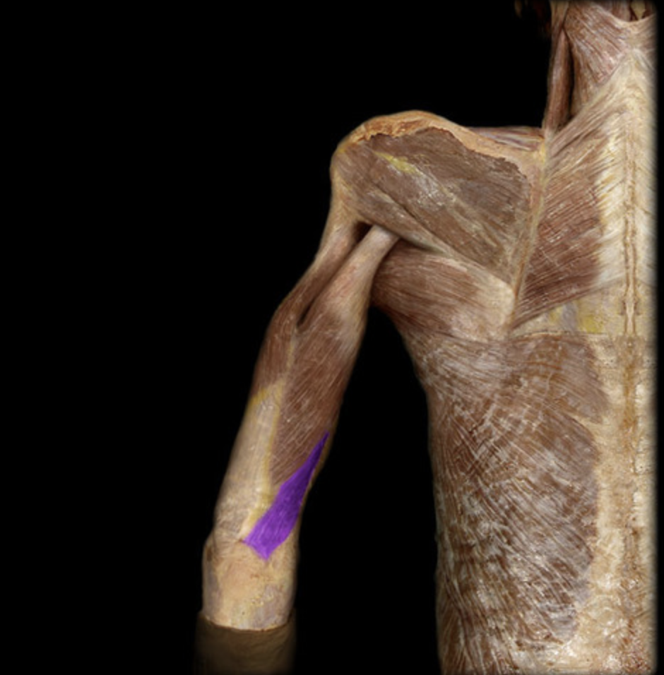
Triceps brachii lateral head
table 1
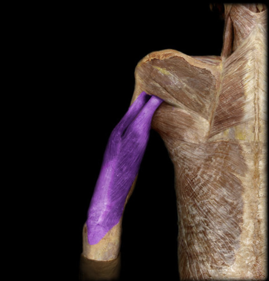
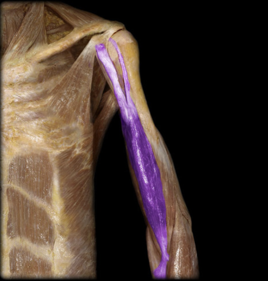
Biceps brachii long head
table 3, 7
O: supraglenoid tubercle above the glenoid cavity of the scapula
I: radial tuberosity
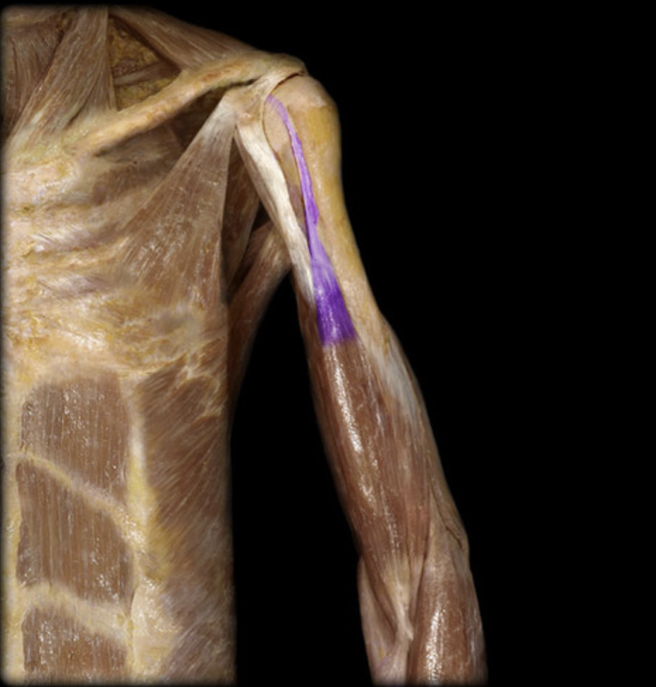
Biceps brachii short head
table 3, 7
O: coracoid process of the scapula
I: radial tuberosity
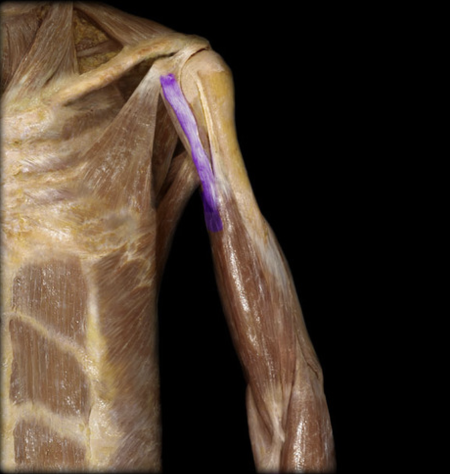
Biceps brachii
table 3, 7, musculocutaneous n (C5-C7), forearm supination/elbow flexion
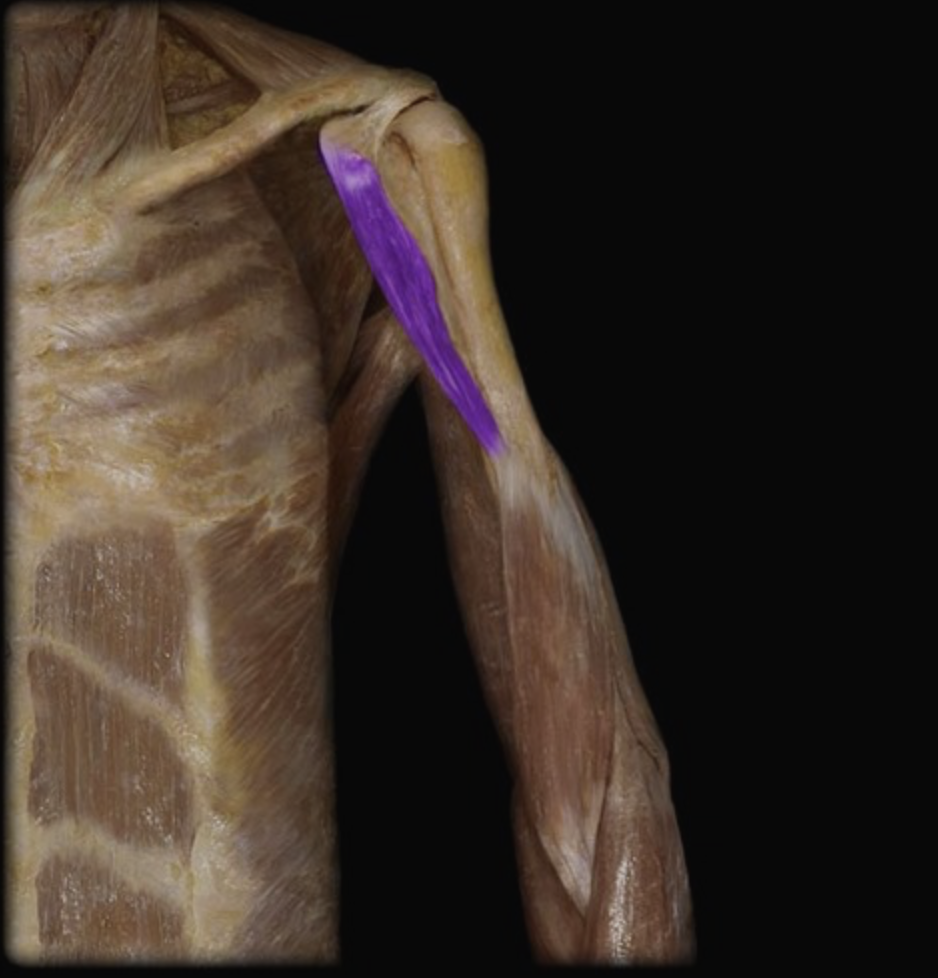
Coracobrachialis
table 7
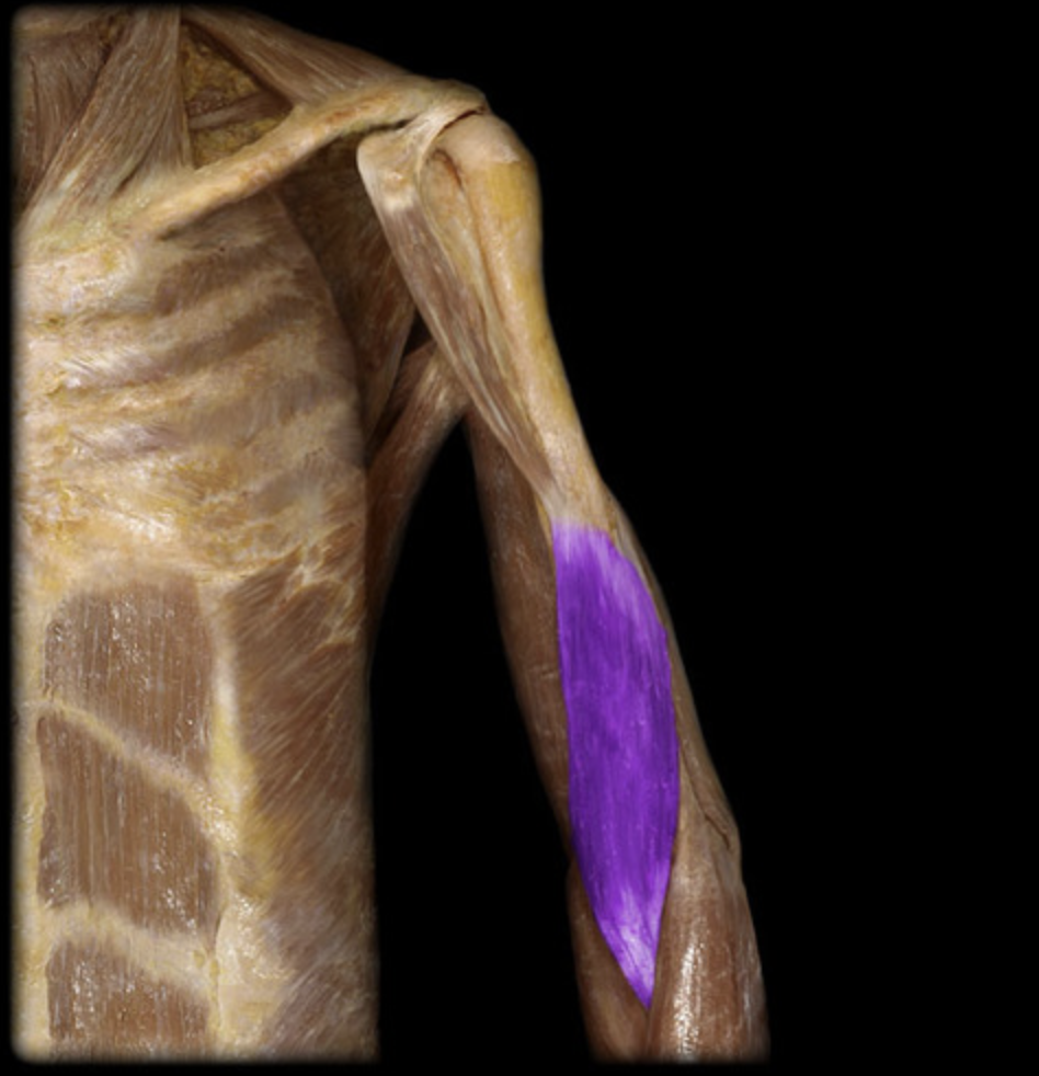
Brachialis
table 3, 7, musculocutaneous n (C5-C7)/radial n (C6-C8), elbow flexion
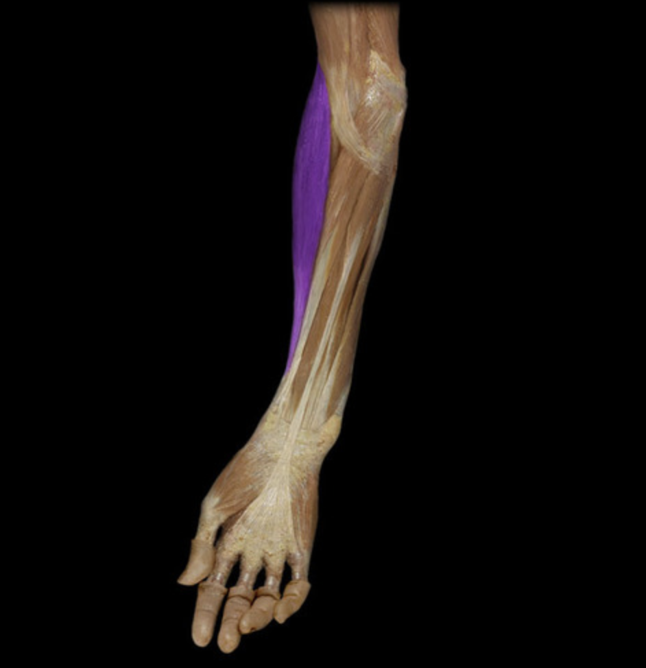
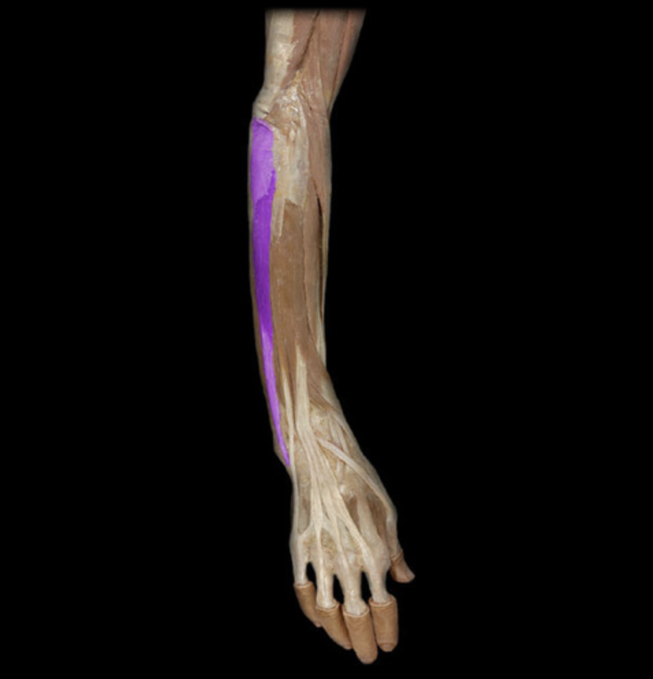
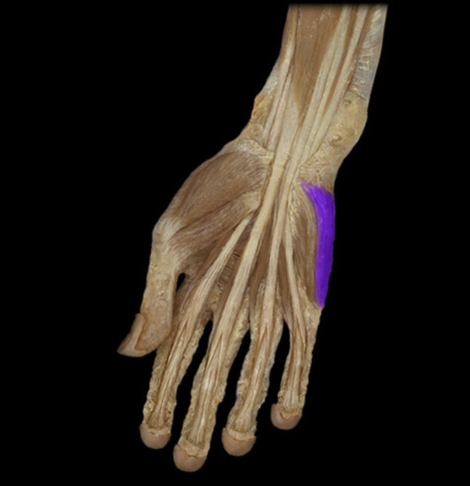
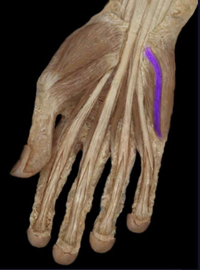
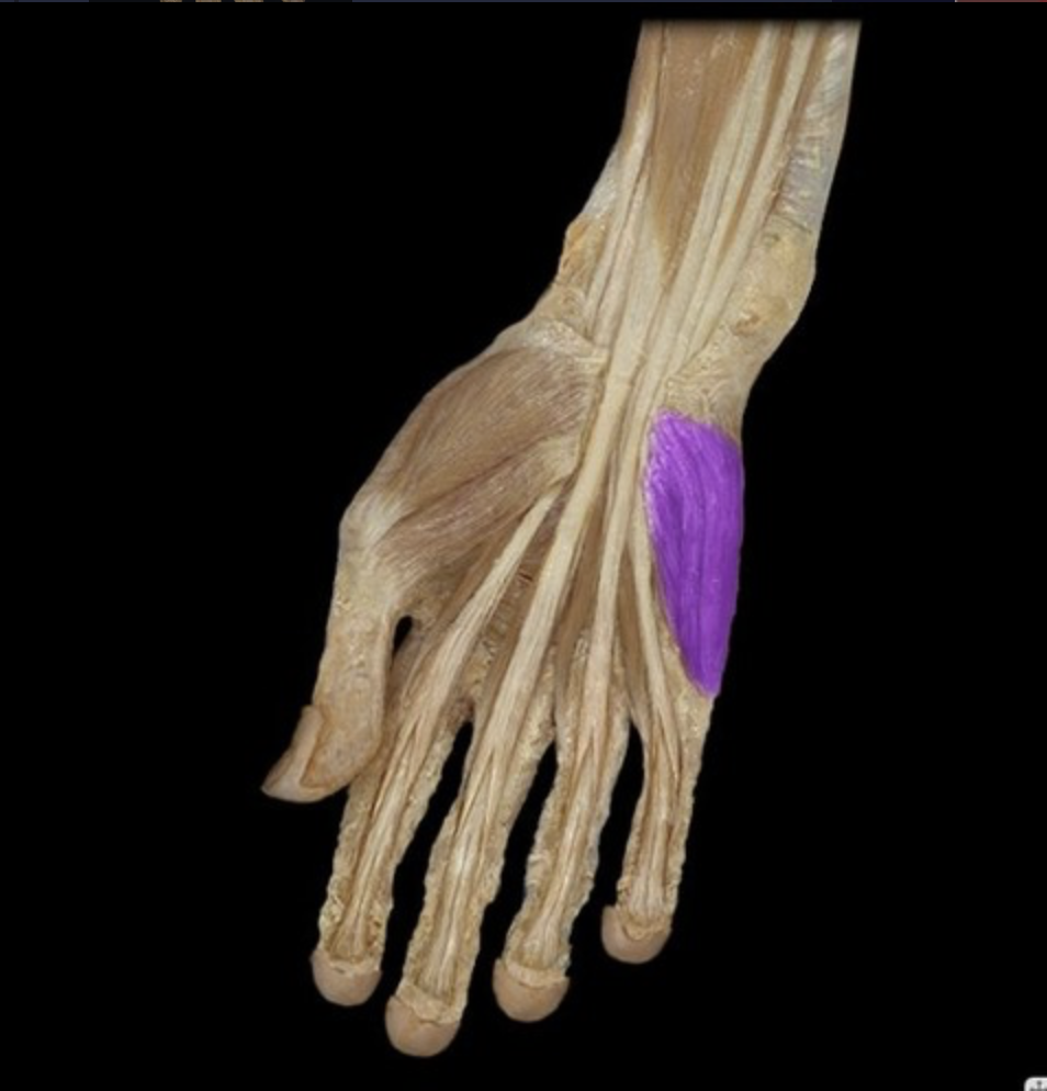
Brachioradialis
table 3, radial n (C5-C6), elbow flexion
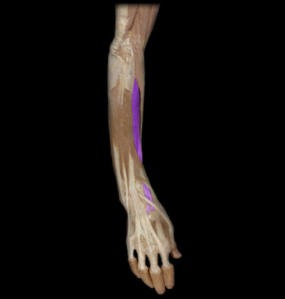
Flexor carpi radialis
table 1
O: medial epicondyle of humerus

Pronator teres
table 1, median n (C6-C7), deep to superficial flexors
O: medial epicondyle of humerus
Palmaris longus
table 3
O: medial epicondyle of humerus
Flexor carpi ulnaris
table 1
O: medial epicondyle of humerus
I: olecranon process of ulna
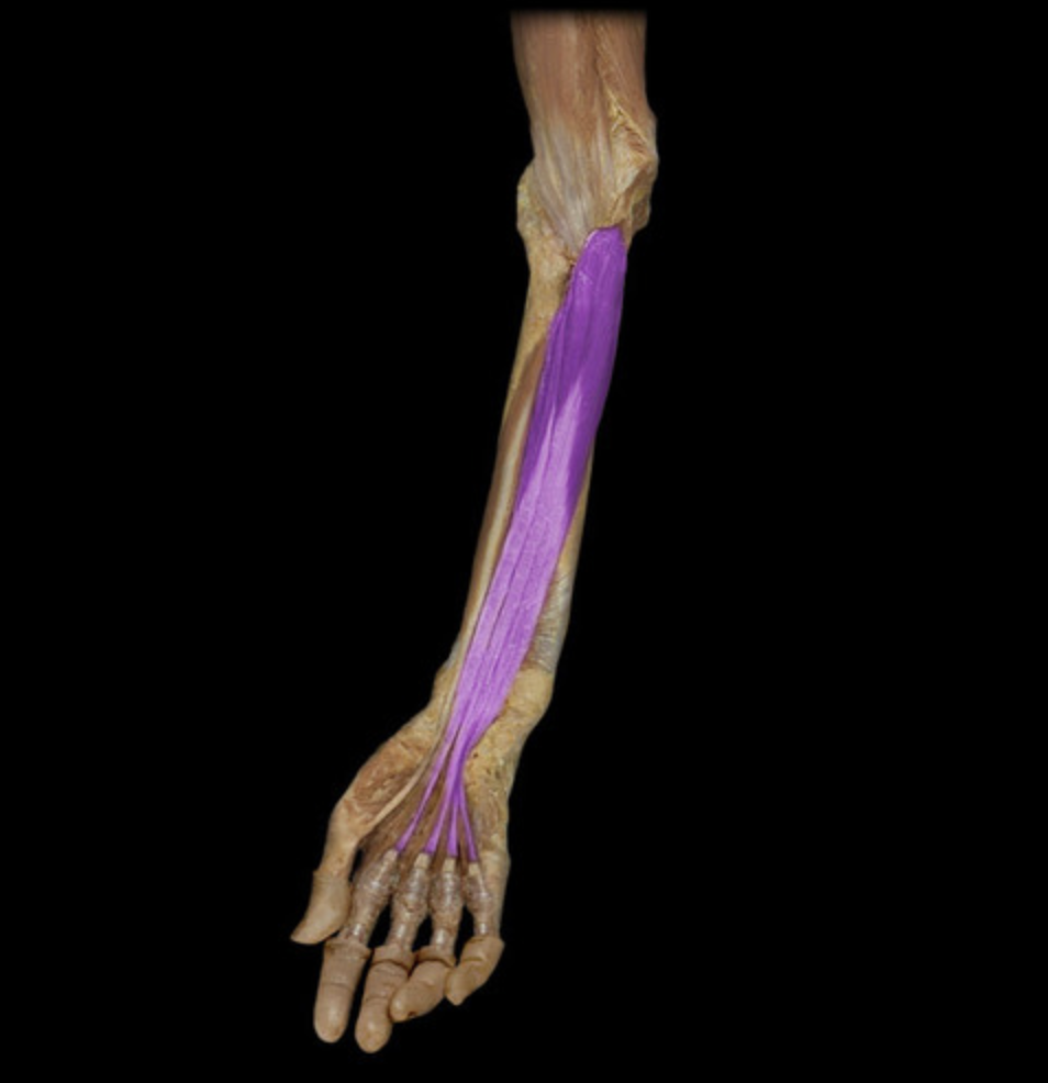
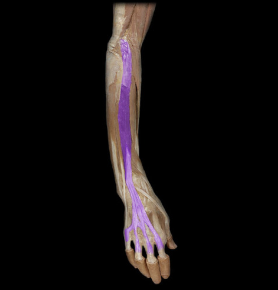
Flexor digitorum superficialis
table 1, median n (C7-T1), PIP/MCP joint flexion digits 2-5
O: medial epicondyle of humerus
Flexor digitorum profundus
table 1, median nerve (digits 2-3), ulnar nerve (digits 4-5); DIP flexion digits 2-5
lies deep to the superficial flexors
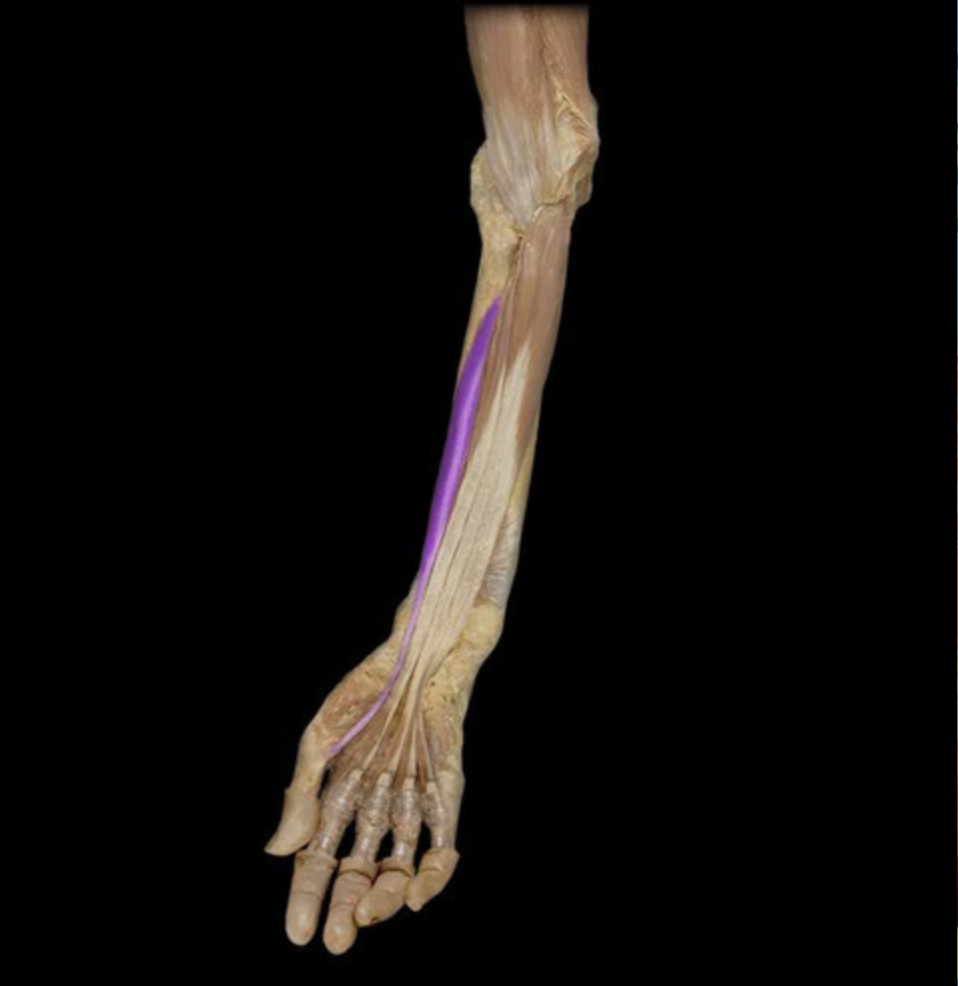
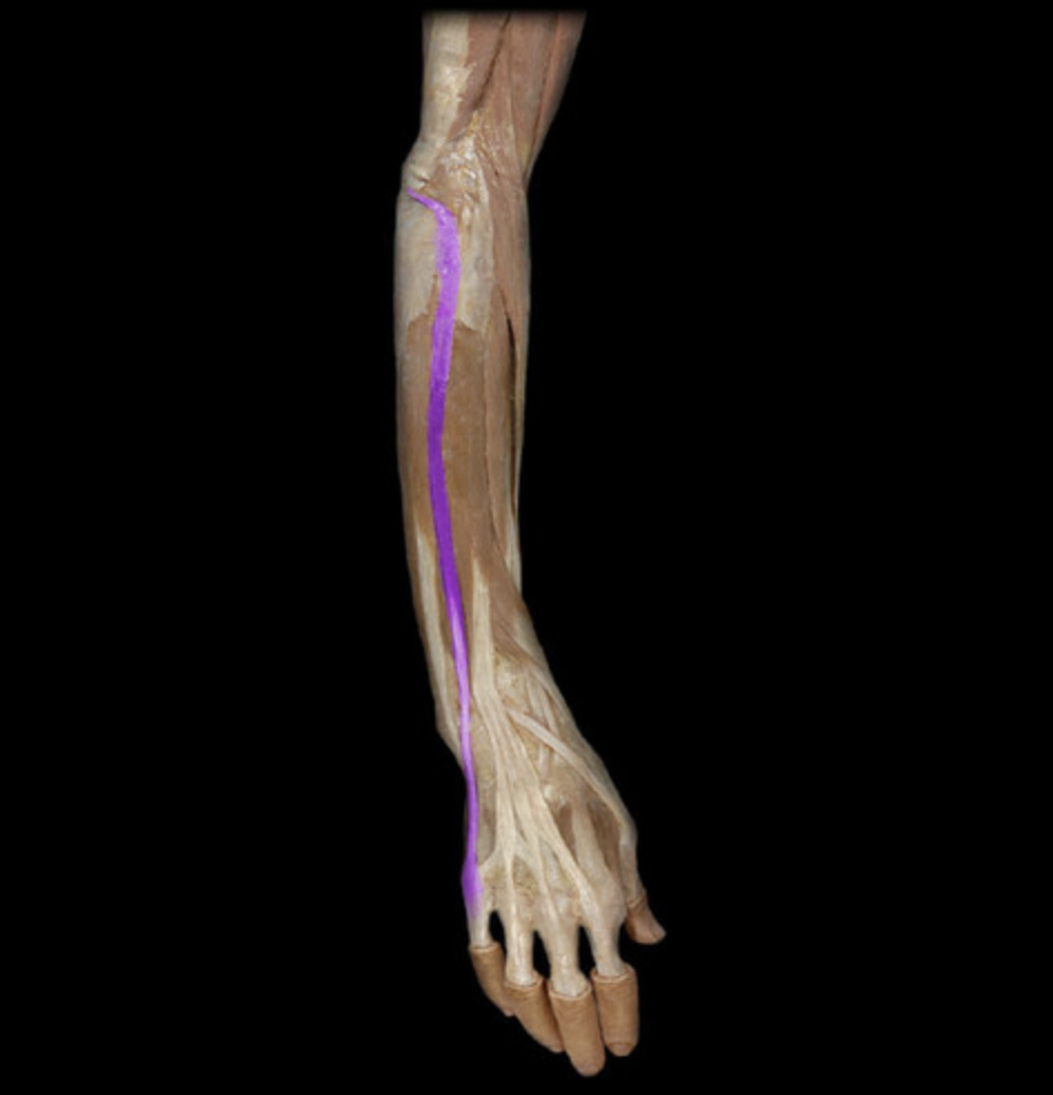
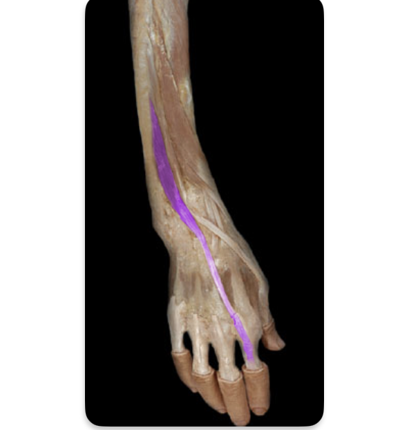
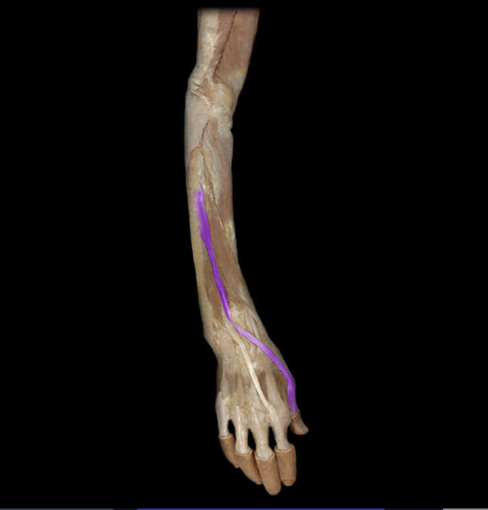
Flexor pollicis longus
table 1, median n (C8-T1), thumb IP joint flexion
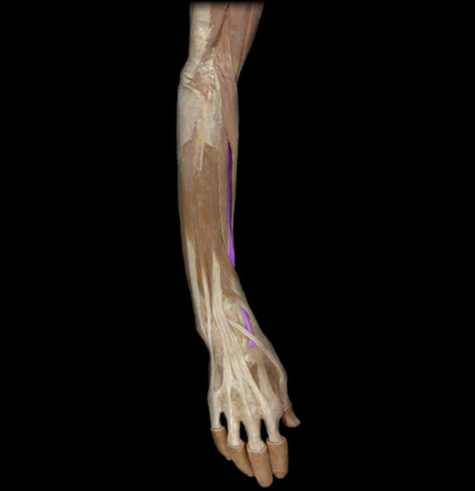
Pronator quadratus
table 1, median n (C8-T1), forearm pronation
lies deep to the flexor tendons
Extensor carpi radialis longus
table 5
Extensor carpi radialis brevis
table 5
O: lateral epicondyle of humerus
Extensor digitorum
table 5, radial n (C7-C8), MCP/DIP extension digits 2-5
O: lateral epicondyle of humerus
Extensor digiti minimi
table 5, radial n (C7-C8), extension 5th digit
O: lateral epicondyle of humerus
Extensor carpi ulnaris
table 5
O: lateral epicondyle of humerus
Extensor indicis
table 5, radial n (C7-C8), deep to extensor digitorum
Extensor pollicis longus
table 5, radial n (C7-C8), extension of thumb IP joint
Extensor pollicis brevis
table 5, radial n (C7-C8), extension of thumb MCP joint
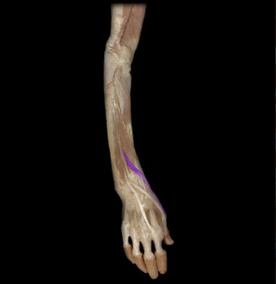
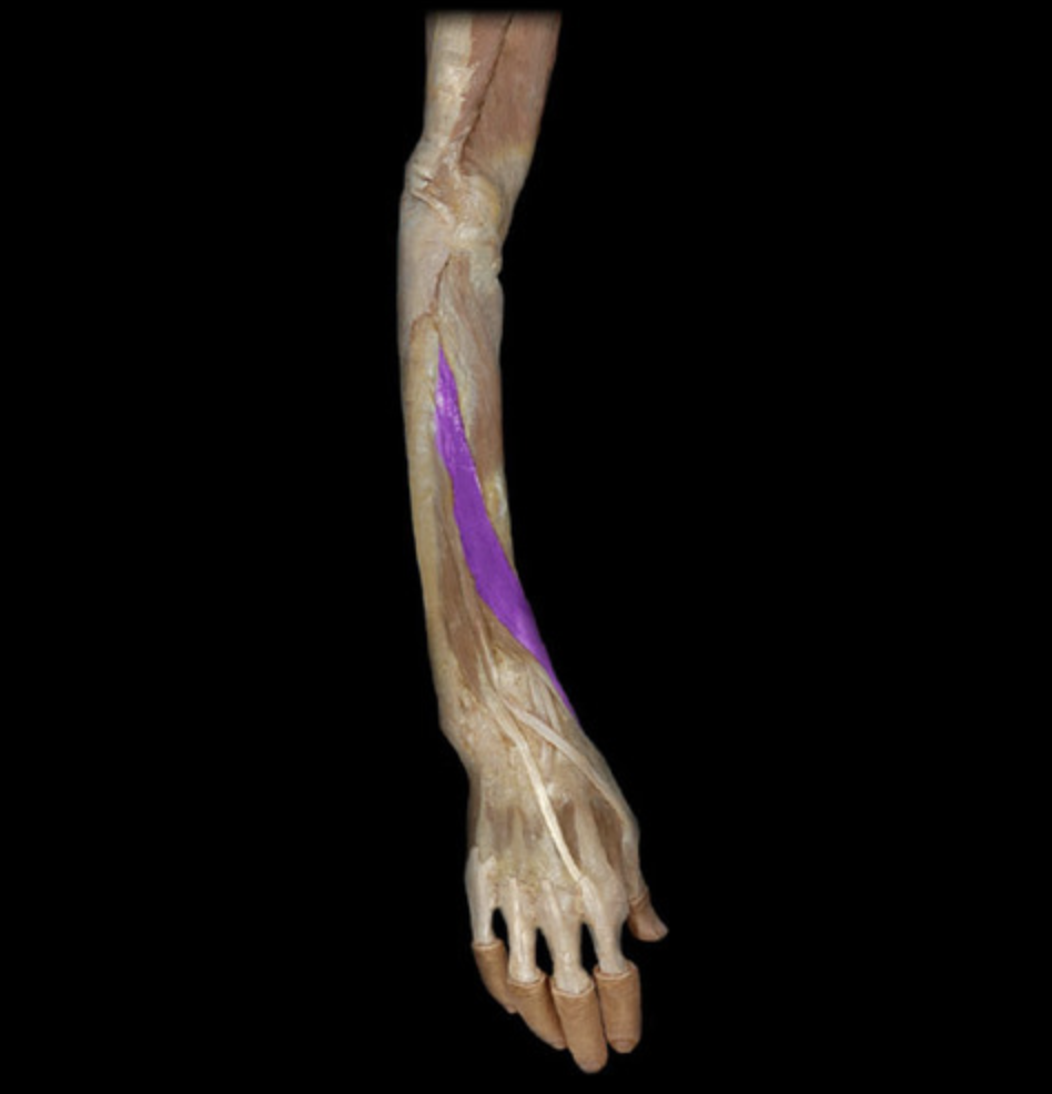
Abductor pollicis longus
table 5, radial n (C7-C8), abduction of thumb CMC joint
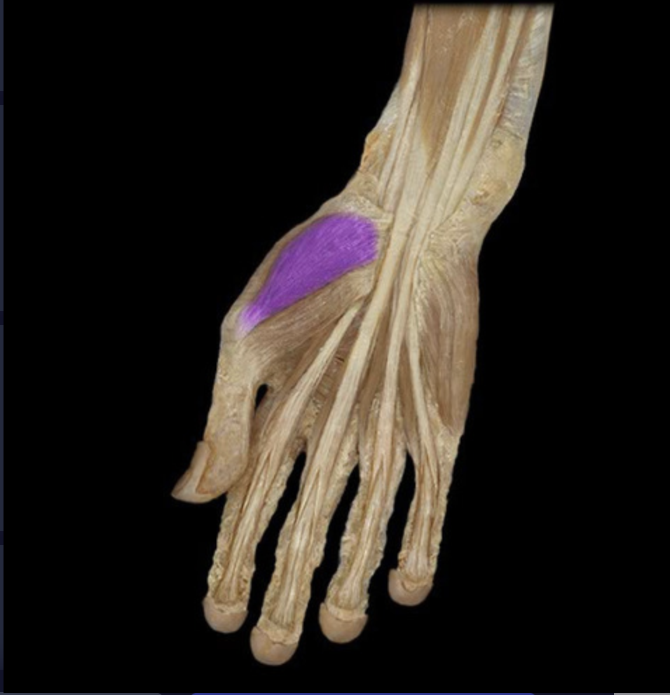
Abductor pollicis brevis
table 5, thenar m, median n
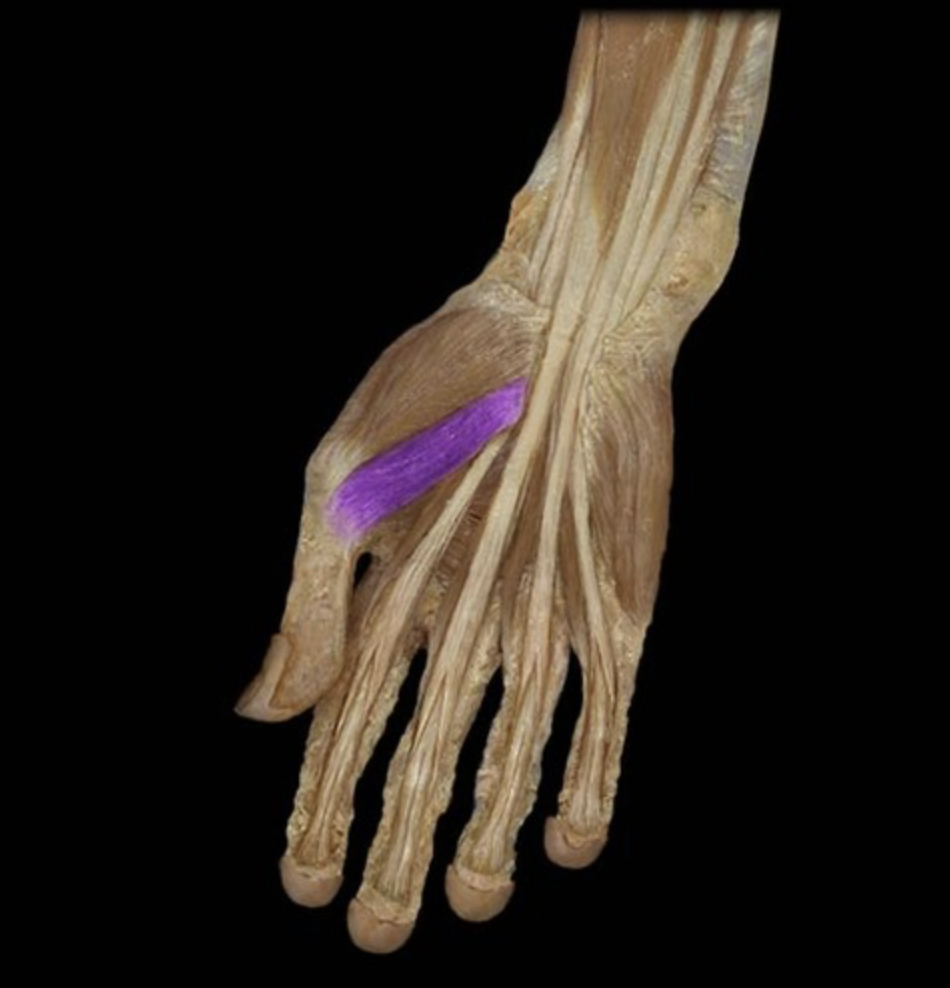
Flexor pollicis brevis
table 7, thenar m, median n
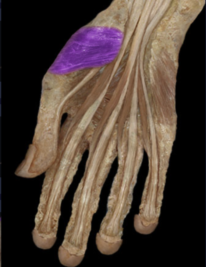
Opponens pollicis
table 7, thenar m, median n
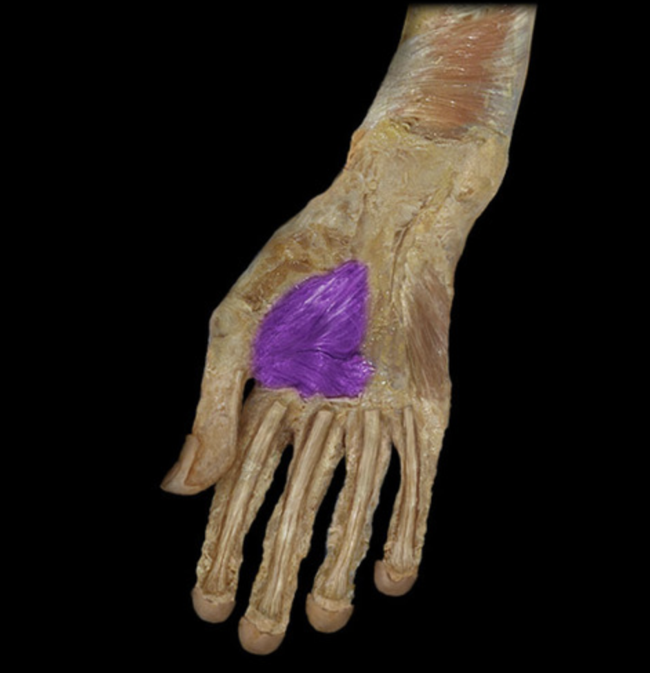
Adductor pollicis
table 7
Abductor digiti minimi
table 7, hypothenar m, ulnar n
Flexor digiti minimi
table 7, hypothenar m, ulnar n
Opponens digiti minimi
table 7, hypothenar m, ulnar n
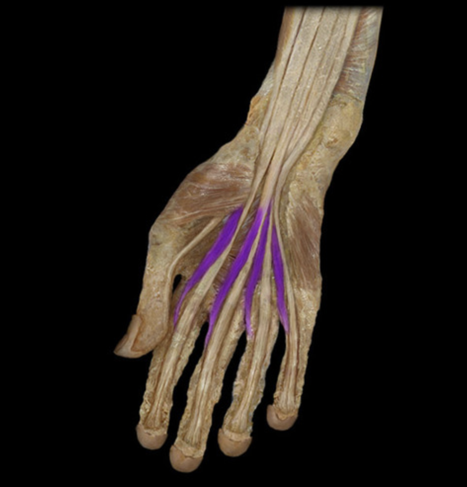
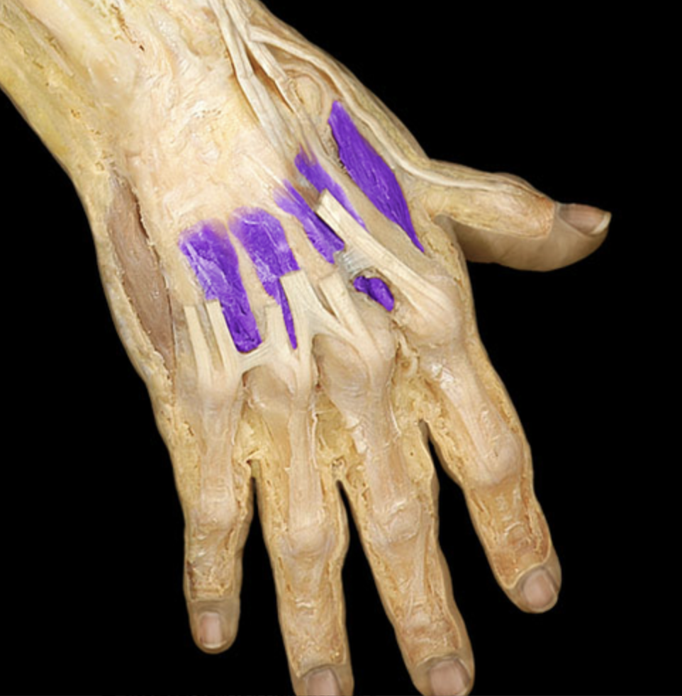
Lumbricals
table 7, number them 1-4
Dorsal interosseous
table 5
number them 1-4
Supinator
table 5, radial n (C6-C7), deep to extensor tendons
O: lateral epicondyle of humerus

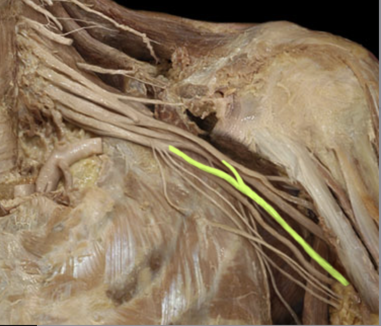
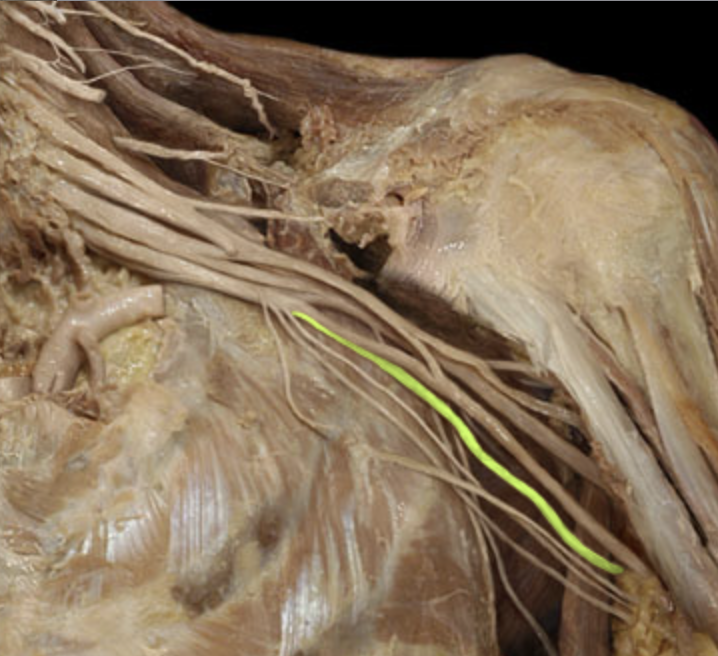
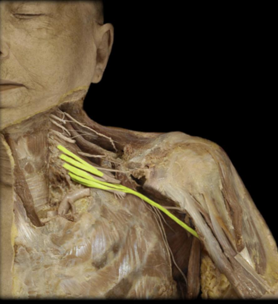
Lateral cord
table 7, terminal branches are musculocutaneous n and median n
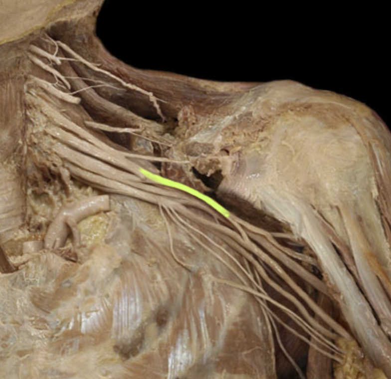
Medial cord
table 7, terminal branches are ulnar n and median n
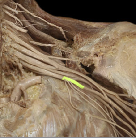
Posterior cord
table 7, terminal branches are axillary n and radial n
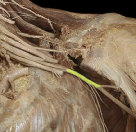
Axillary n
table 7, terminal branch of posterior cord into armpit
supplies deltoid and teres minor; C5-C6 ventral rami contributions only
Axillary a
table 7, crosses through brachial plexus
Axillary v
table 7
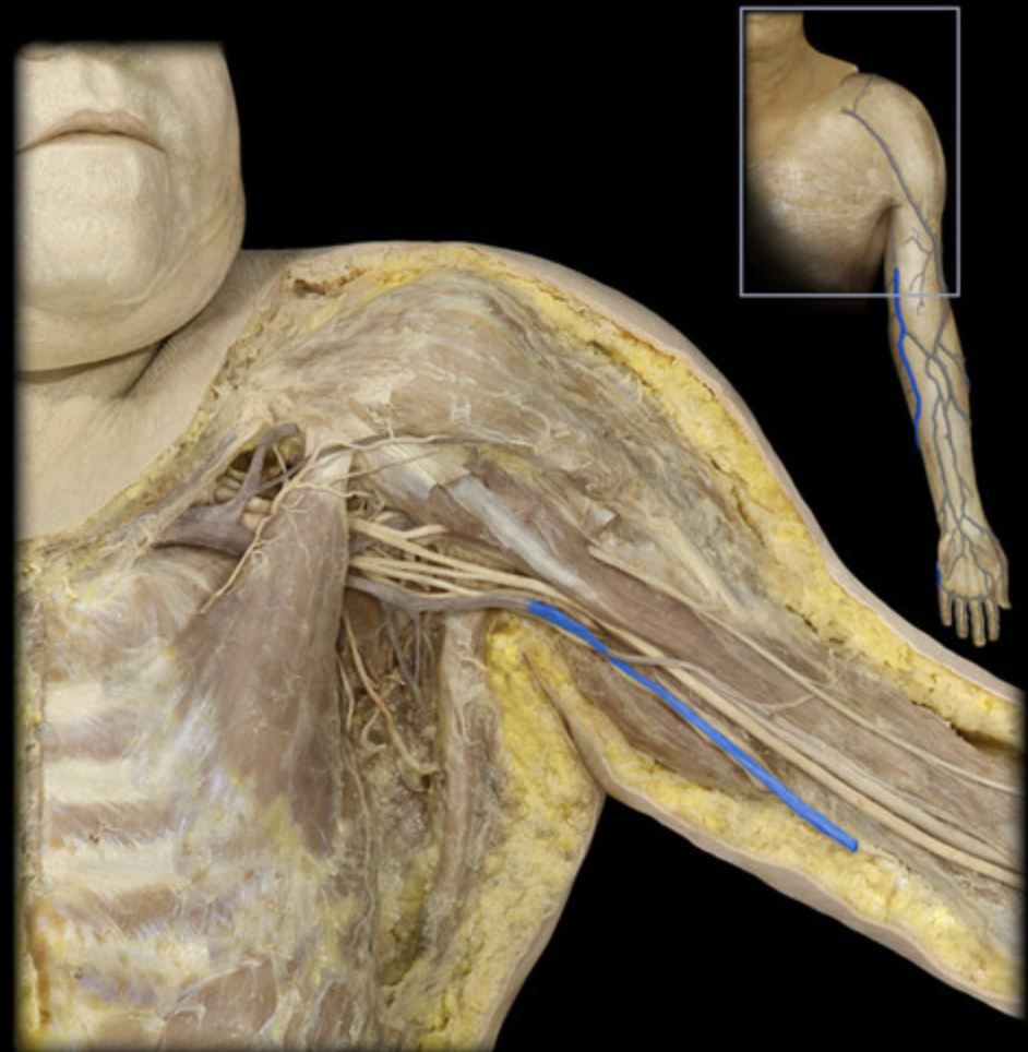
Brachial a
table 7
Brachial v
table 7
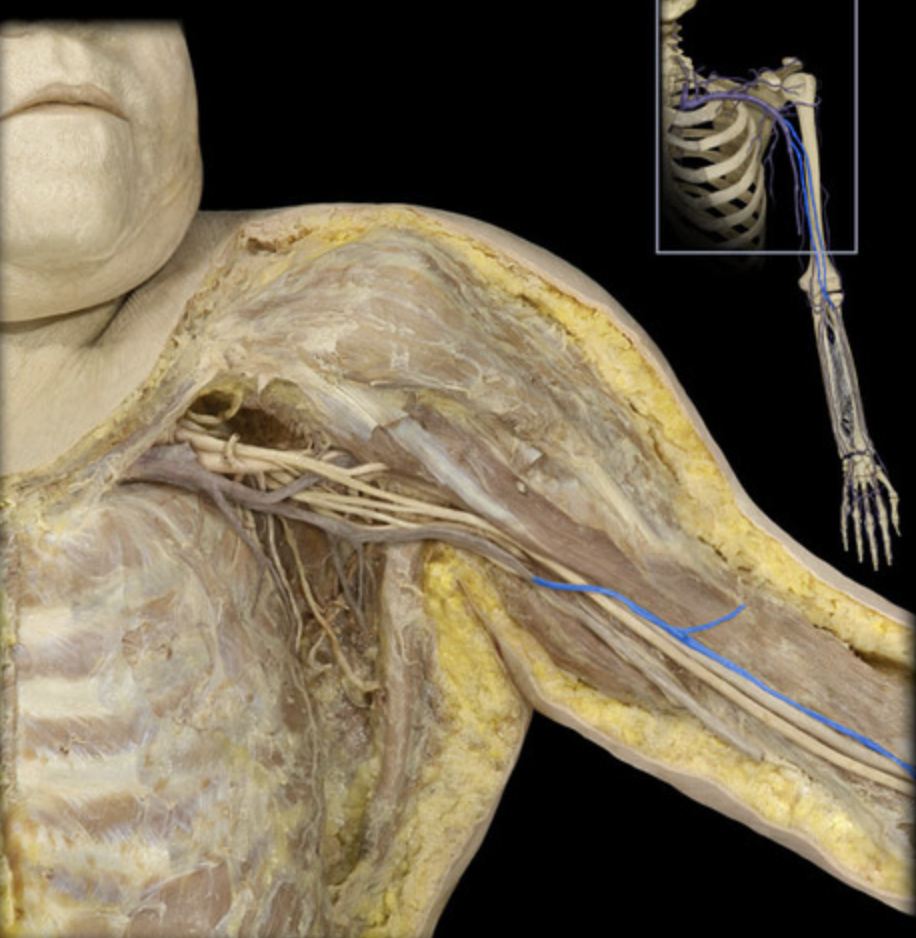
Radial a
table 7
Ulnar a
table 7
Cephalic v
table 5
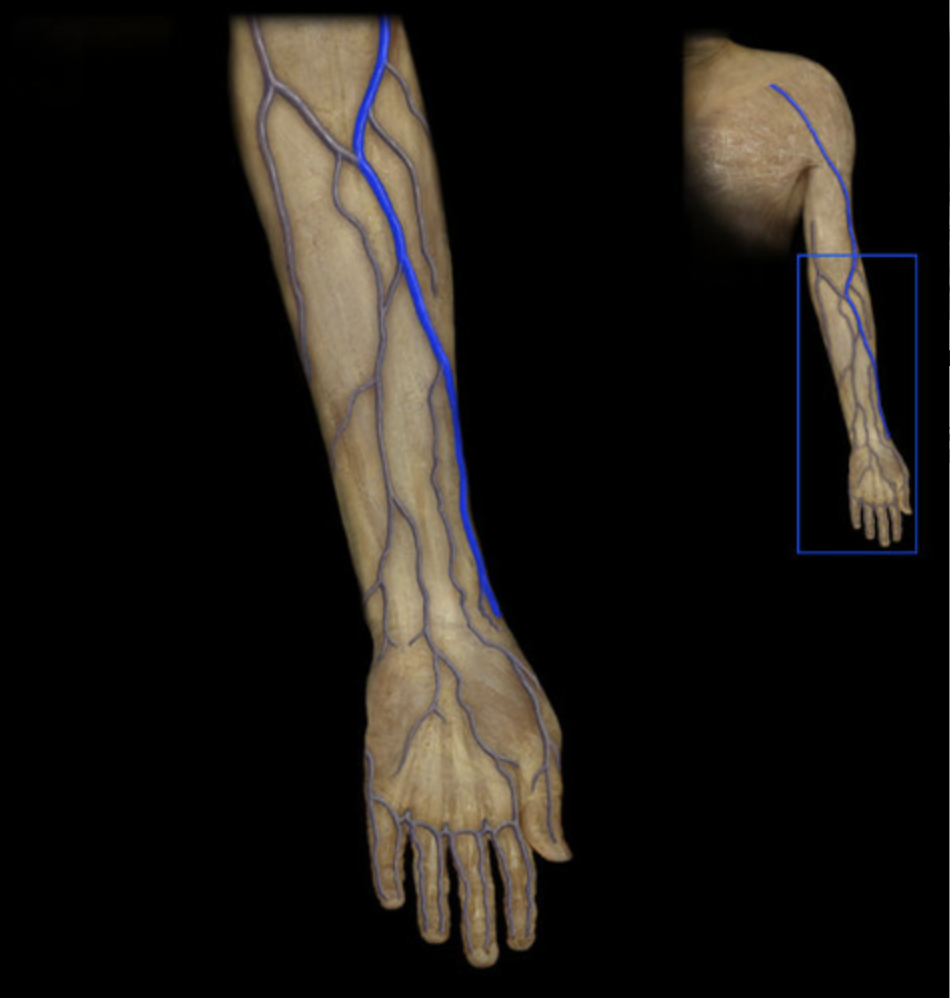
Basilic v
table 5, closer to body
Musculocutaneous n
table 5, lateral cord terminal branch, supplies flexor compartment of arm
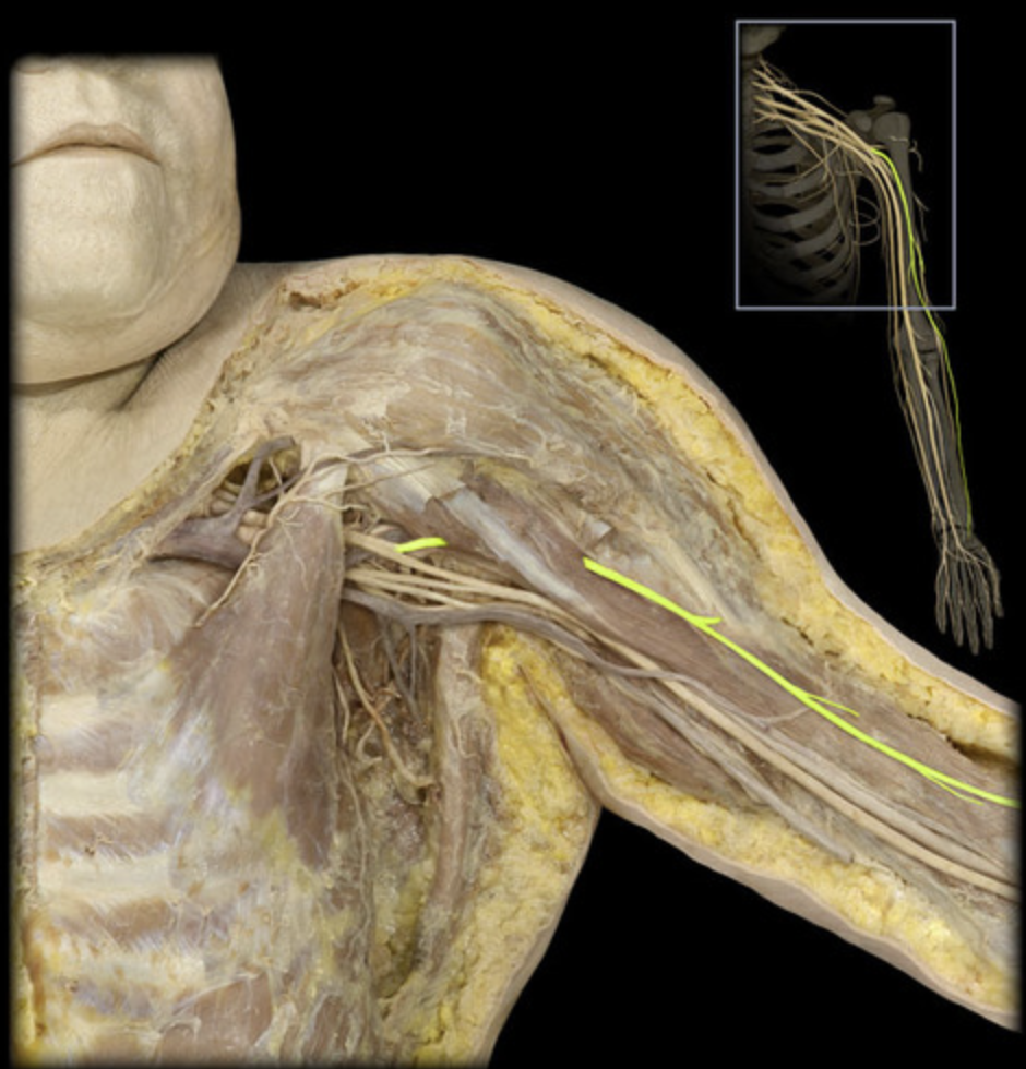
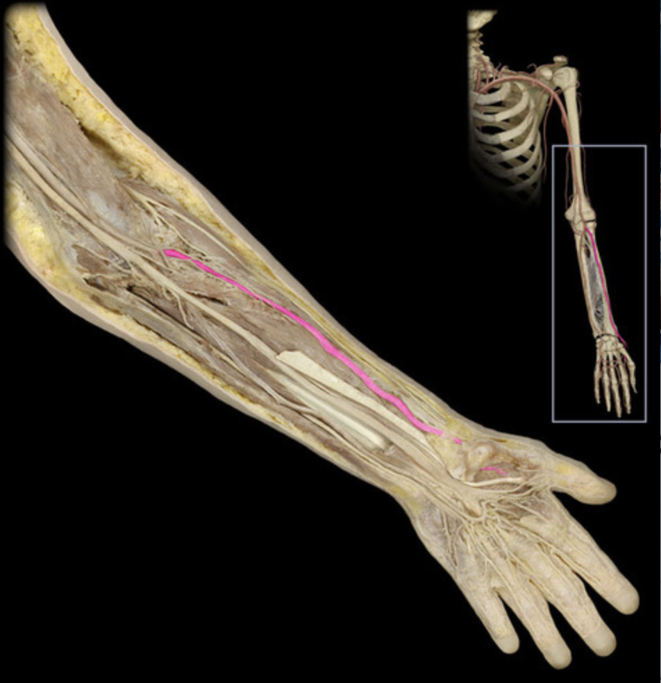
Median n
table 7, medial/lateral cord terminal branch
supplies flexor compartment of forearm, thenar mm, lumbricals 1 & 2
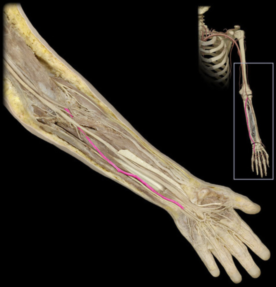
Ulnar n
table 7, medial cord terminal branch
supplies flexor carpi ulnaris, FDP tendons 3 & 4, rest of intrinsic hand mm
Radial n
table 7, posterior cord terminal branch
supplies all extensors of arm and forearm
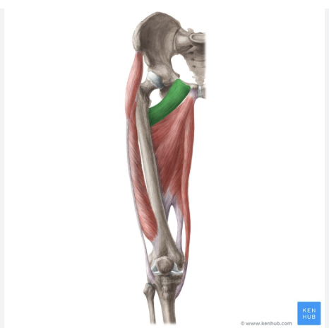
Tensor fasciae latae
table 5, 7
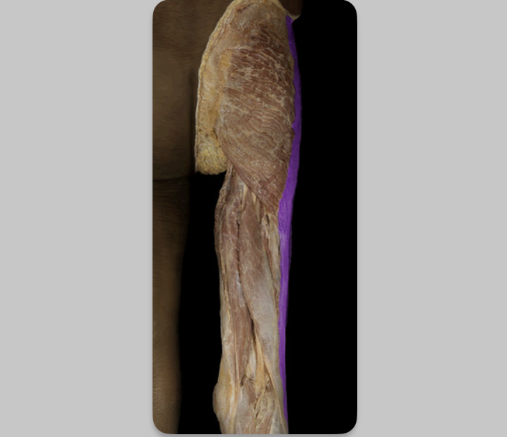
Iliotibial tract
table 5, 7
Sartorius
table 5, 7
Piriformis
table 3
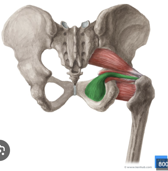
Obturator internus
table 3, below superior gemellus under sciatic n
Superior gemellus
table 3, below piriformis, under sciatic n
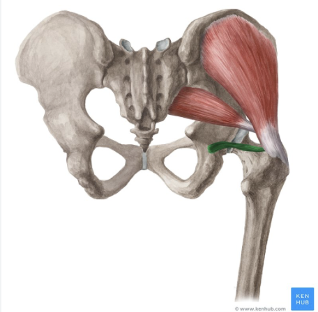
Inferior gemellus
table 3, below obturator internus
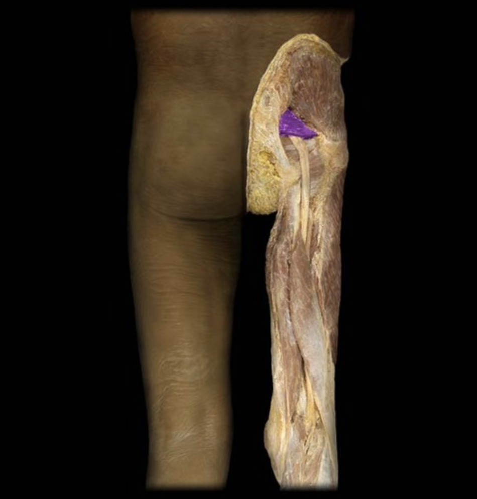
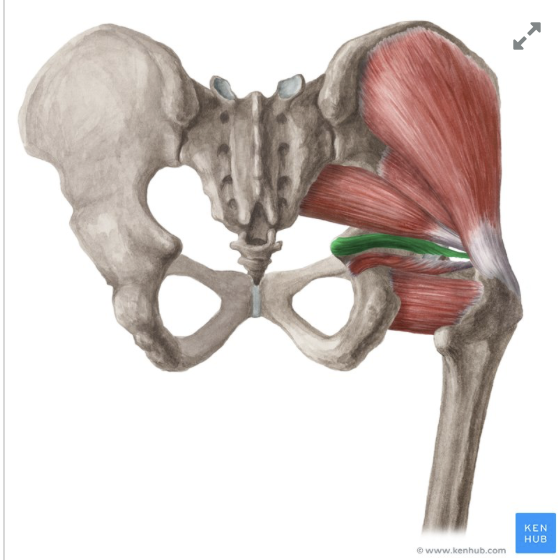
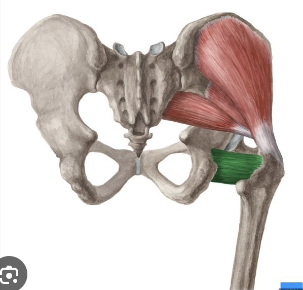
Quadratus femoris
table 3, below inferior gemellus
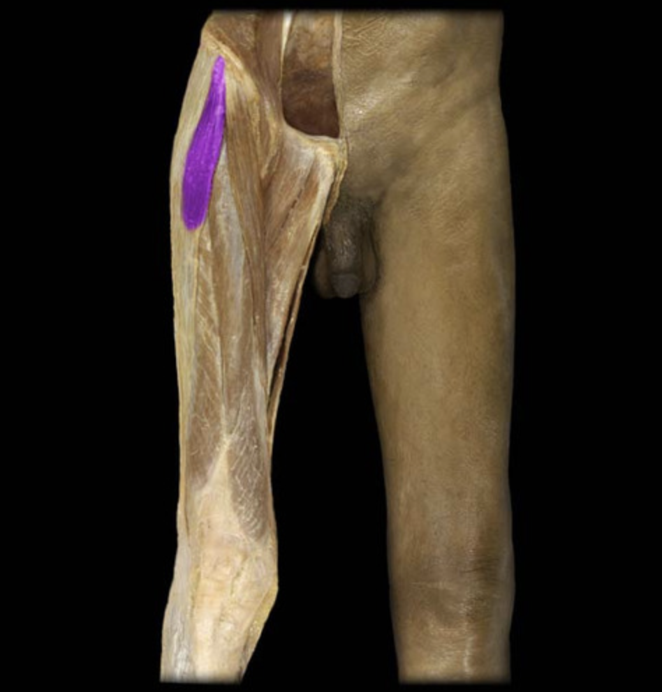
Rectus femoris
Quadriceps muscle, table 5,7
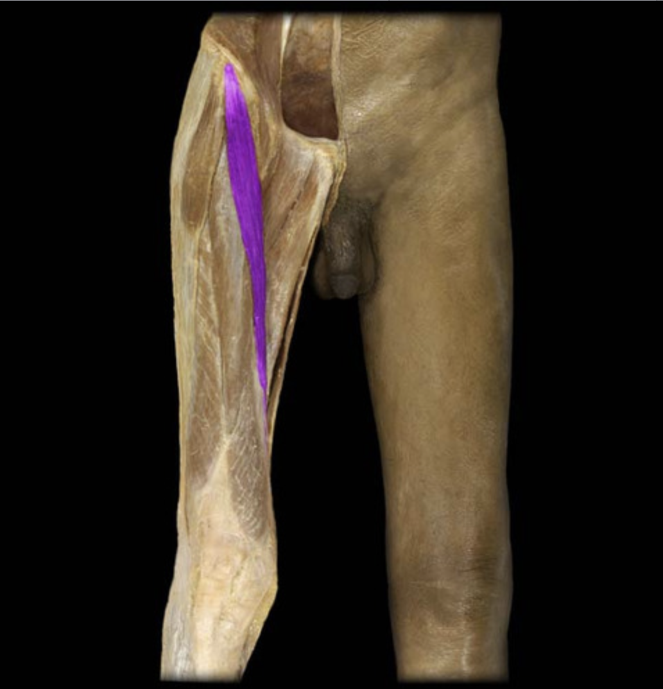
Vastus lateralis
Quadriceps muscle, table 5, 7
Vastus intermedius
Quadriceps muscle, lies beneath rectus femoris, table 5,7
Vastus medialis
Quadriceps muscle, table 5, 7
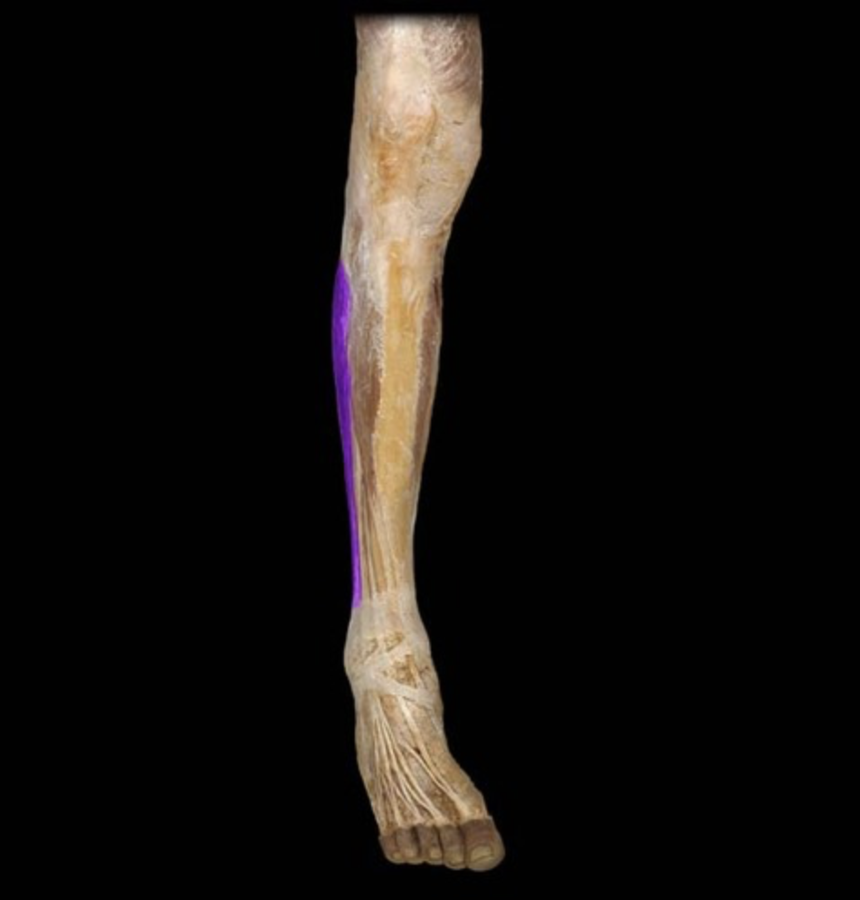
Tibialis anterior
table 7
Extensor hallucis longus
table 7, next to tibialis anterior
Extensor digitorum longus
table 7, next to extensor hallucis longus
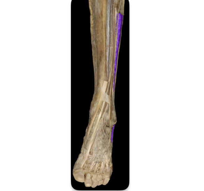
Fibularis longus
table 7, (peroneus)
Fibularis brevis
table 7, deep to fibularis (peroneus) longus
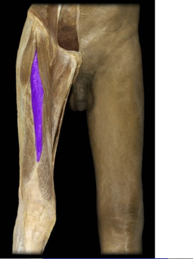
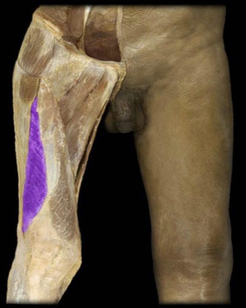
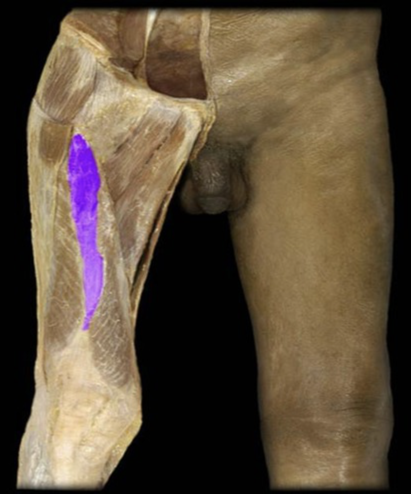
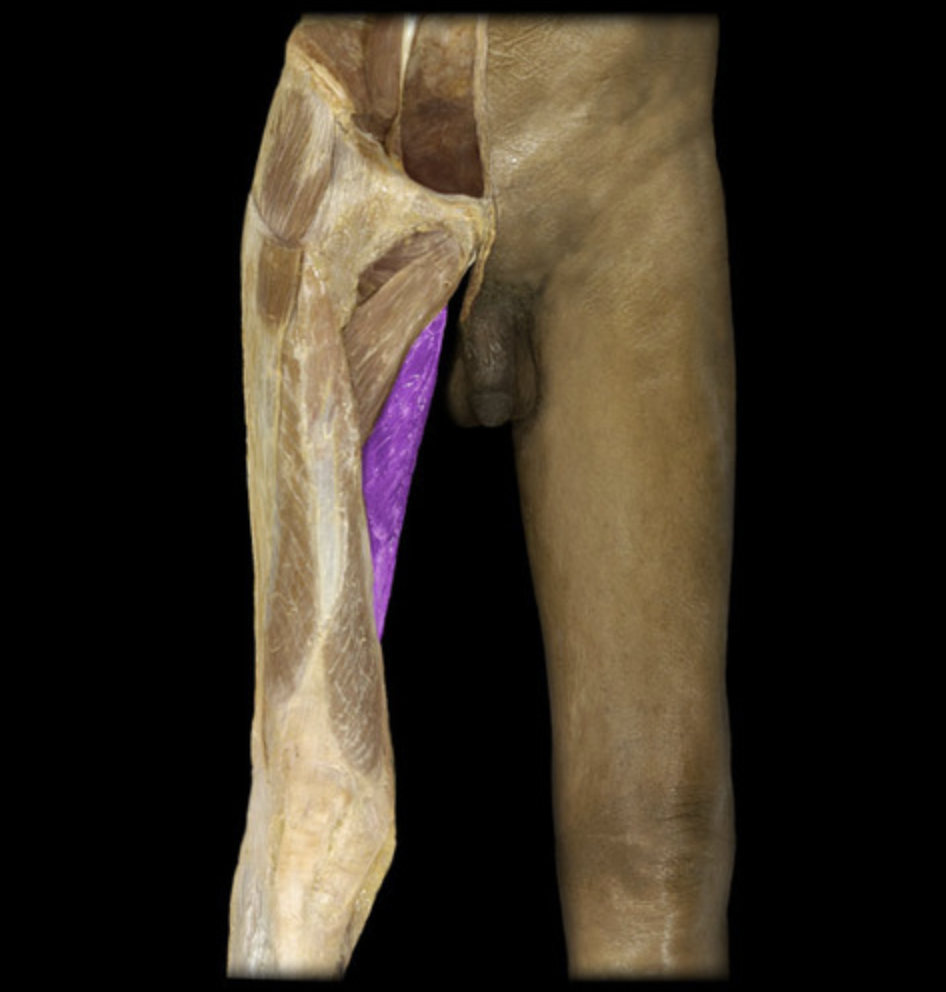
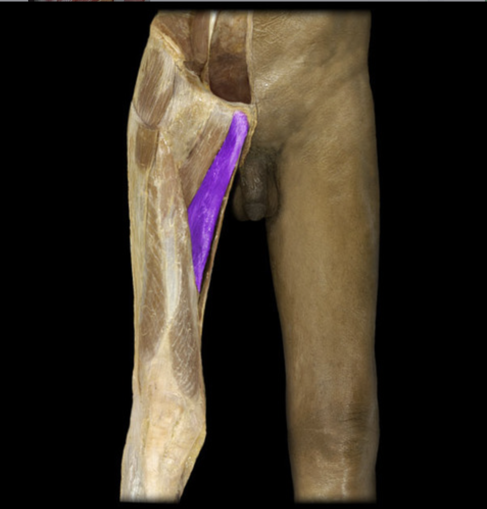
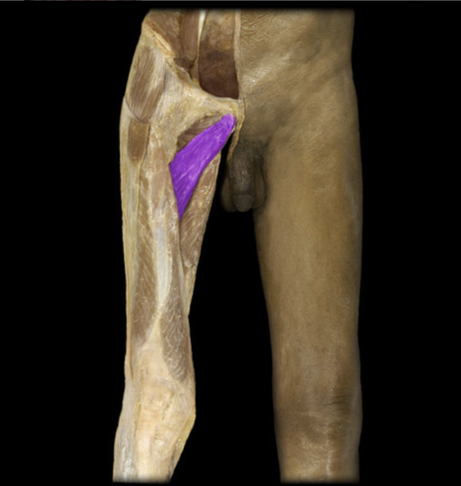
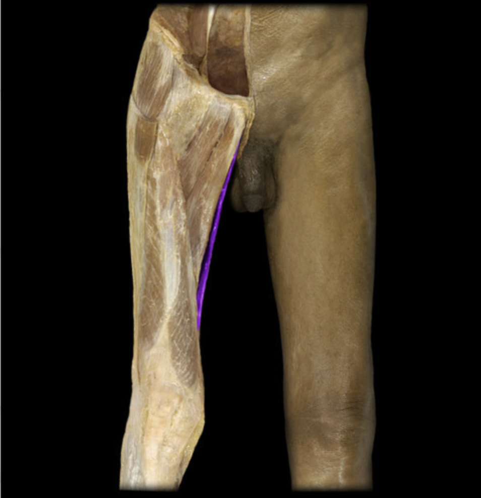
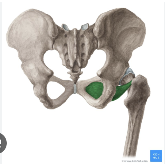
Pectineus
table 7
Adductor magnus
table 5, 7, deepest muscle next to gracilis
Adductor longus
table 5, 7
Adductor brevis
table 5, 7
Gracilis
table 5, 7, most medial
Obturator externus
table 5, 7, deep, more medial next to pectineus
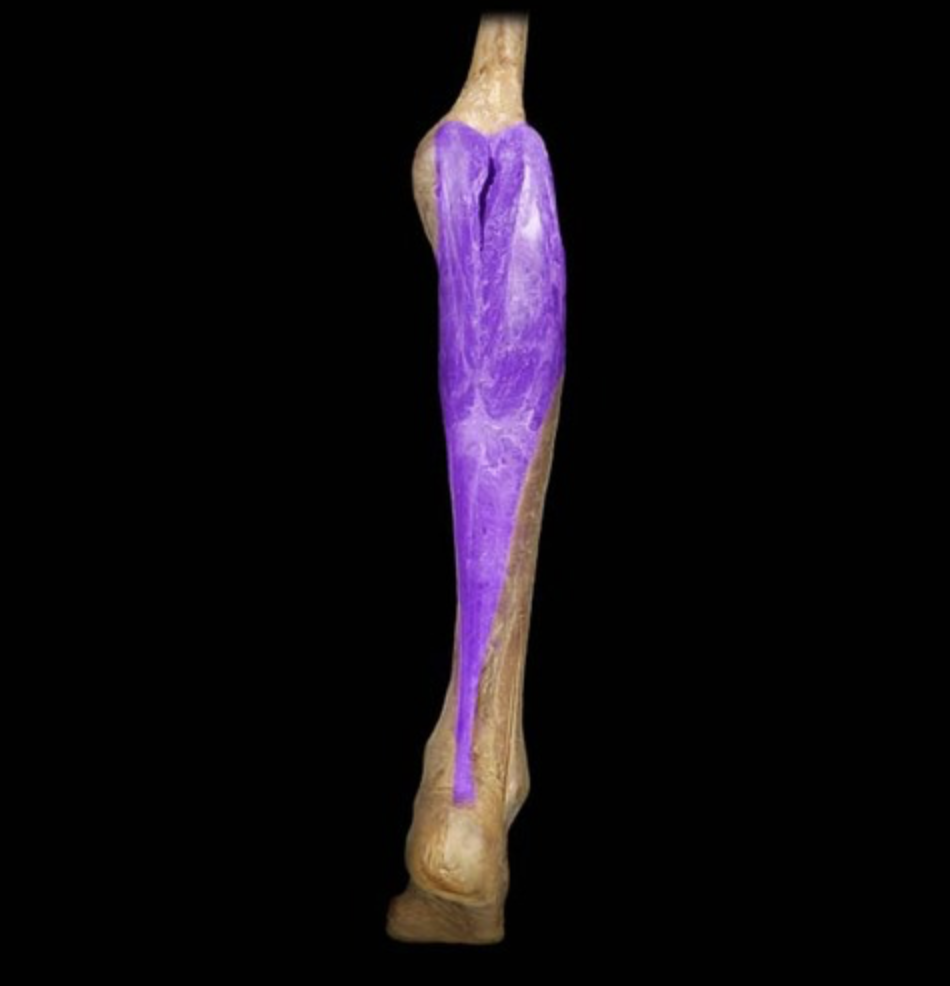
Gastrocnemius
table 3, superficial, has 2 heads