Checklist of Superior Mediastinum and Heart Structures
1/89
There's no tags or description
Looks like no tags are added yet.
Name | Mastery | Learn | Test | Matching | Spaced | Call with Kai |
|---|
No analytics yet
Send a link to your students to track their progress
90 Terms
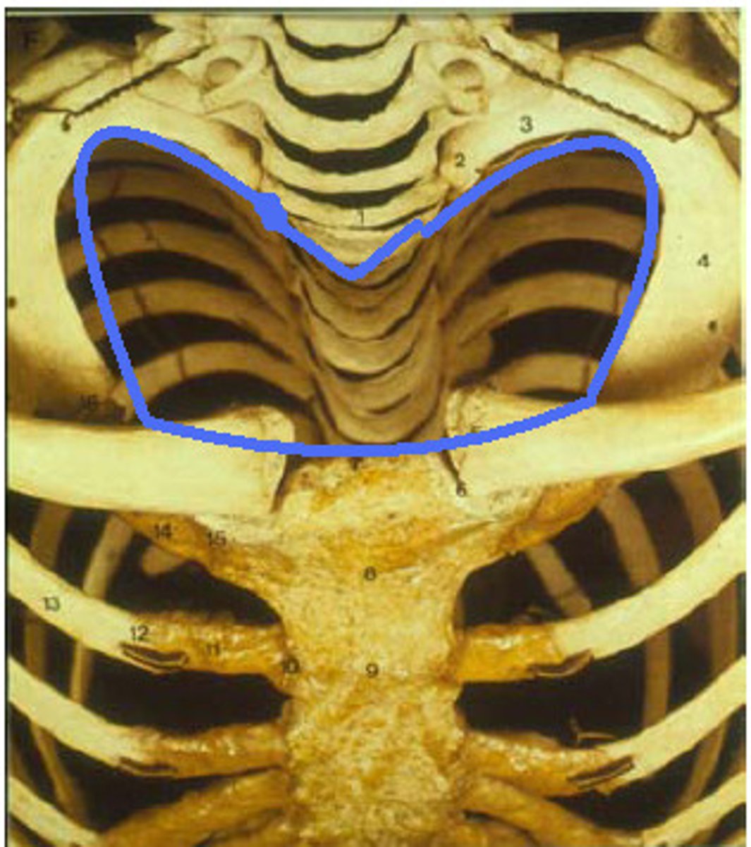
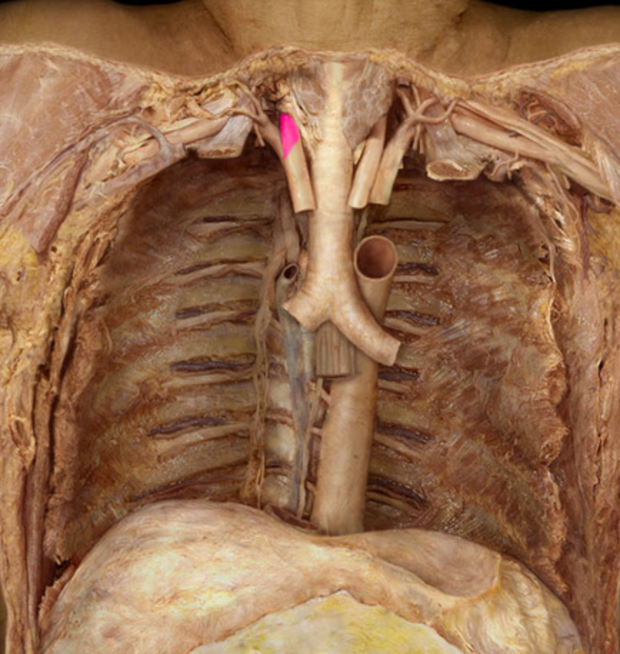
Superior thoracic aperture
Opening at the top of the thoracic cavity.
right subclavian artery
left subclavian artery
left common carotid artery
right common carotid artery
C3,4,5
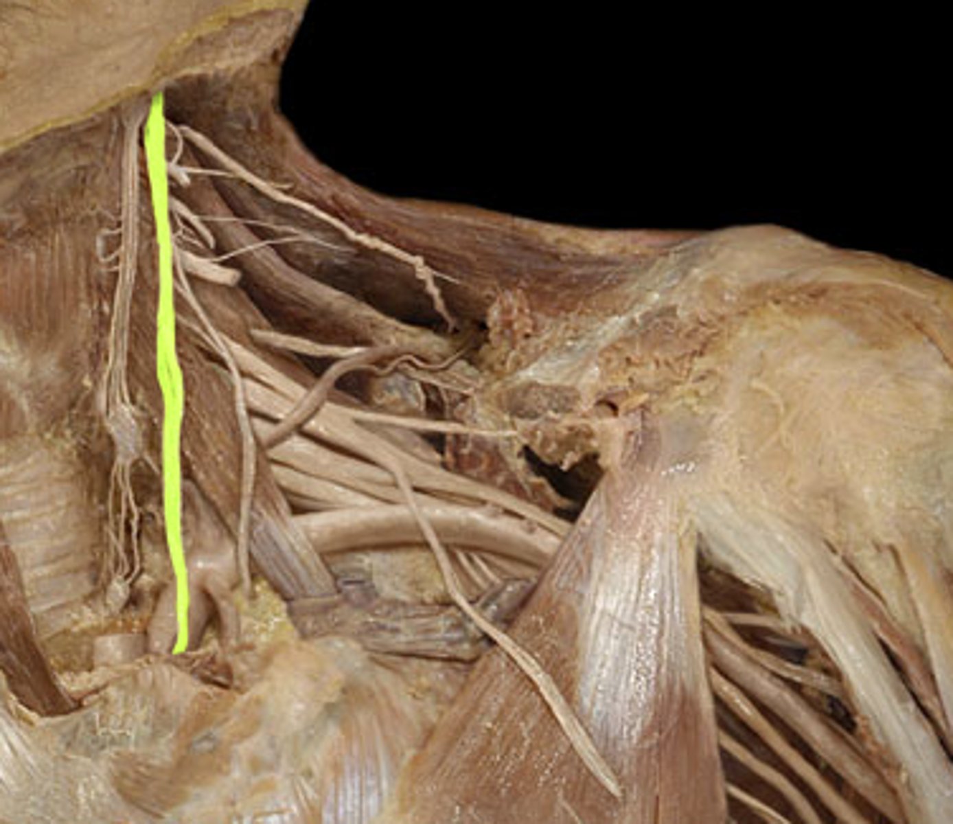
origin of phrenic nerve
parasympathetic to the body, slow heart rate
function of vagus nerve
aortic arch
A

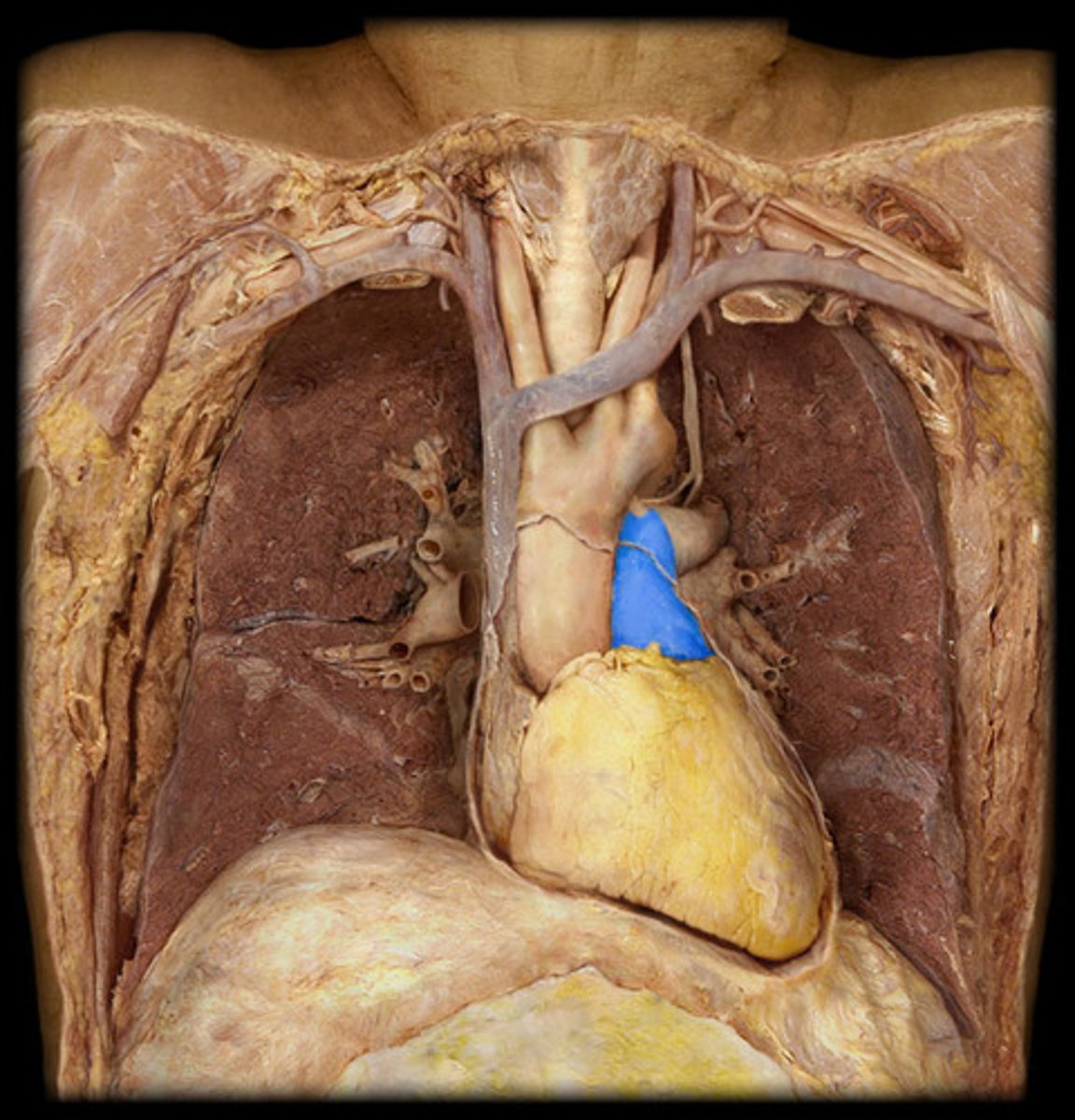
main pulmonary artery
B
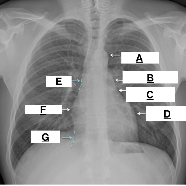
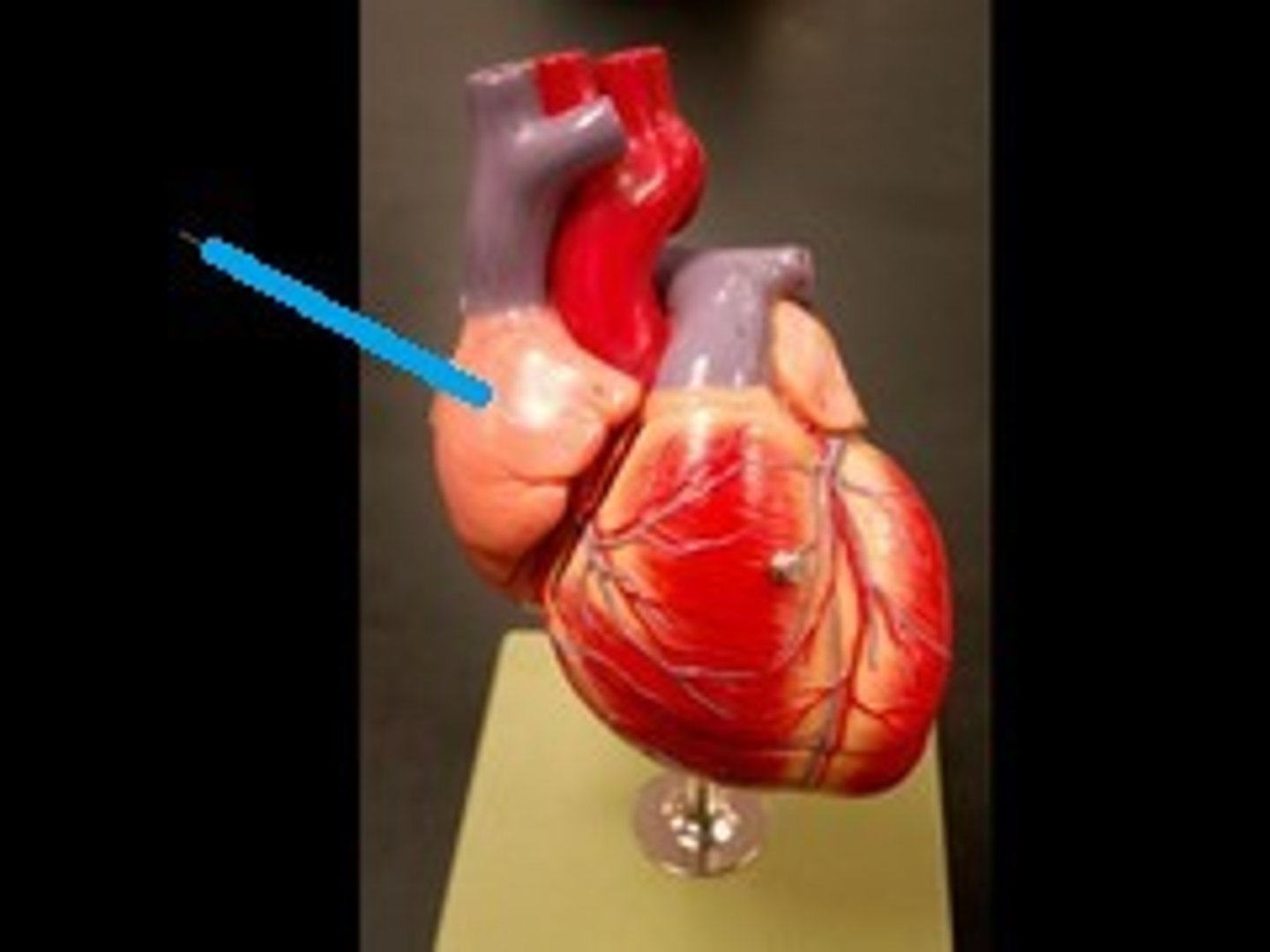
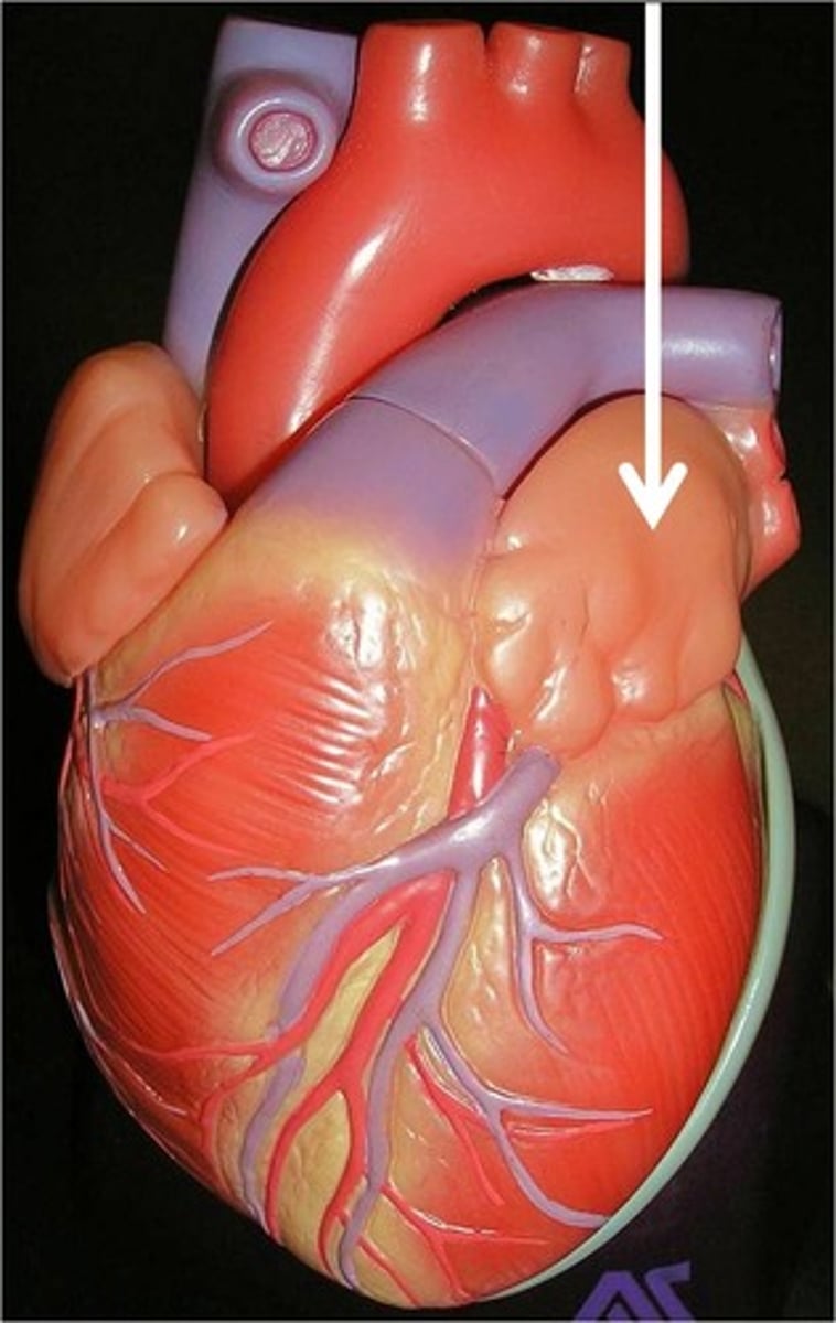
left atrial appendage
C

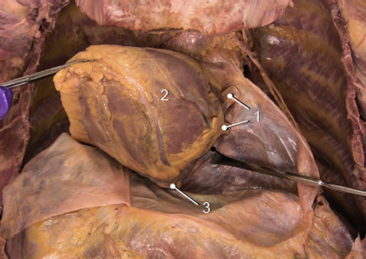
left ventricle
D

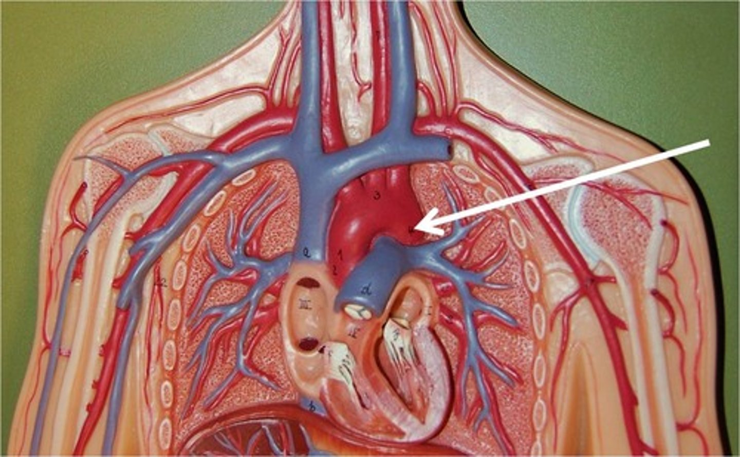
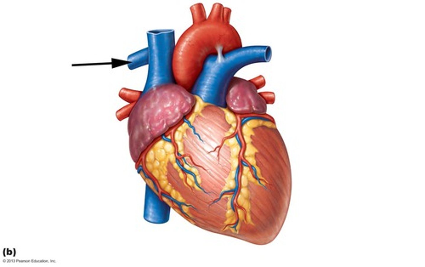
superior vena cava
E

right atrium
F

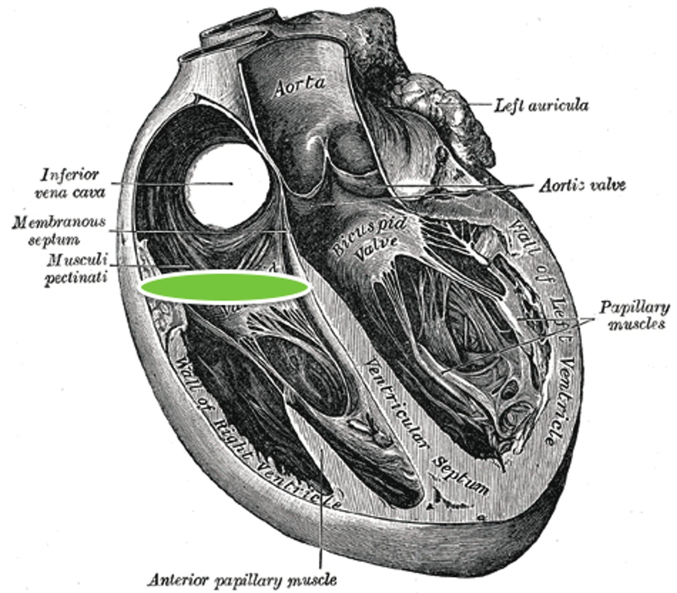
inferior vena cava
G

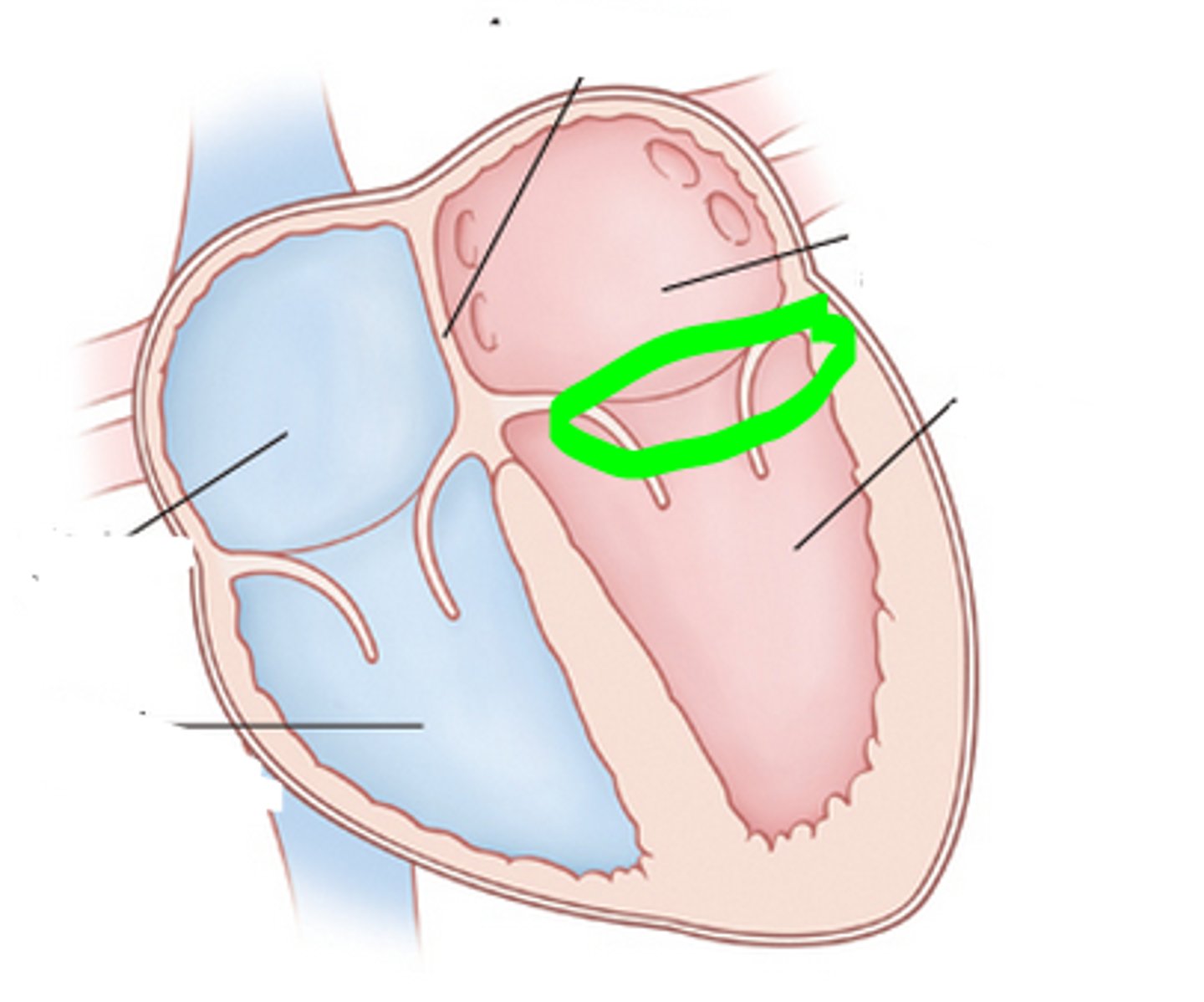
foramen ovale
embryologic structure that becomes fossa ovalis in a mature heart, allowing blood flow between atria.
blood bypasses right ventricle and lungs
function of foramen ovale
ductus arteriosus
embryonic structure that becomes ligamentum arteriosum in mature heart
allows blood to bypass the fetal lungs by connecting the pulmonary artery to the aorta
function of ductus arteriosus
somatic (voluntary) innervation of laryngeal muscles (except cricothyroid)
function of recurrent laryngeal nerve
Innervates the diaphragm, somatic (voluntary) breathing
function of phrenic nerve
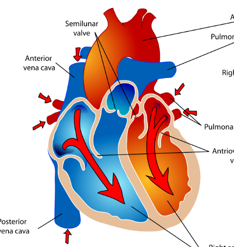
diastole
what is happening in this picture
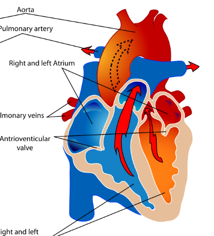
systole
what is happening in this picture
right and left AV valves
what valves are open during diastole
pulmonary and aortic valves
what valves are open during systole
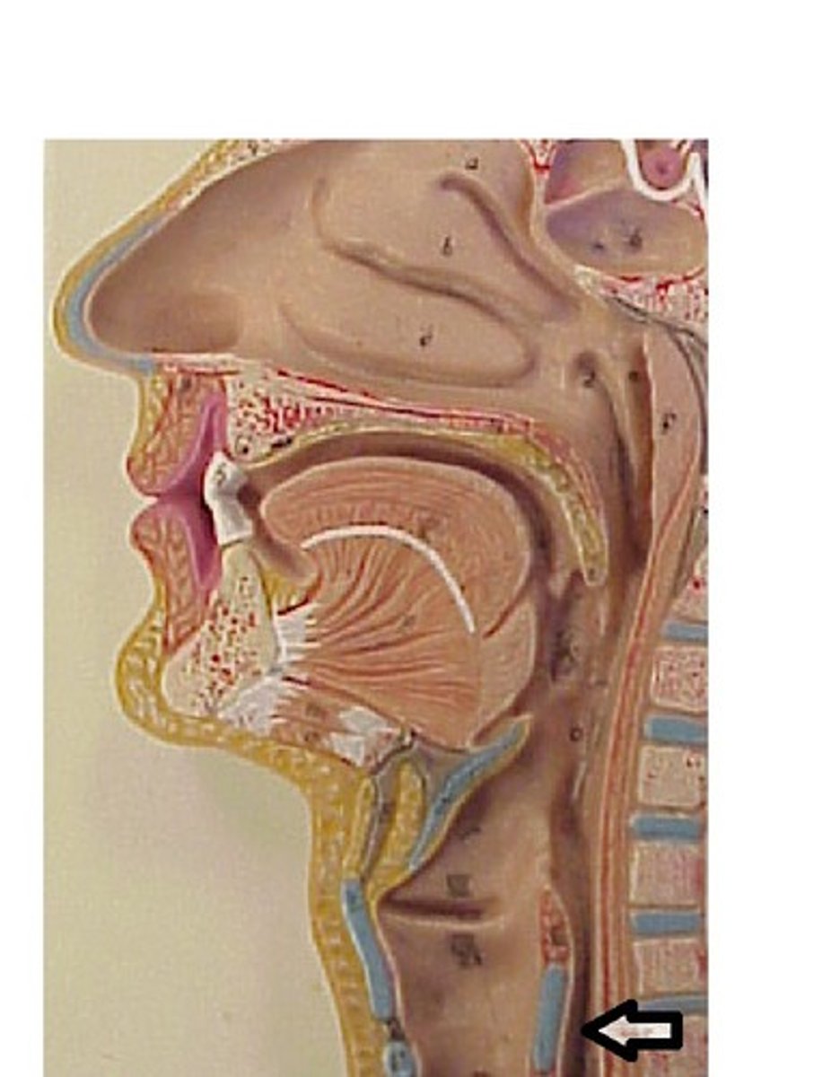
Trachea
Windpipe connecting larynx to bronchi.
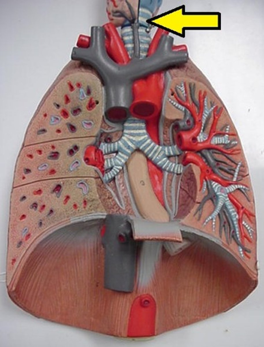
Esophagus
Tube transporting food to the stomach.
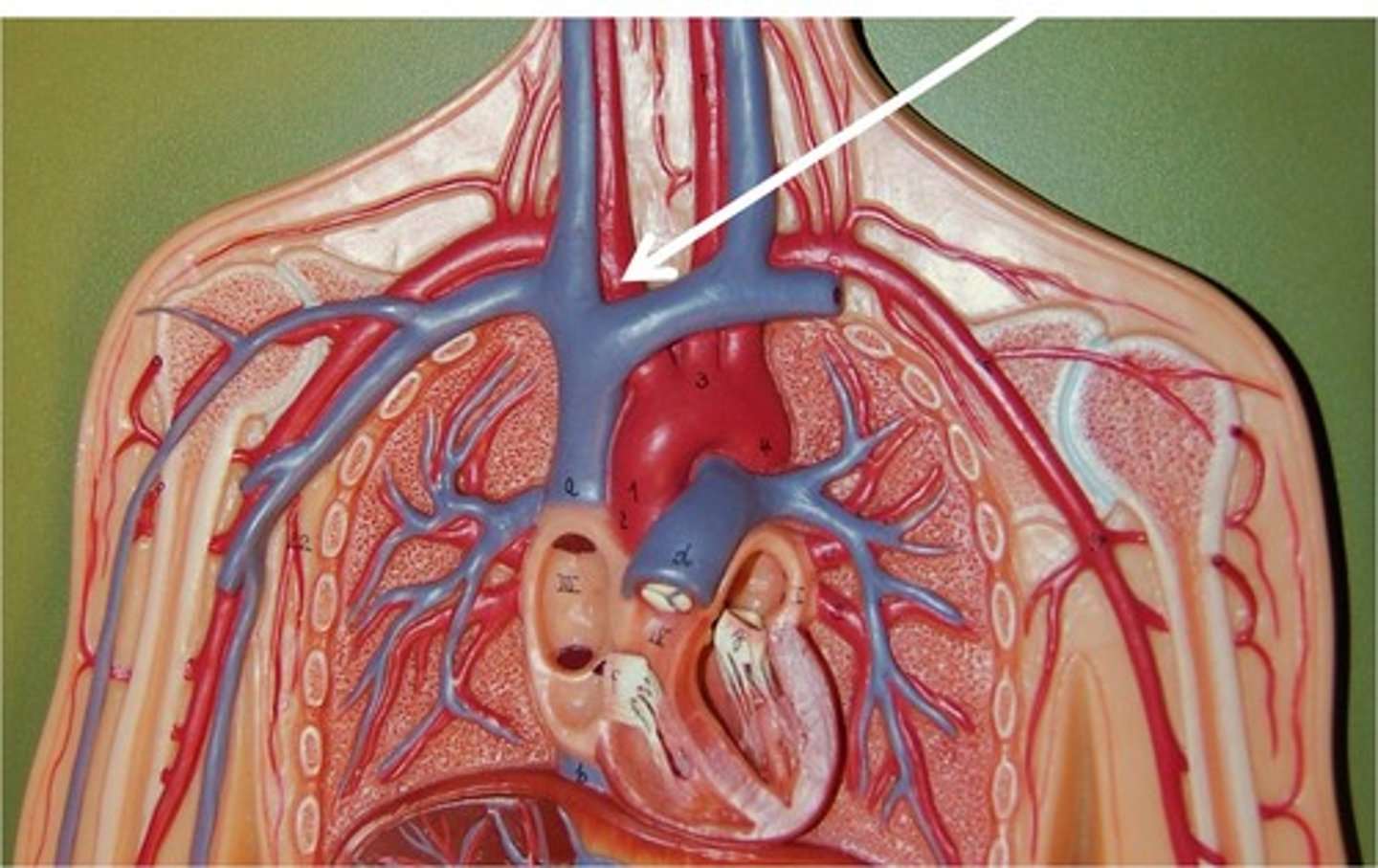
Subclavian veins (right or left)
Veins draining blood from upper limbs.
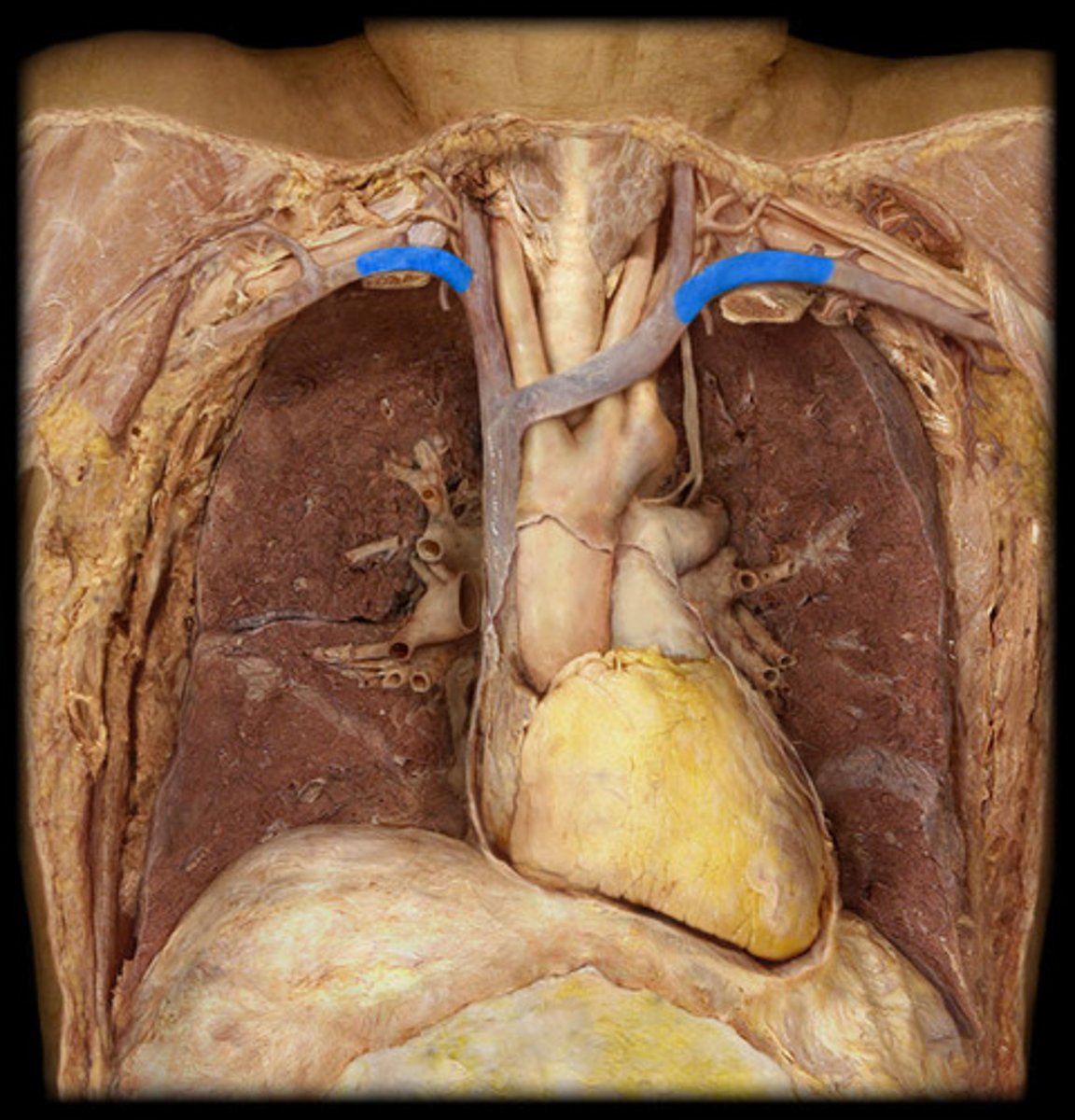
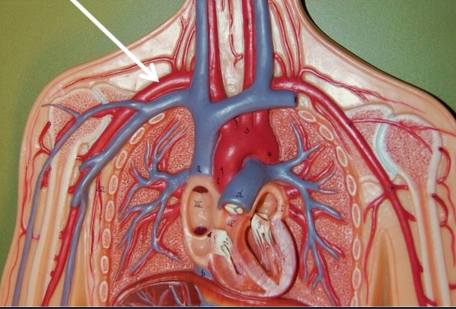
Brachiocephalic veins (right or left)
Veins formed by merging subclavian and internal jugular.
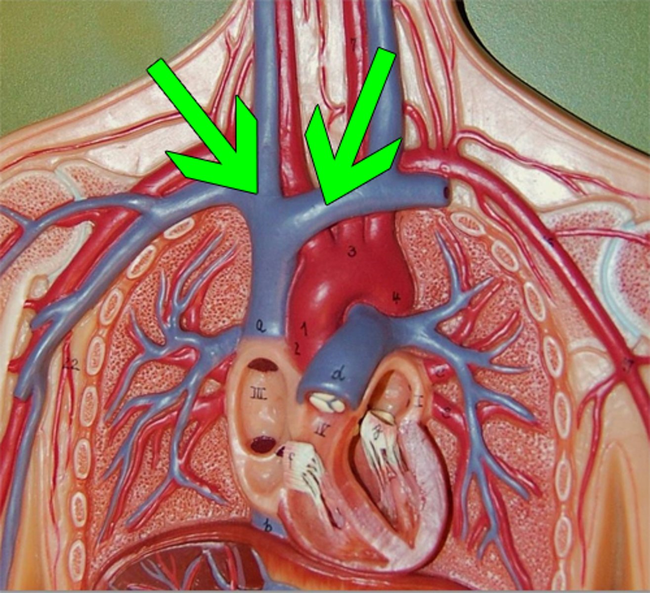
Superior vena cava
Large vein carrying deoxygenated blood to heart.
Arch of azygos vein
Vein arching over the right main bronchus.
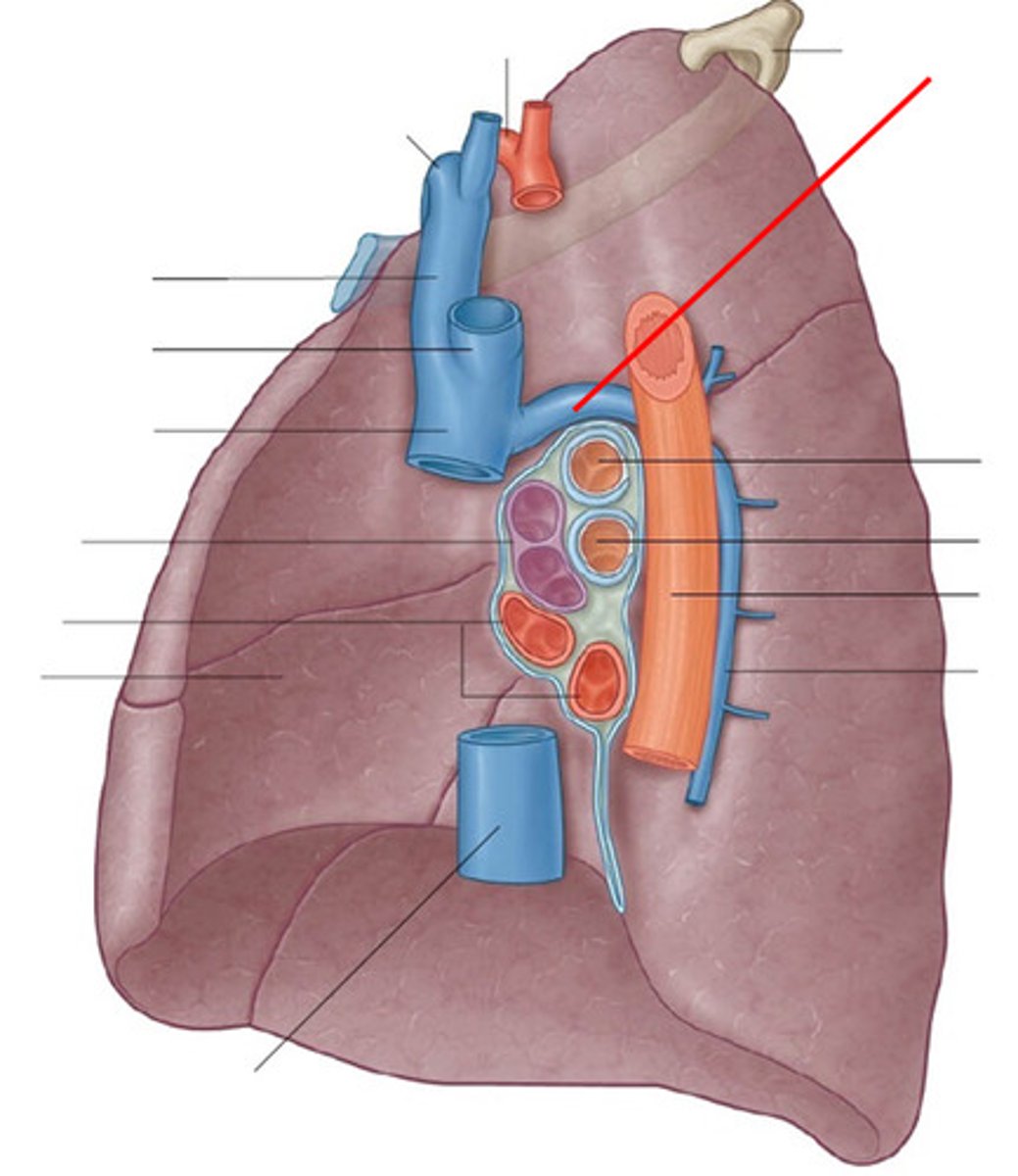
ascending Aorta
Main artery distributing oxygenated blood from heart.
aortic arch
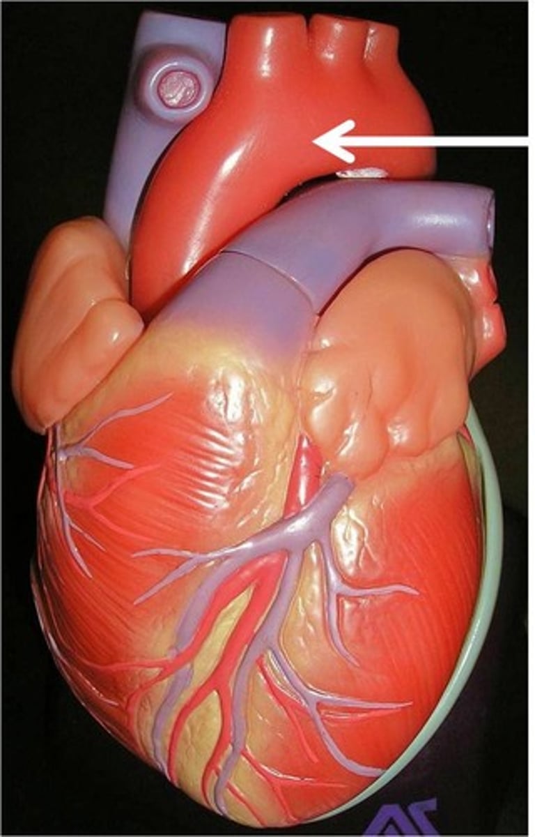
descending aorta
part of the aorta that branches into the thoracic and abdominal aortae
Brachiocephalic artery
First branch of the aortic arch.
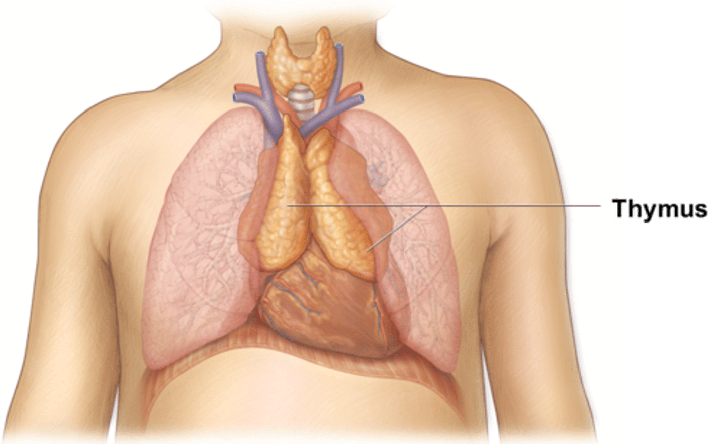
Thymus
Gland involved in immune system development.
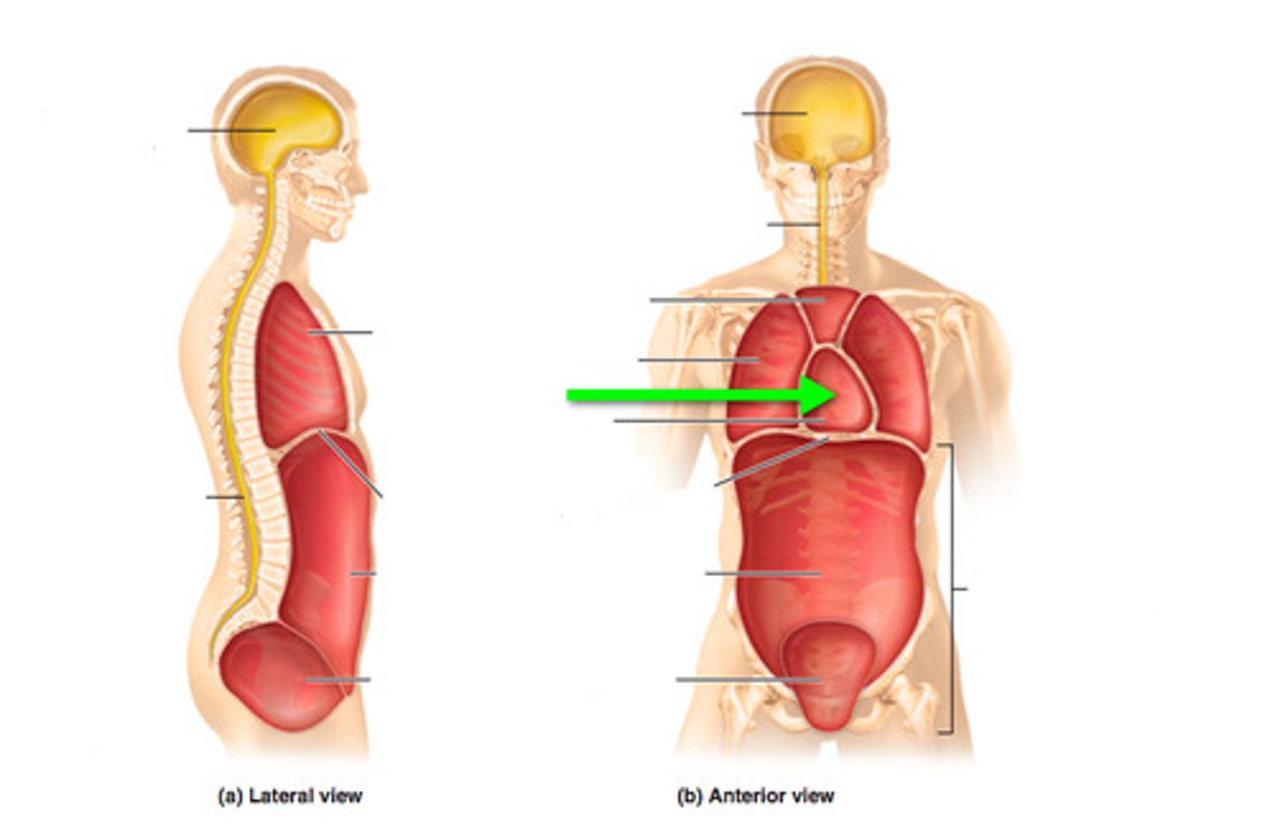
Phrenic nerves
innervates fibrous pericardium
Vagus nerve
Nerves regulating autonomic functions in the body.
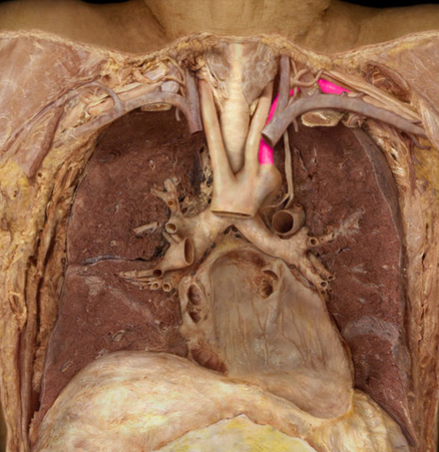
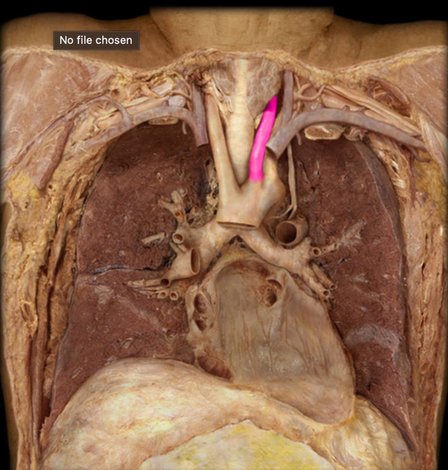
Recurrent laryngeal nerves
Nerves supplying muscles of the larynx.
Pericardium
Double-walled sac surrounding the heart.
Fibrous pericardium
Outer layer of the pericardium.
parietal serous pericardium
lines the inner surface of the fibrous pericardium
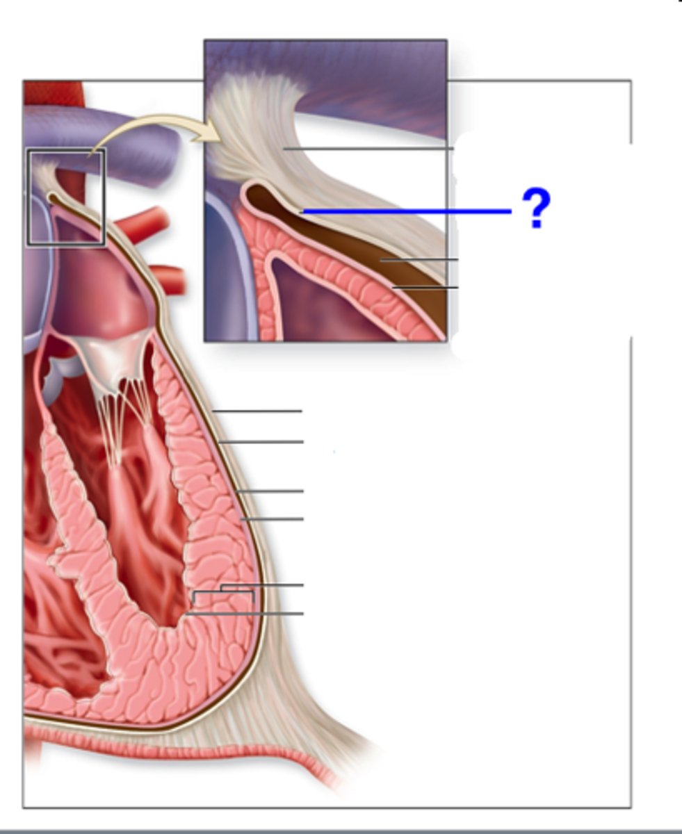
visceral serous pericardium
Pericardial cavity
Space between the pericardial layers containing fluid.
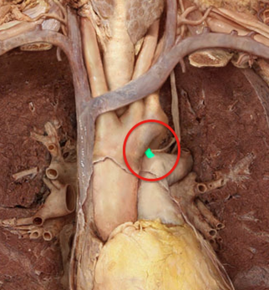
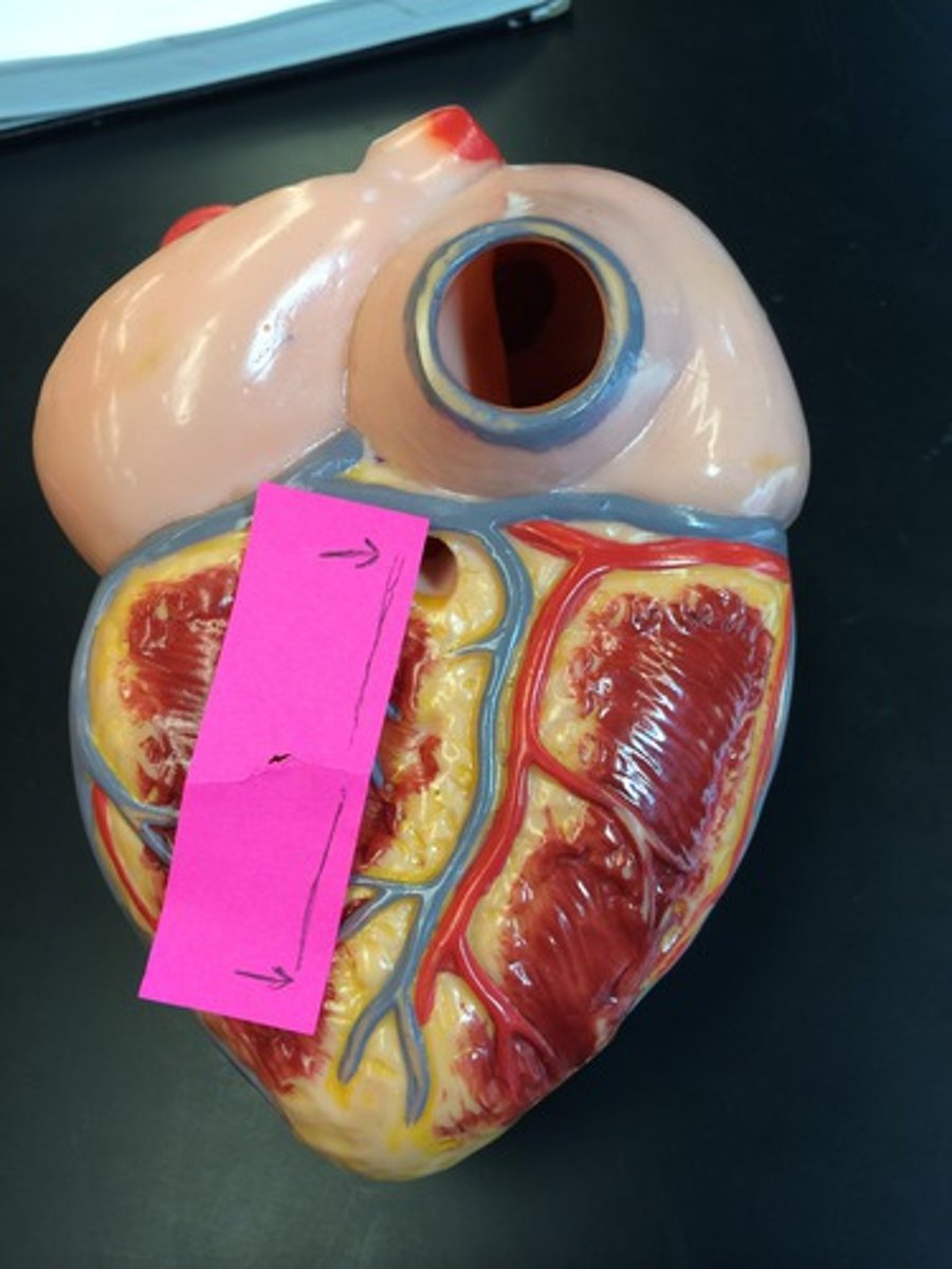
transverse sinus
space posterior to ascending aorta and pulmonary trunk and anterior to superior vena cava

oblique sinus
blind recess on posterior aspect of heart caused by pericardial reflections off the pulmonary veins and inferior vena cava
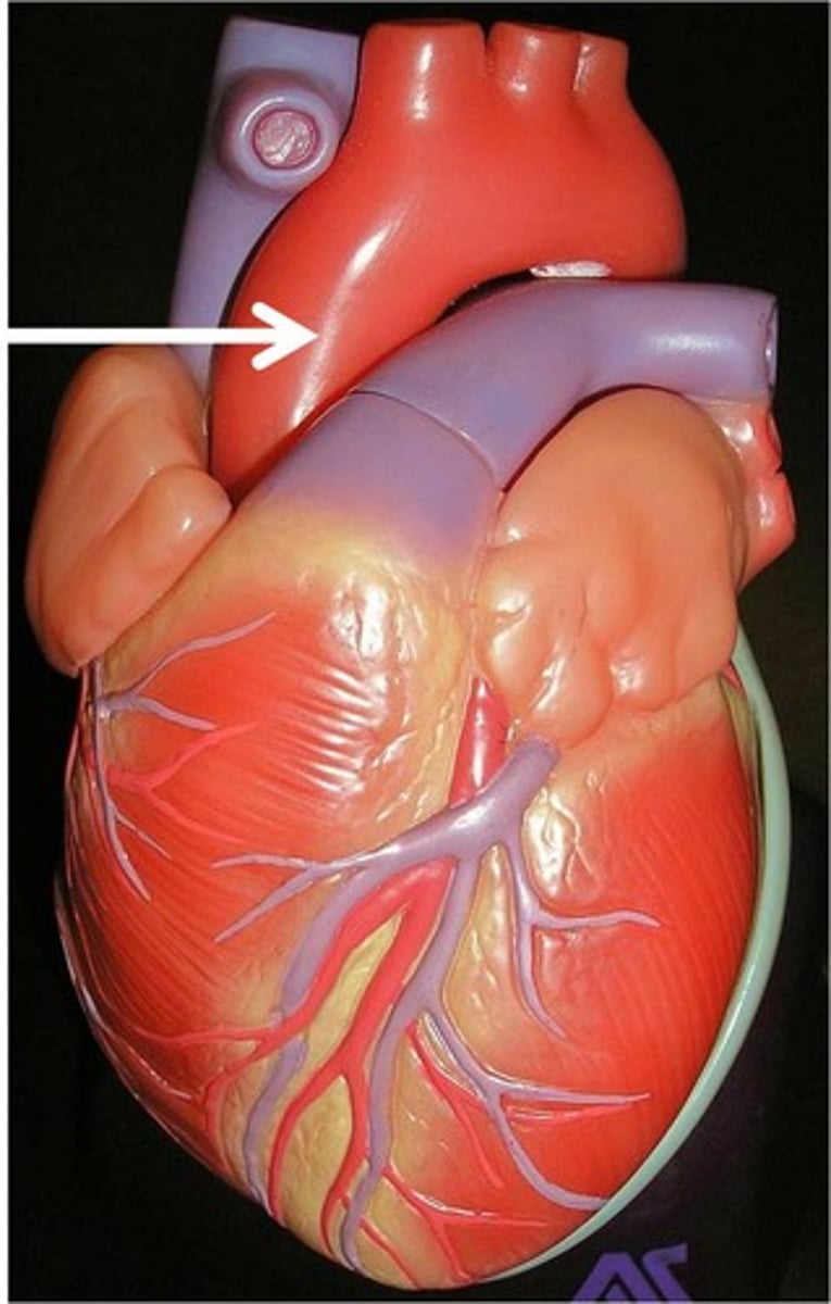
ligamentum arteriosum
remnant of ductus arteriosus
pulmonary trunk
carries blood from right ventricle to pulmonary arteries
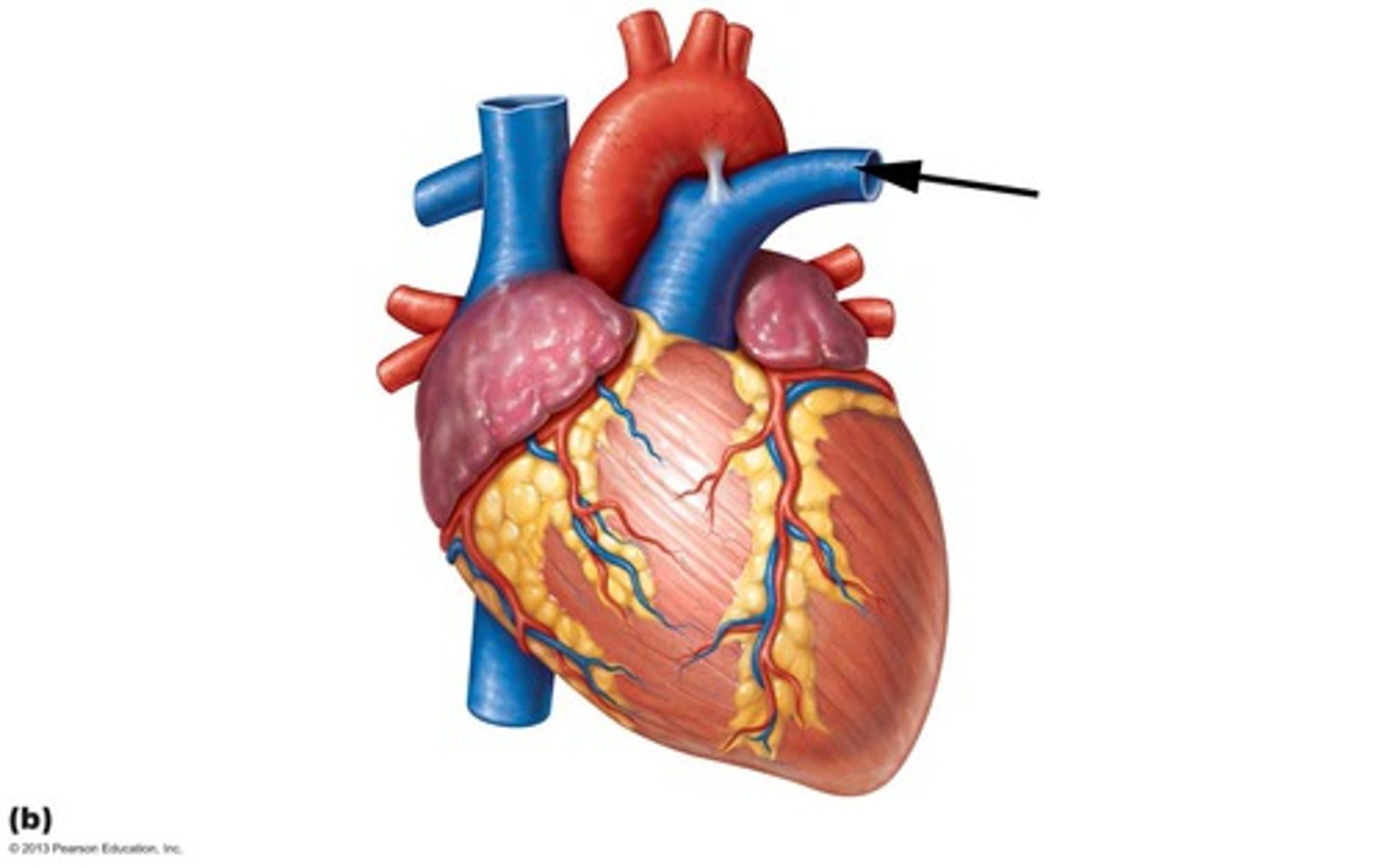
right pulmonary artery
takes blood from the right ventricle to the right lung
left pulmonary artery
carries poor oxygenated blood from the right ventricle to the left lung.
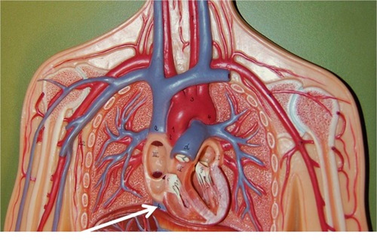
inferior vena cava
carries blood from lower regions of the body to right atrium
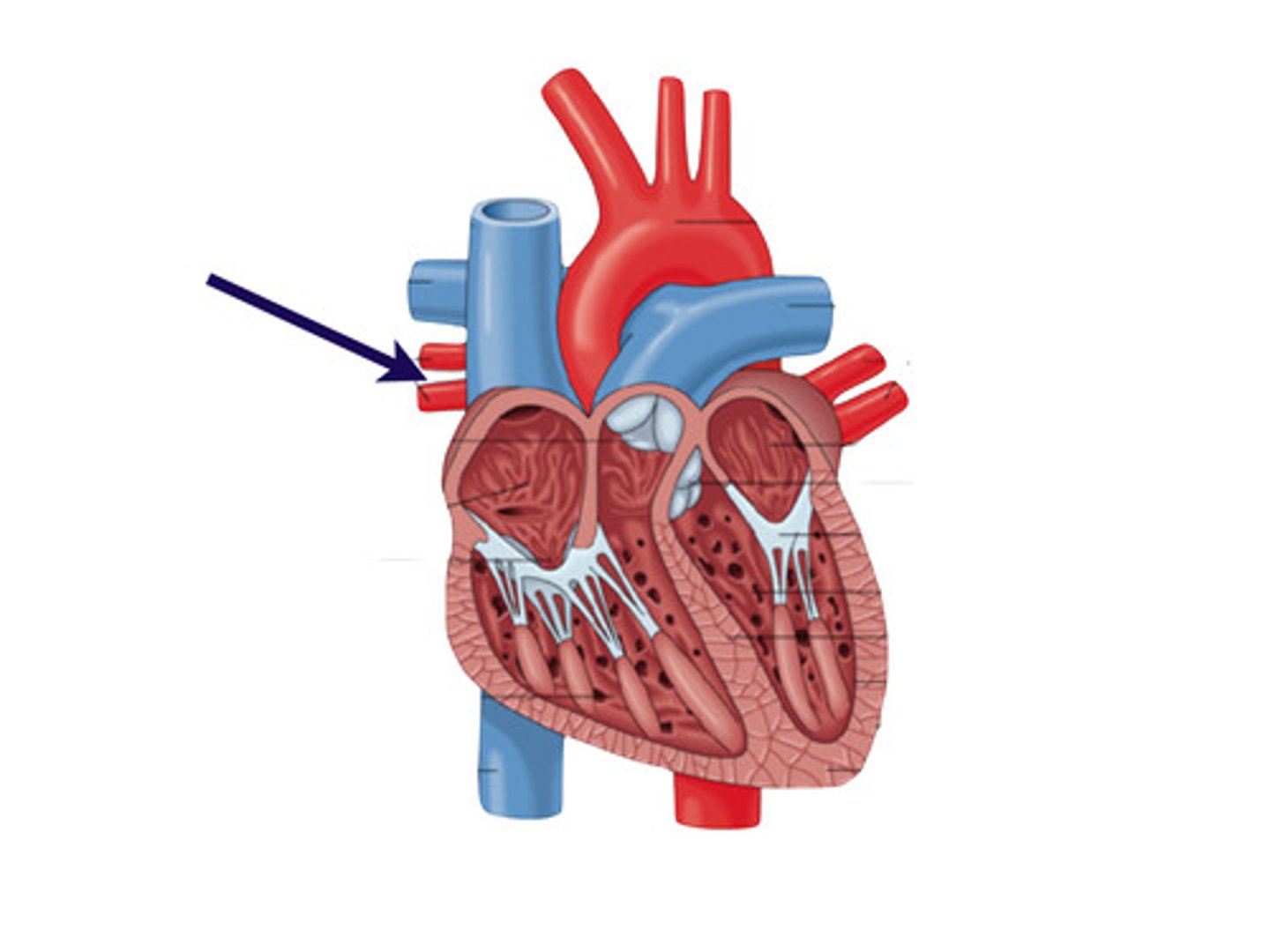
left pulmonary vein (superior and inferior)
transports oxygenated blood from the left lung to the left atrium
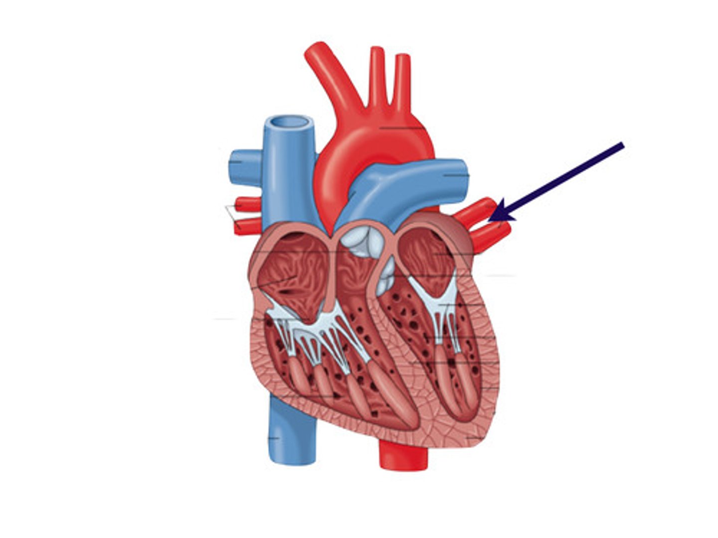
right pulmonary vein (superior and inferior)
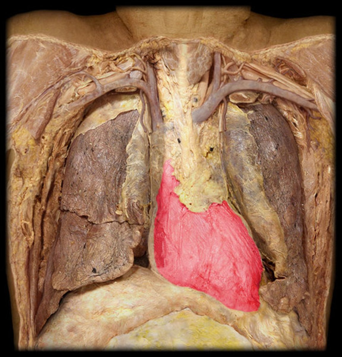
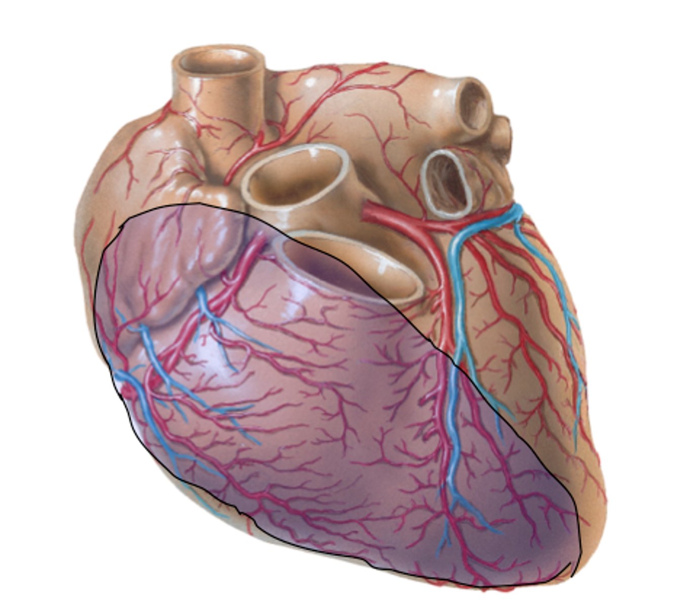
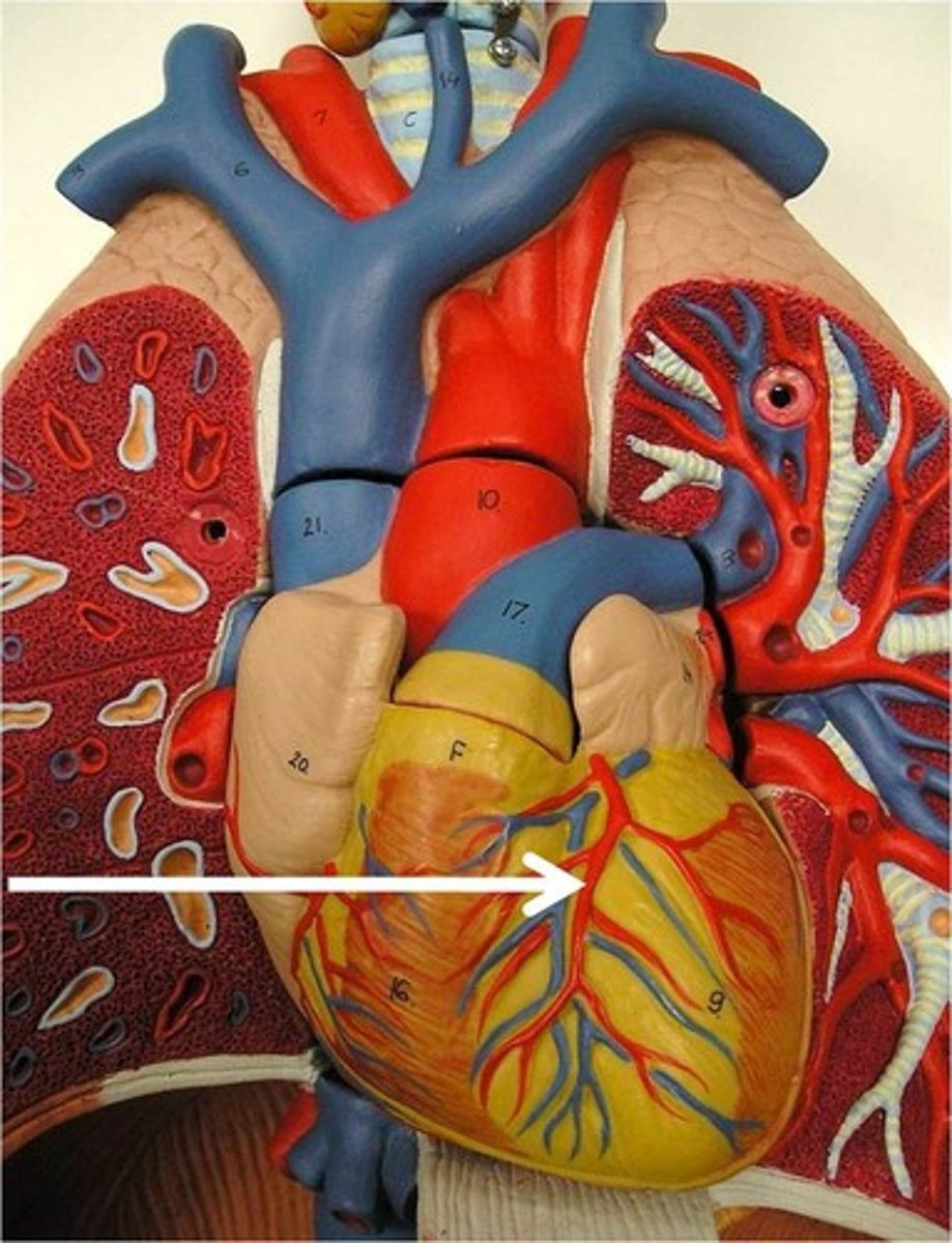
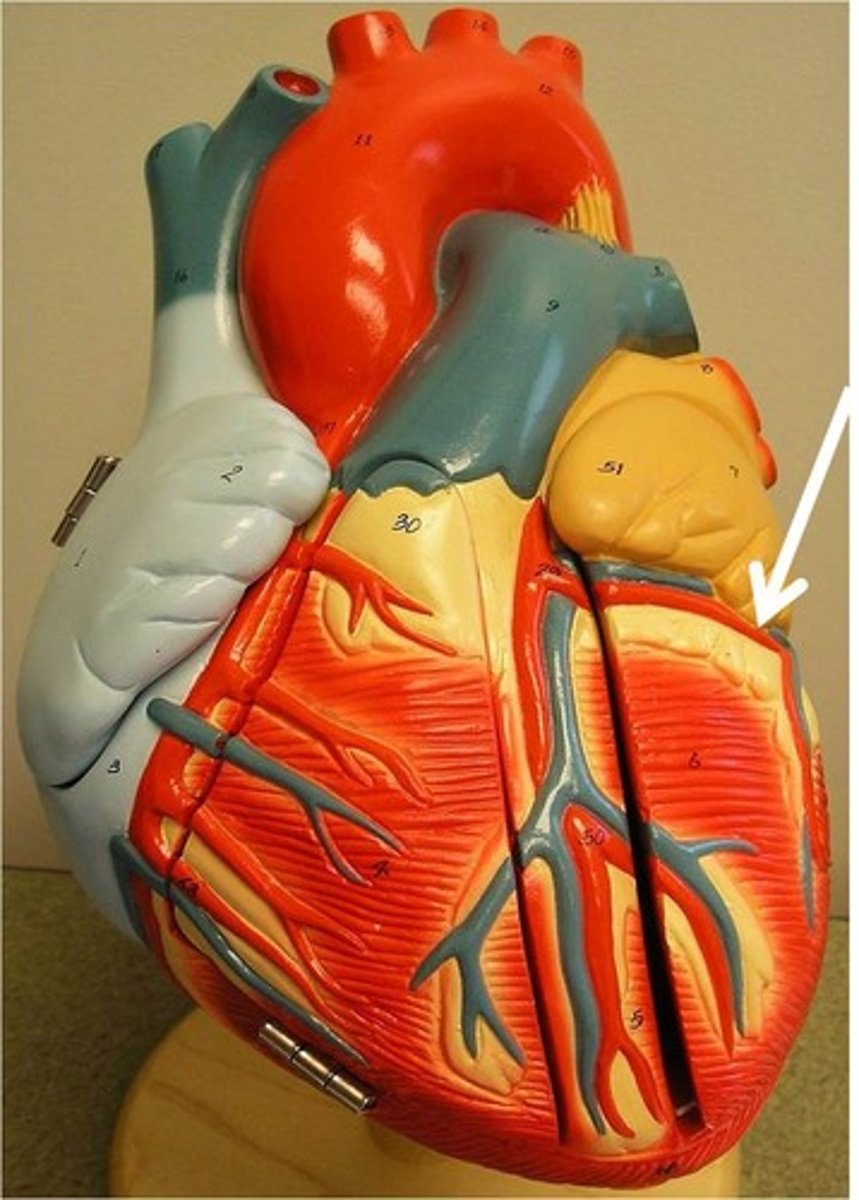
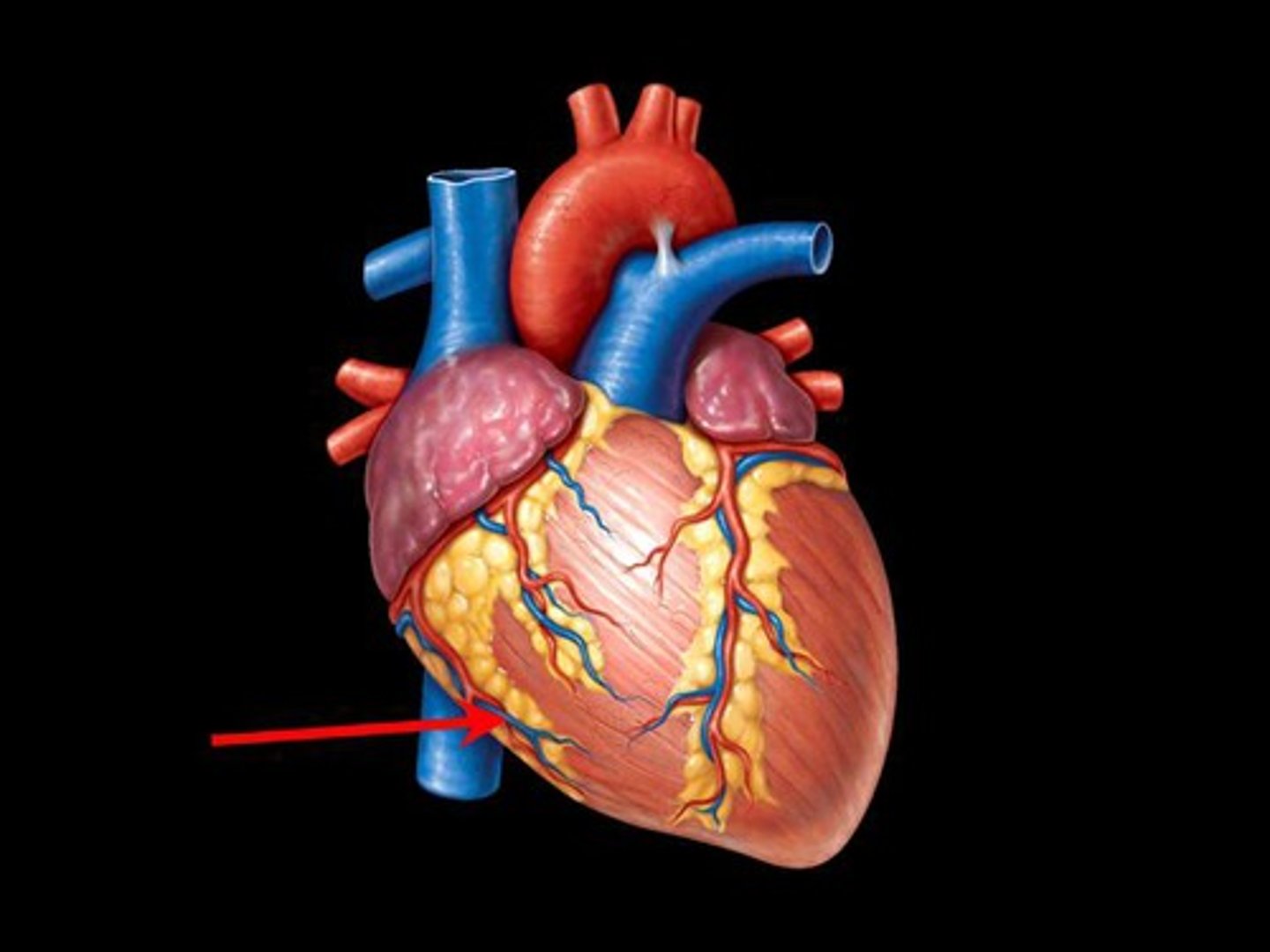
anterior surface of heart
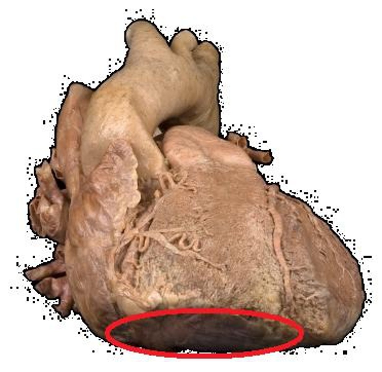
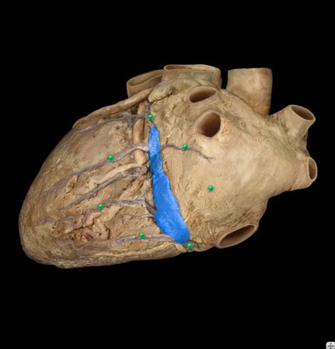
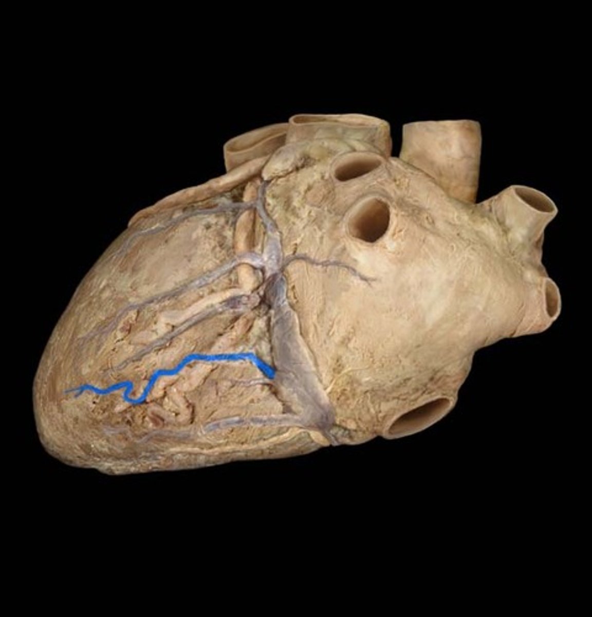
diaphragmatic surface of the heart
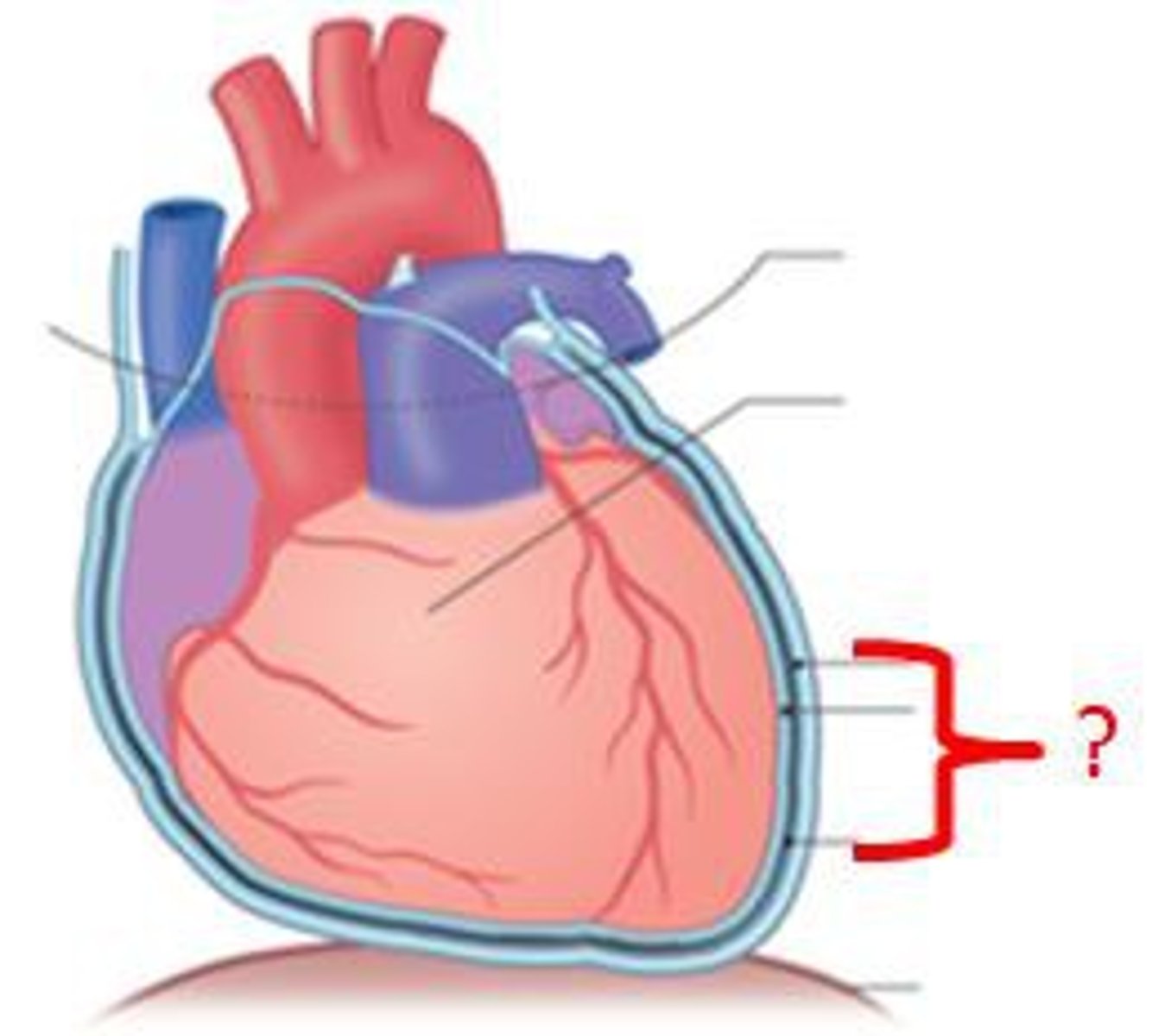
Coronary sulcus
Groove separating atria from ventricles.
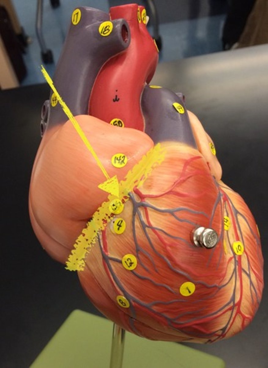
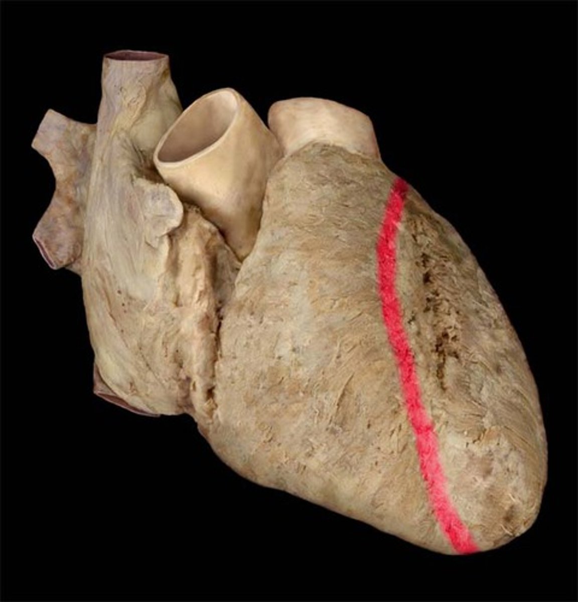
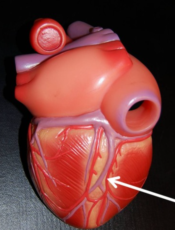
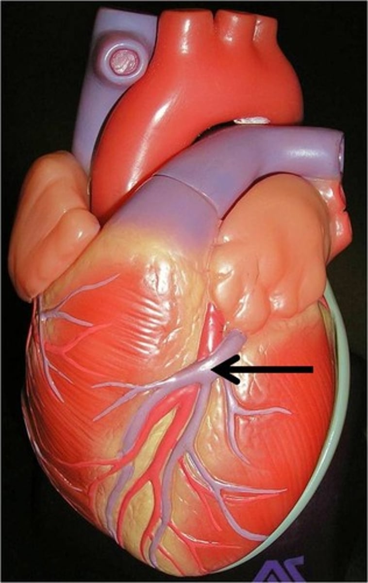
anterior interventricular sulcus
separates ventricles of the heart on the anterior side
posterior interventricular sulcus
separates ventricles of the heart on the posterior side
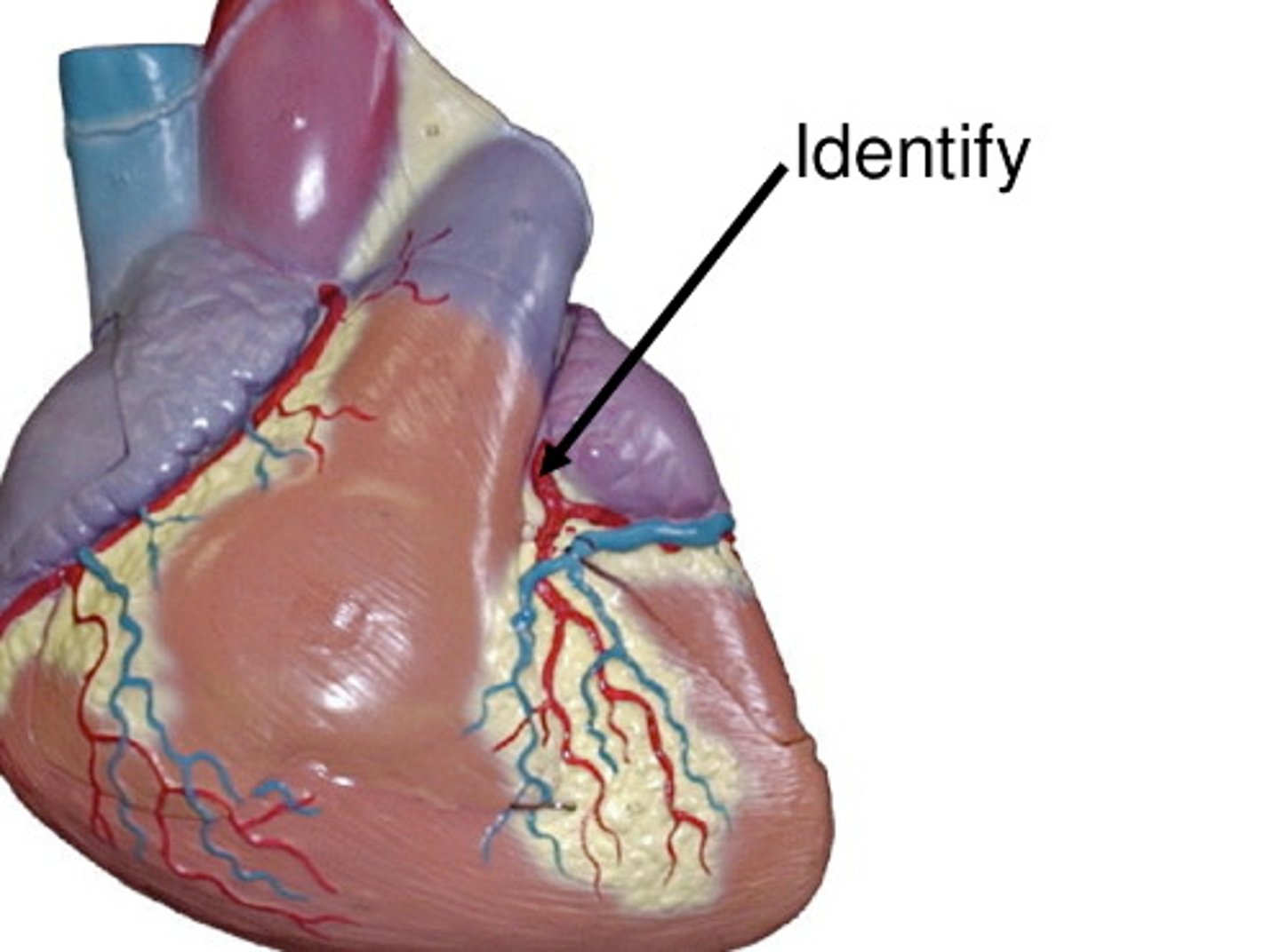
right coronary artery
artery vascularizing the right side of the heart
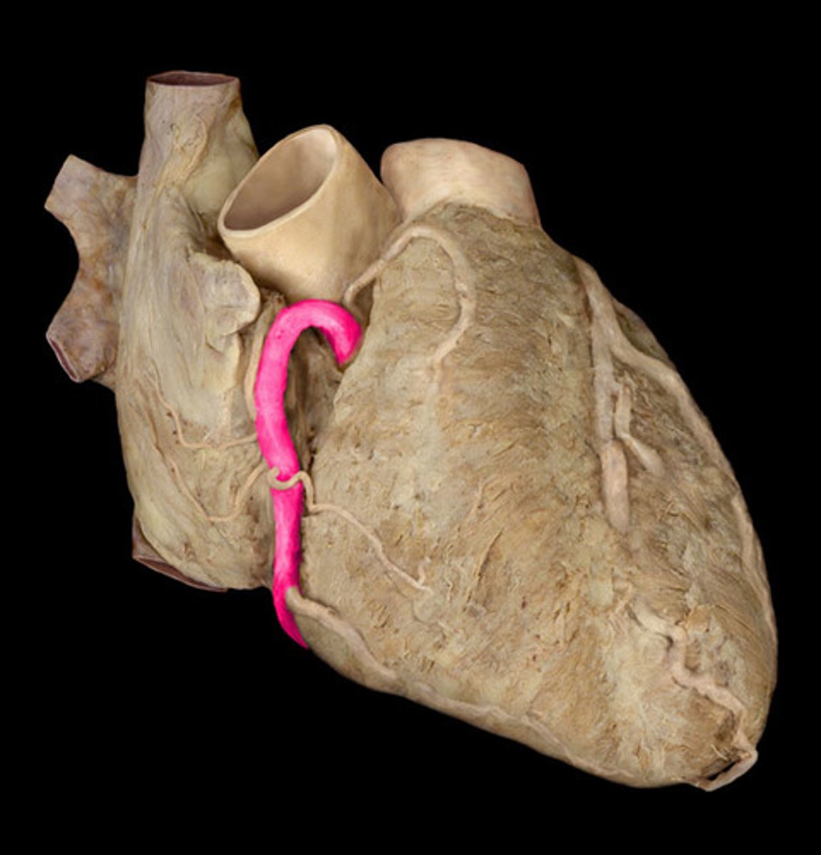
right marginal artery
supplies the right border of the heart
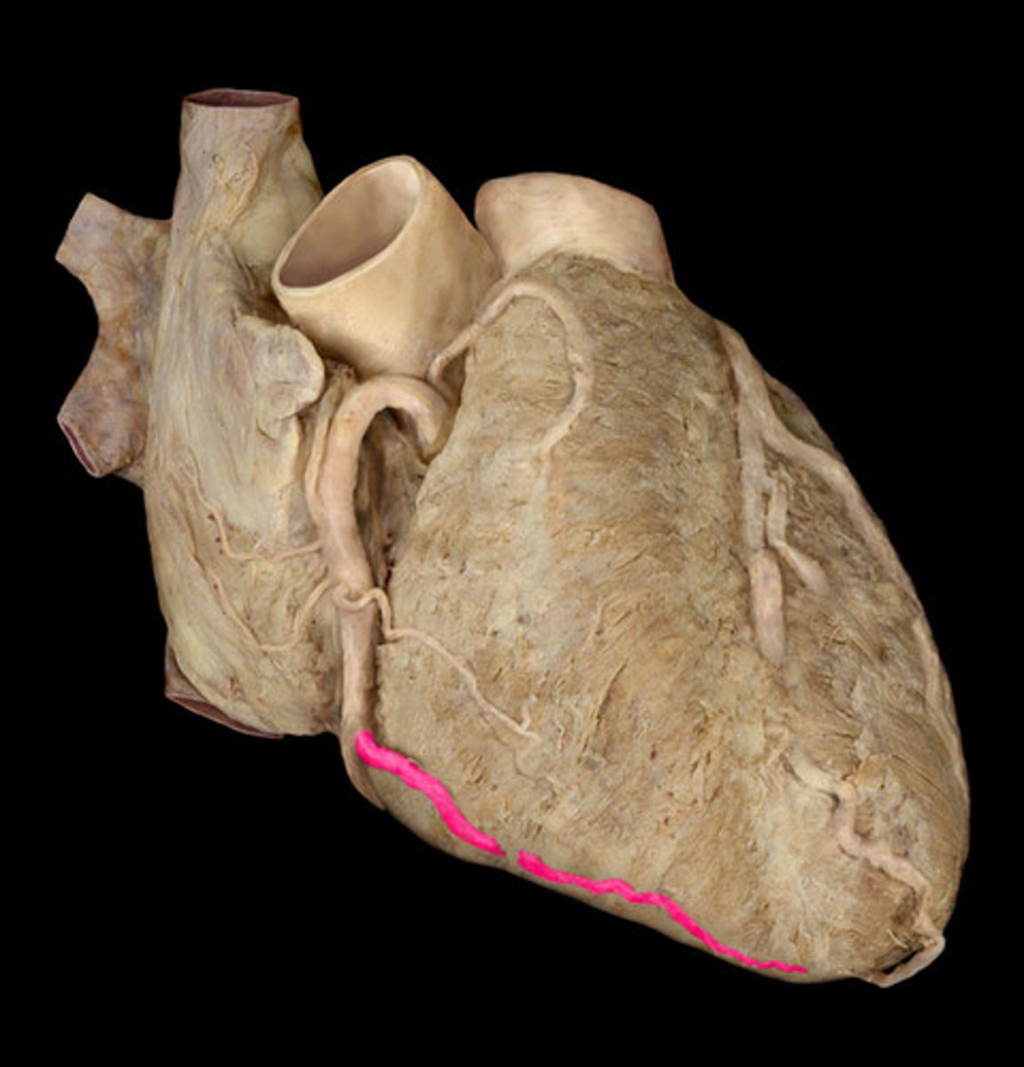
posterior descending artery (PDA)
Inferior wall. feeds back side of heart.
left coronary artery
supplies blood to the left ventricle, left atrium, and interventricular septum
left anterior descending artery
supplies blood to the front and bottom of the left ventricle and the front of the septum
circumflex artery
left anterior descending artery and circumflex artery
terminal branches of left coronary artery
coronary sinus
enlarged vessel on the posterior aspect of the heart that empties blood into the right atrium
great cardiac vein
runs alongside the anterior interventricular artery (LAD)
middle cardiac vein
runs alongside the posterior interventricular artery (PDA)
small cardiac vein
Vein that travels along side the right marginal artery.
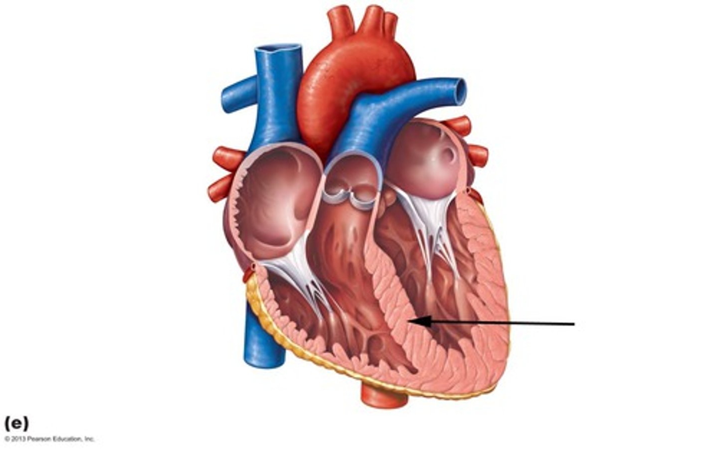
Interventricular septum
Muscular wall separating left and right ventricles.
interatrial septum
separates atria
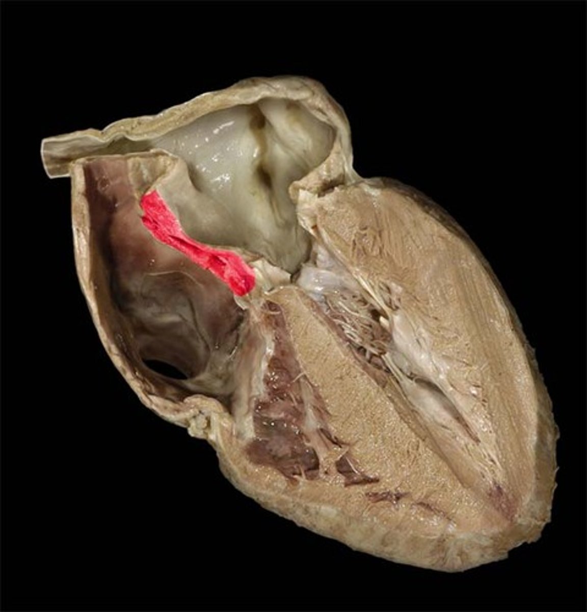
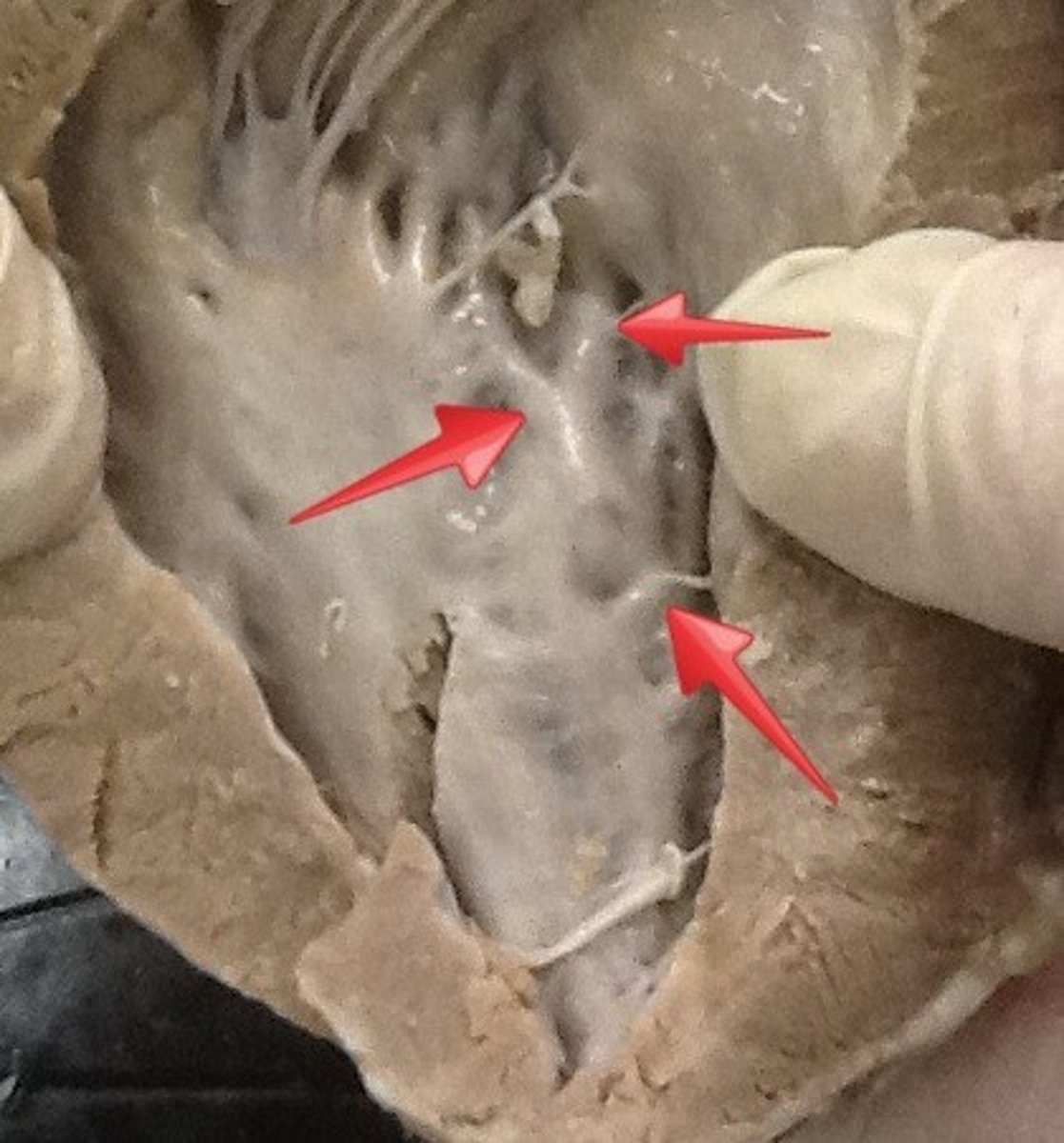
fossa ovalis
remnant of foramen ovale of fetal heart
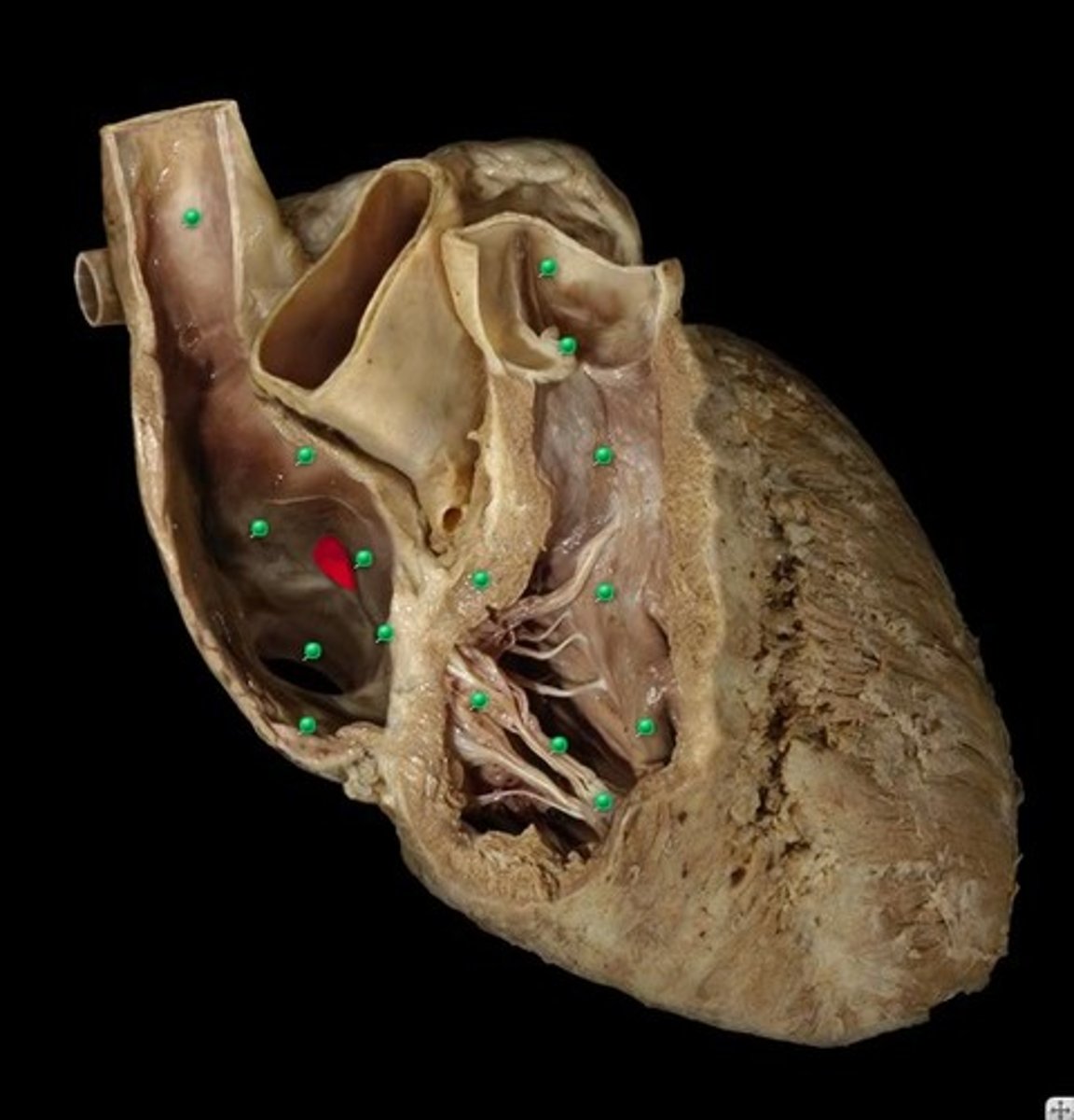
limbus fossae ovalis
border of oval fossa
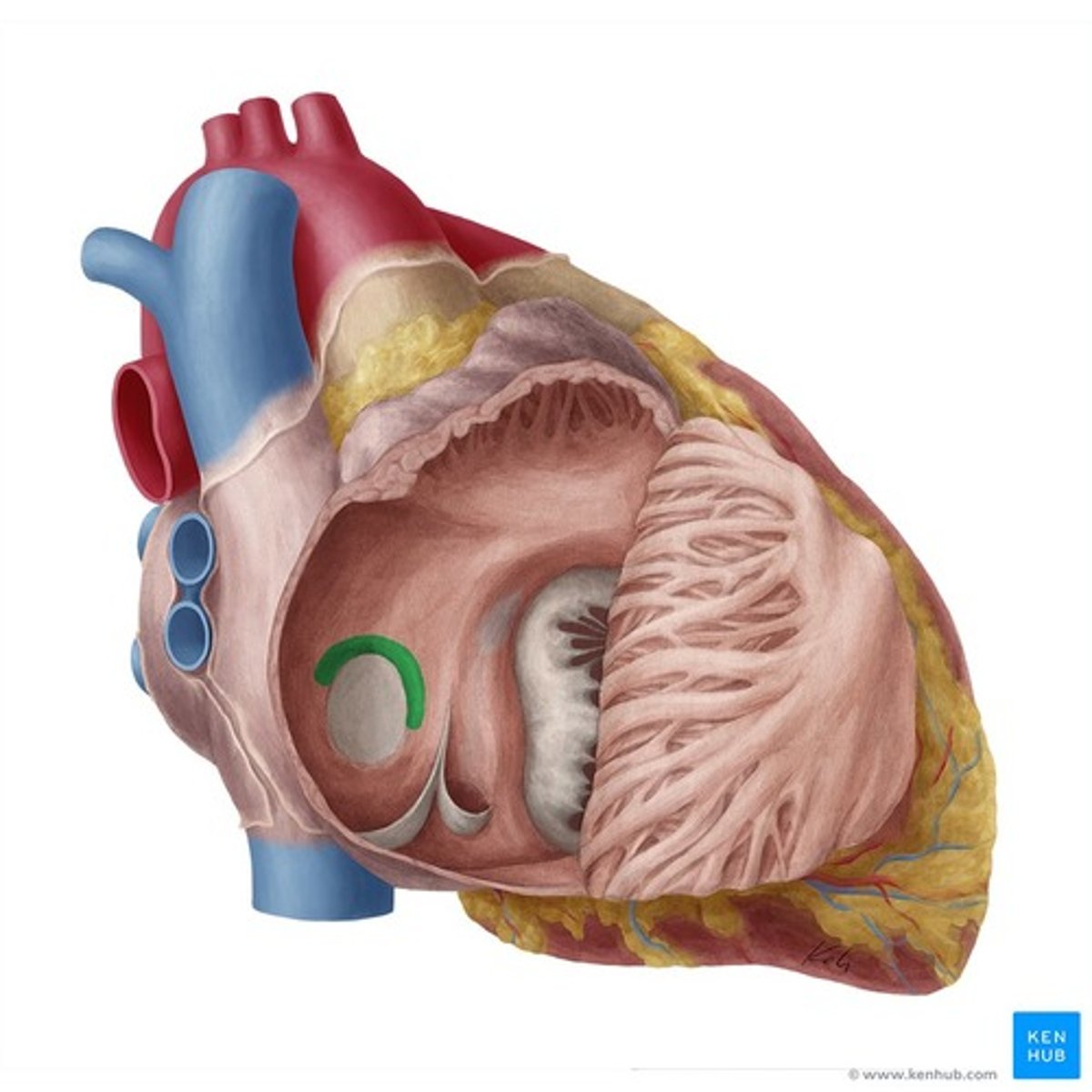
opening of coronary sinus
returns venous coronary circulation to right atrium
sinus venarum
Smooth part of right atrium
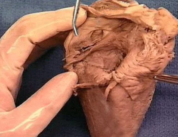
right auricle
Identify the flap.
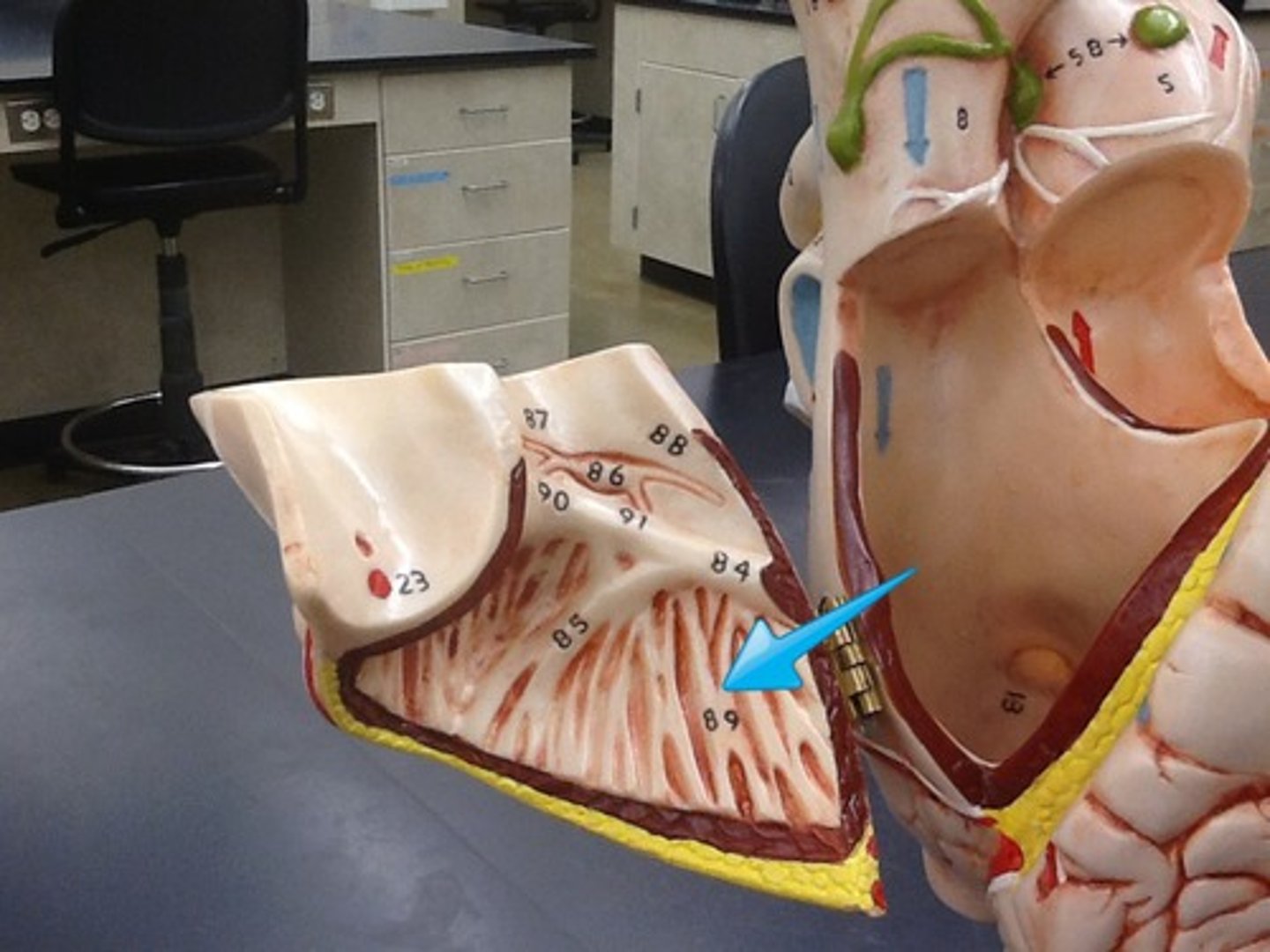
pectinate muscle
prominent ridges lining the surface of the atria
right atrioventricular/ tricuspid valve
Valve preventing backflow of blood into right atrium when the ventricles contract
right atrioventricular orifice
opening from right atrium to right ventricle
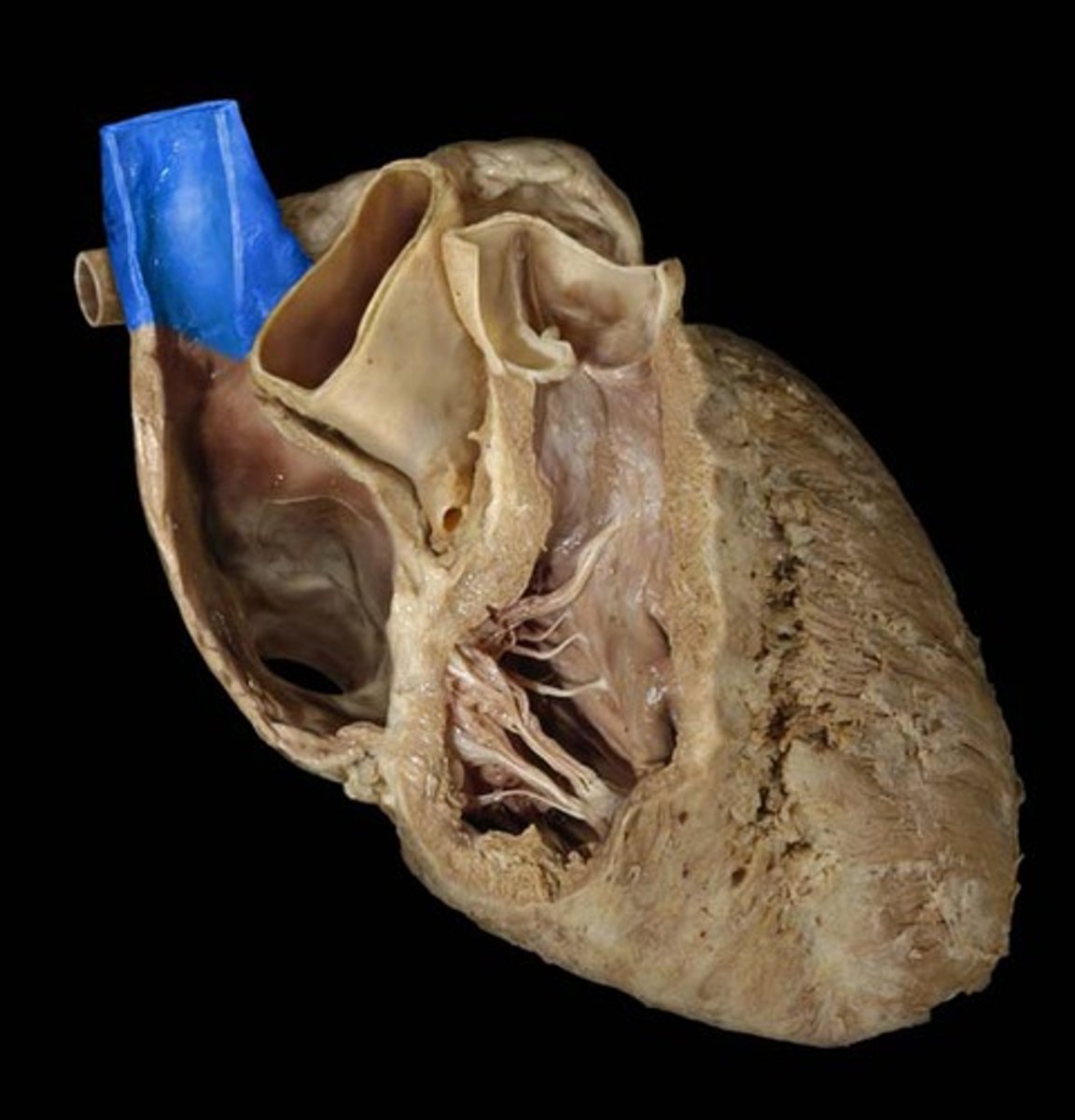
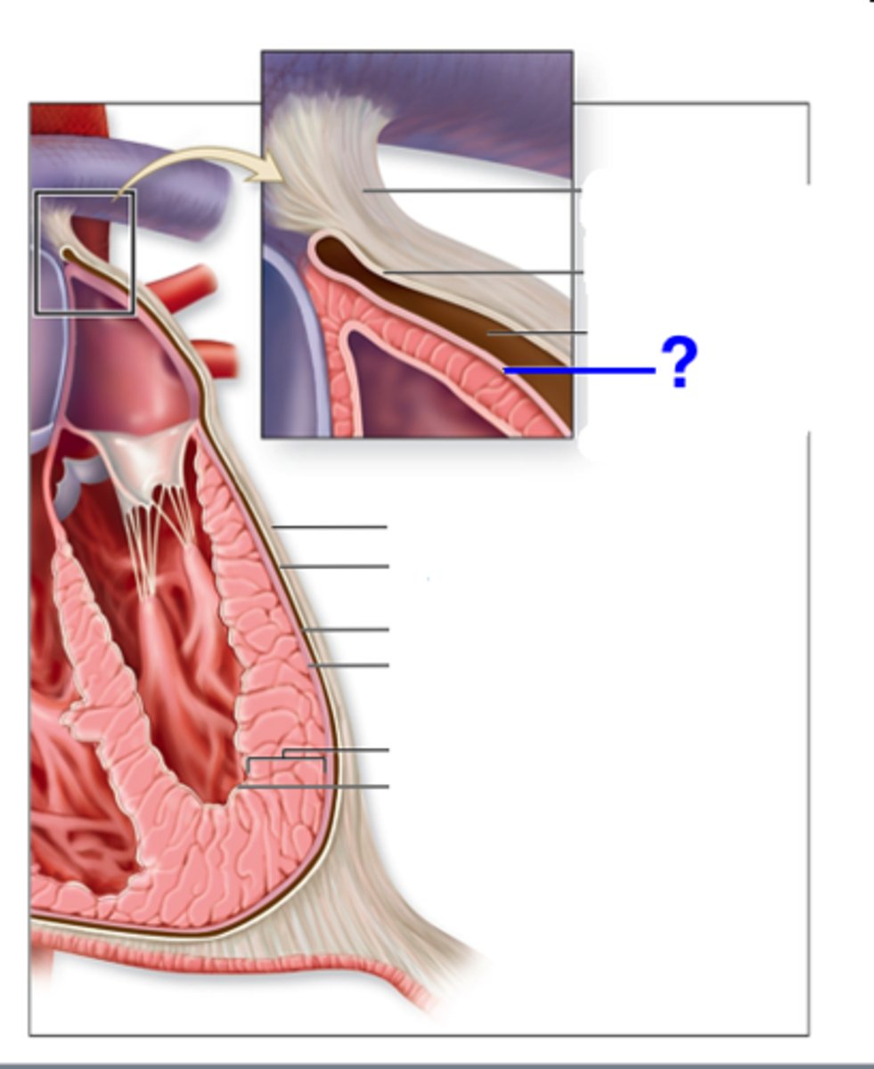
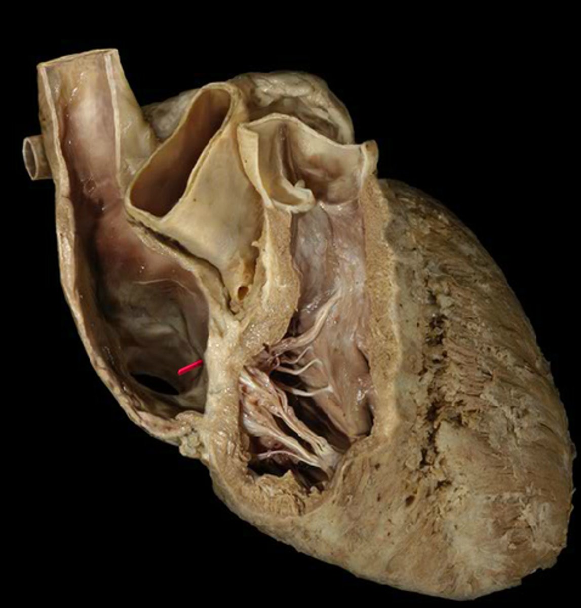
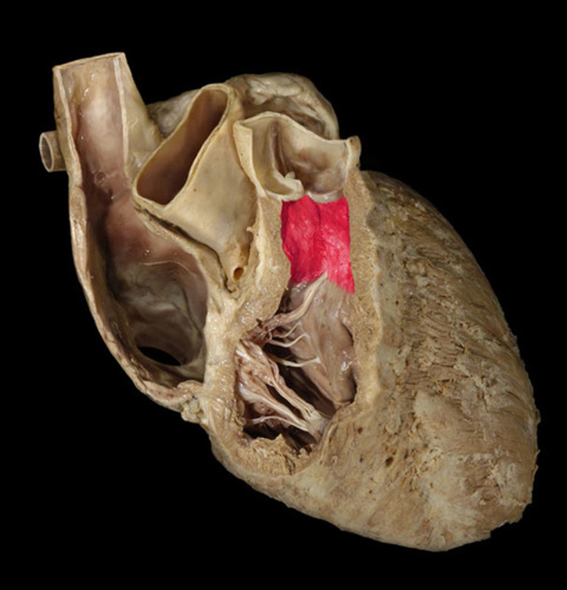
trabeculae carneae
muscular ridges on the internal surface of the ventricles
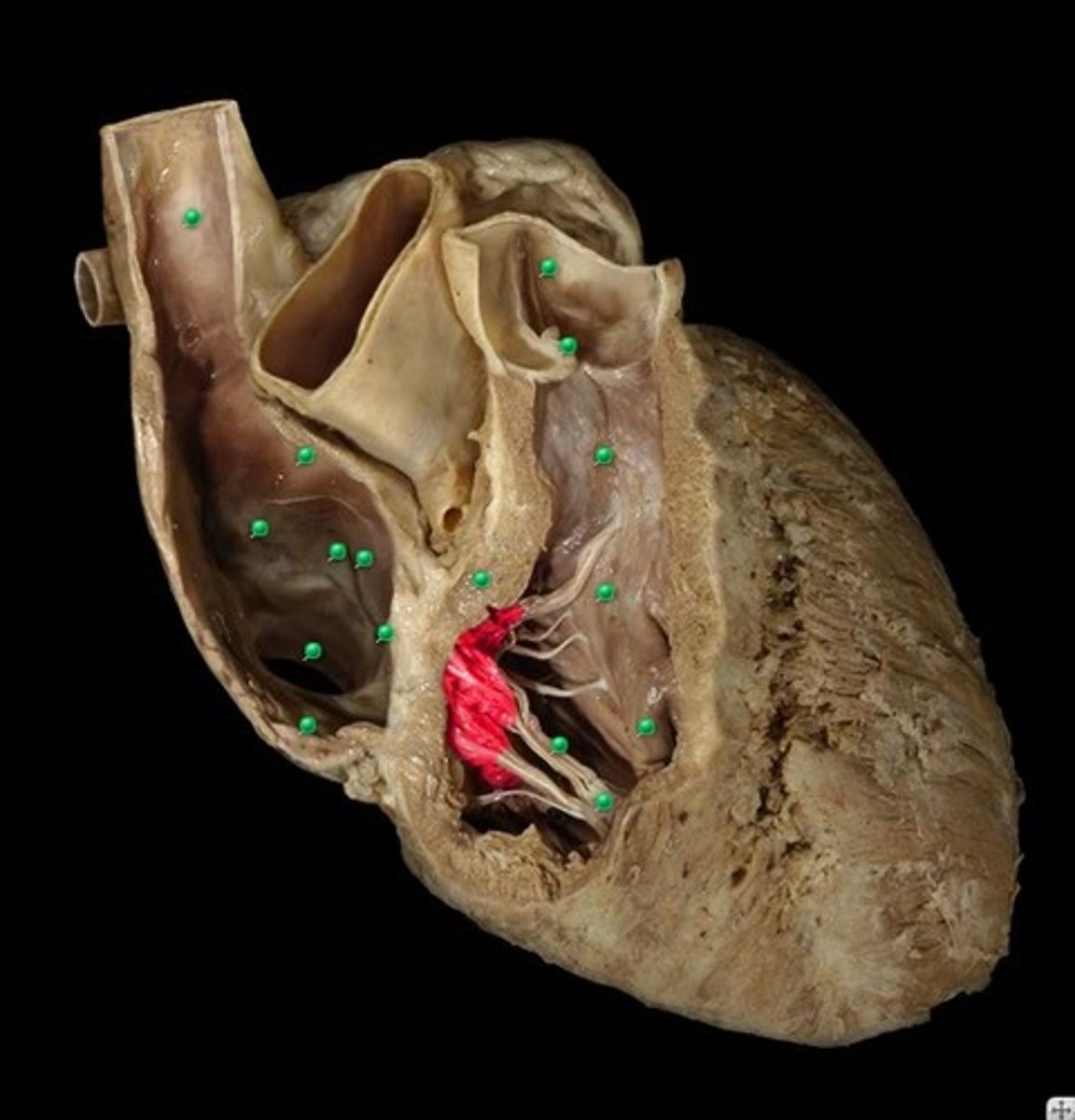
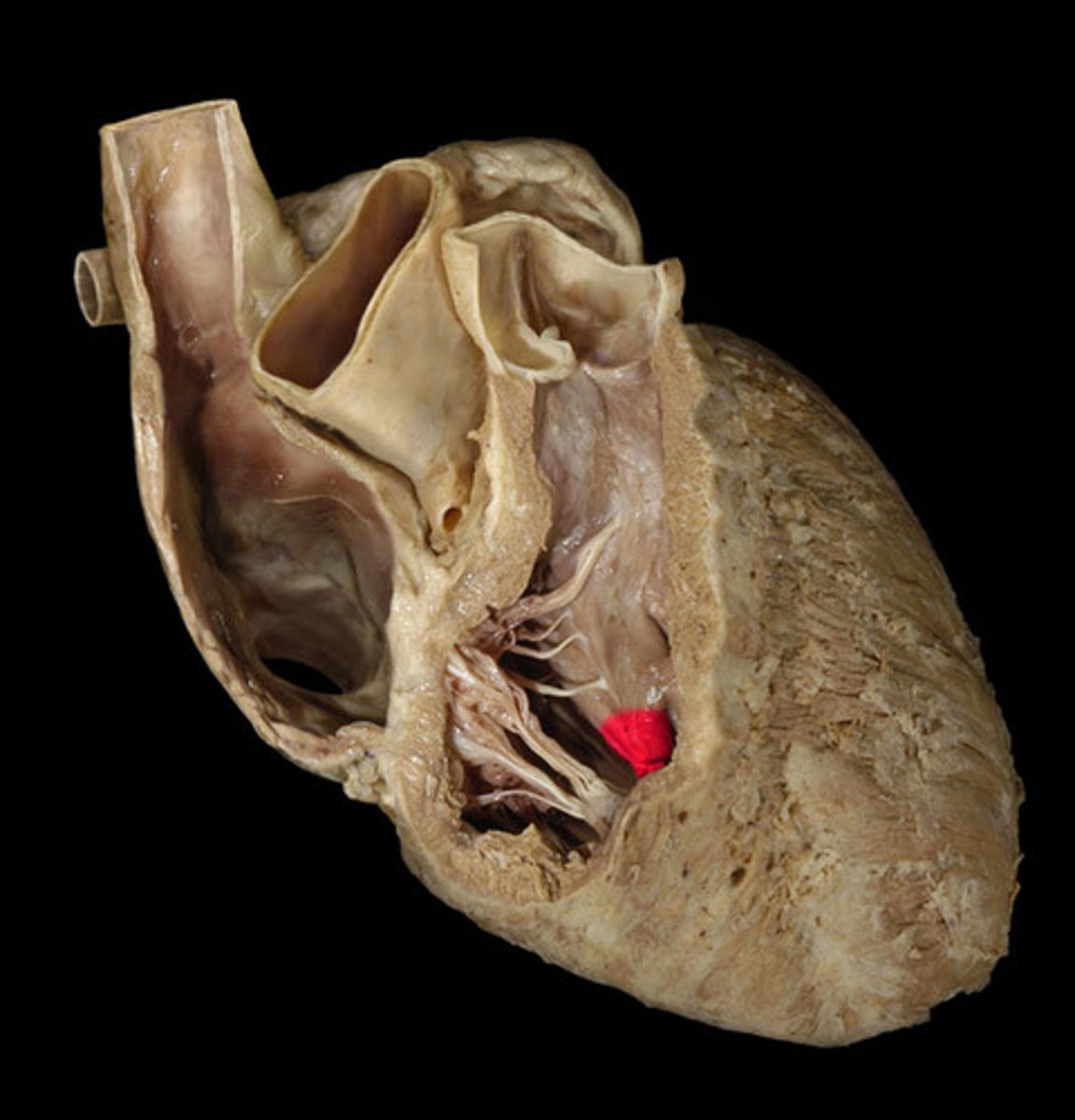
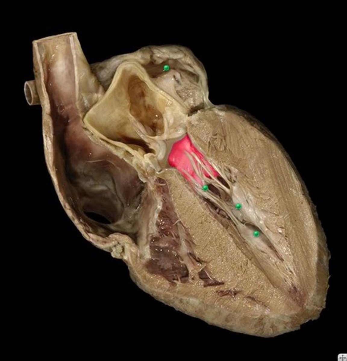
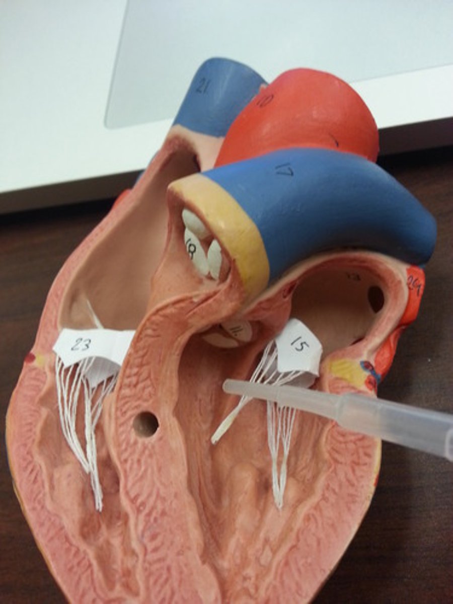
papillary muscles
prevent prolapse of AV valves
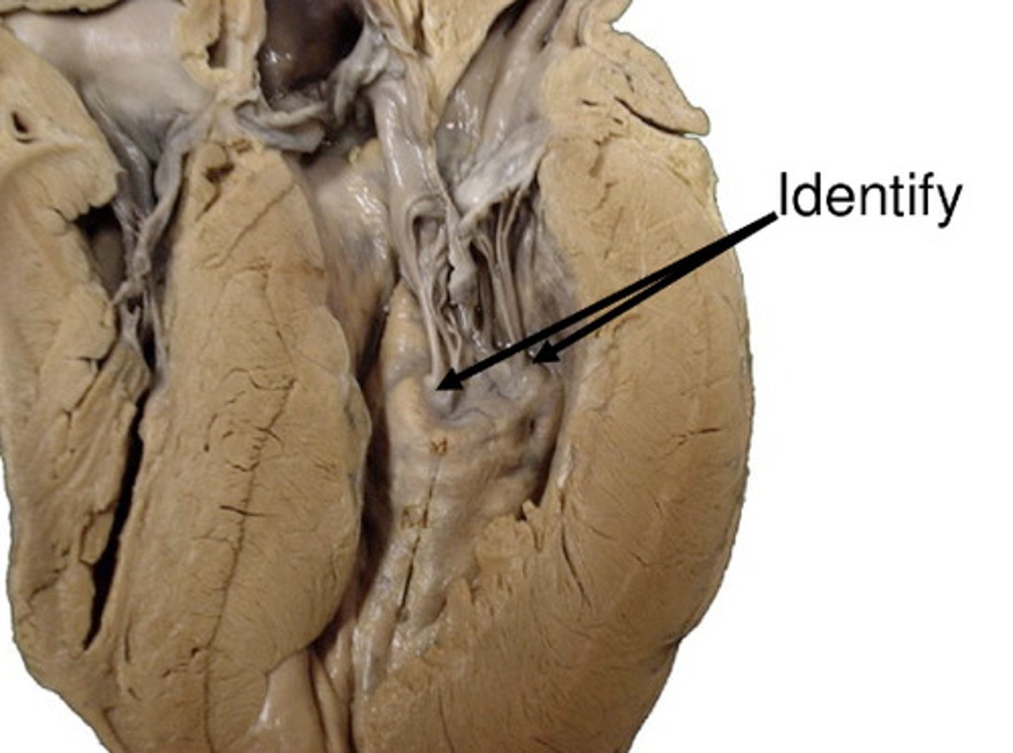
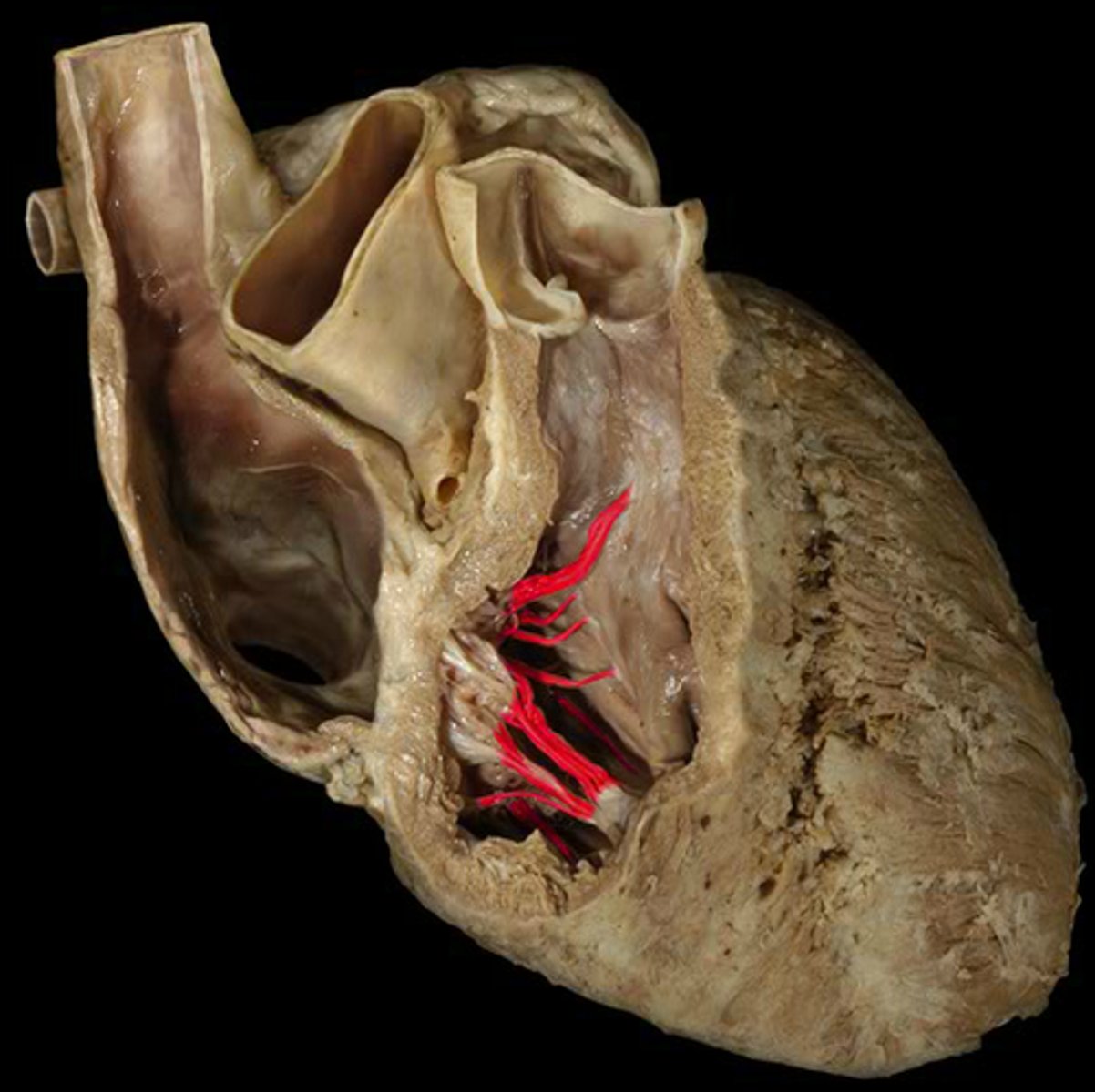
tendinous cords
String-like tendons used to attach the atrioventricular valves of the heart to the sides of the ventricle wall. Sometimes called heart strings.
septomarginal trabecula/moderator band
Carries part of the right branch of the AV bundle and prevents overdistension
conus arteriosus/infundibulum
the upper smooth-walled portion of the right ventricle, which leads to the pulmonary trunk
left auricle
Identify the structure (be specific).
left atrioventricular orifice
opening between left atrium and left ventricle
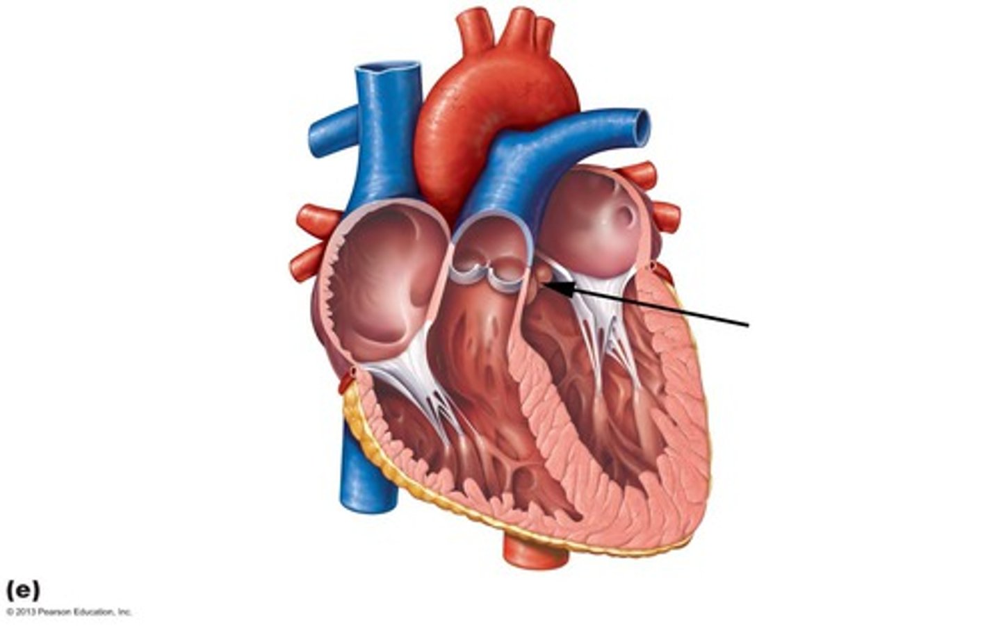
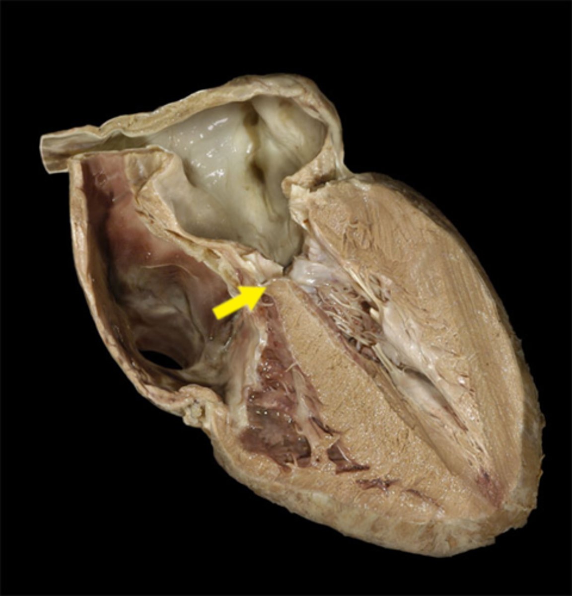
left atrioventricular valve/ bicuspid/ mitral valve
valve between the left atrium and the left ventricle.
aortic vestibule
smooth area of the Left Ventricle that leads into the aortic semilunar valve and the ascending aorta
aortic valve (left, right, and posterior cusps)
The semilunar valve separating the aorta from the left ventricle
membranous portion of interventricular septum
Pulmonary valve
Valve controlling blood flow from right ventricle to pulmonary trunk.
