Anatomy
 Call Kai
 Learn
Learn Practice Test
 Spaced Repetition
 Match
Match Flashcards
 Knowt Play
1/162

There's no tags or description
Looks like no tags are added yet.
Name | Mastery | Learn | Test | Matching | Spaced | Call with Kai | Chat |
|---|
No analytics yet
Send a link to your students to track their progress
163 Terms
superior
towards the head
inferior
towards the feet
medial
towards the midline
lateral
away from the midline
proximal
close to another structure
distal
away from another structure
transverse plane
horizontal
frontal plane
vertical and splits the front and back
sagittal plane
vertical and splits left and right
oblique plane
any angled plane
anterior
also ventral
towards the belly
posterior
also dorsal
towards the back
bone functions
support - framework for the attachment of soft tissue; muscles - movement
protection of the soft tissue: cranium, vertebral column, rib cage, pelvis
haematopoisesis
storage - fat (medullary cavity, yellow bone marrow) and mineral (calcium and phosphorus)
bone classifications
long
short
flat
irregular
sesamoid
long bones
longer than wide
main components of limbs
sustain weight
provide levers for muscles, often curved
humerus, radius, ulna, metacarpals, femur, tibia, fibula, metatarsals, phalanges

diaphysis
shaft of the long bone
cylinder of compact bond around the inner medullary cavity, outer surface surrounded by a layer connective tissue - periosteum
nutrient foramen
medullary cavity
contains yellow bone marrow
lined with thin layer of connective tissue - endosteum
epiphysis
end of the bond, spongy tissue, red bone marrow; articular cartilage
metaphysis
between the diaphysis and epiphysis, in growing bone contains epiphyseal (growth) plate of hyaline cartilage (longitudinal growth), becomes epiphyseal line
short bones
cube-shaped spongy bone covered by a thin layer of compact bone
transfer forces of movement
carpal (wrist) and tarsal (ankle) bones
flat bones
broad surfaces
offer protection and provide wide areas for muscle attachment
irregularly placed articular surfaces
cranial bones
ribs
scapulae
between two surface layers of compact bone and spongy bone (dipploe)
irregular bones
varied shape and often very complex and with peculiar forms for the functions they perform
many surface features for muscle attachment or articulation
variable amount of spongy and compact bone
vertebrae, calcaneus and some skull bones
sesamoid bones
develop in tendons as a response to physical stress
no always completely ossified
patella, pisiform
axial
consists of the bones that provide the axis of the body and support and protect the organs of the head, neck and trunk
skull, auditory ossicles, hyoid bone, vertebral column, rib cage
appendicular
consists of the bonds of the free upper and lower limbs and the girdles that connect them to the axial skeleton
pectoral girdle, free upper limb, pelvic girdle, free lower limb

1
frontal bone
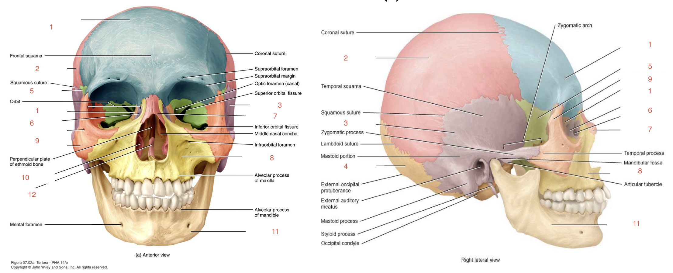
2
parietal bone

3
temporal bone
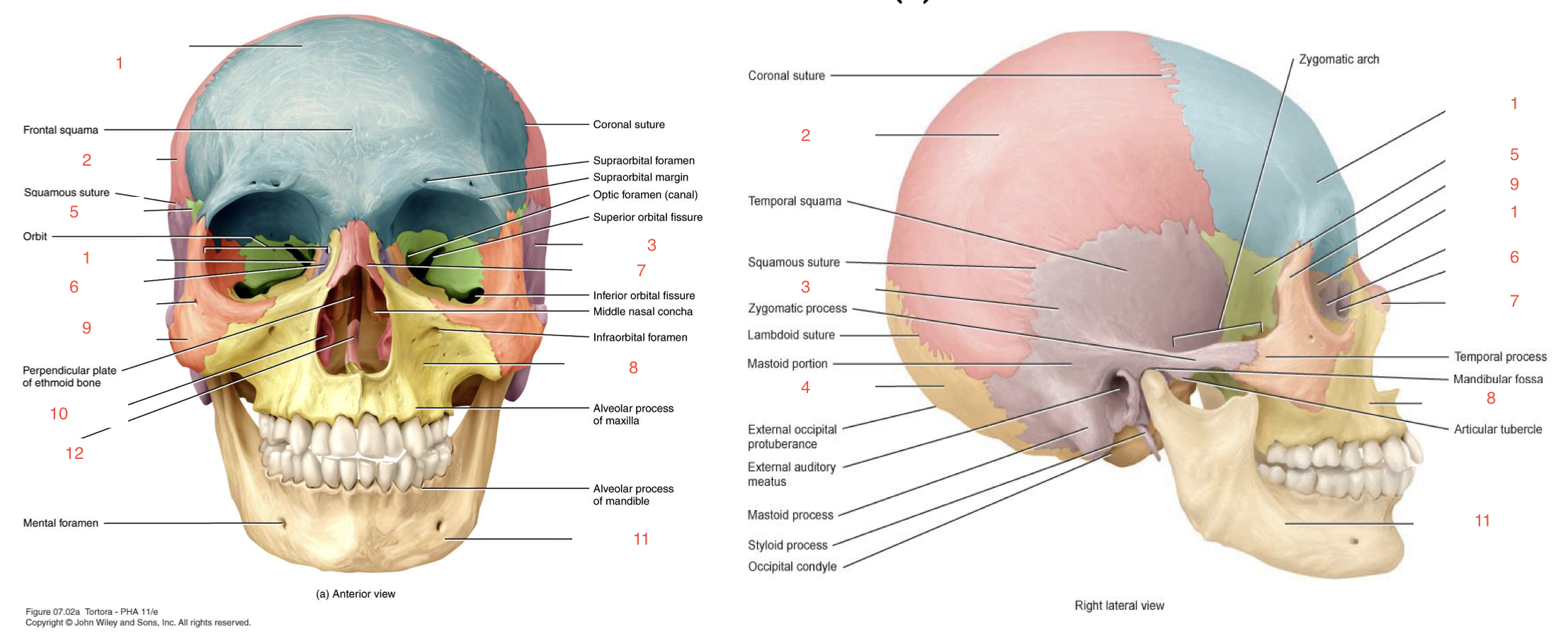
4
occipital bone
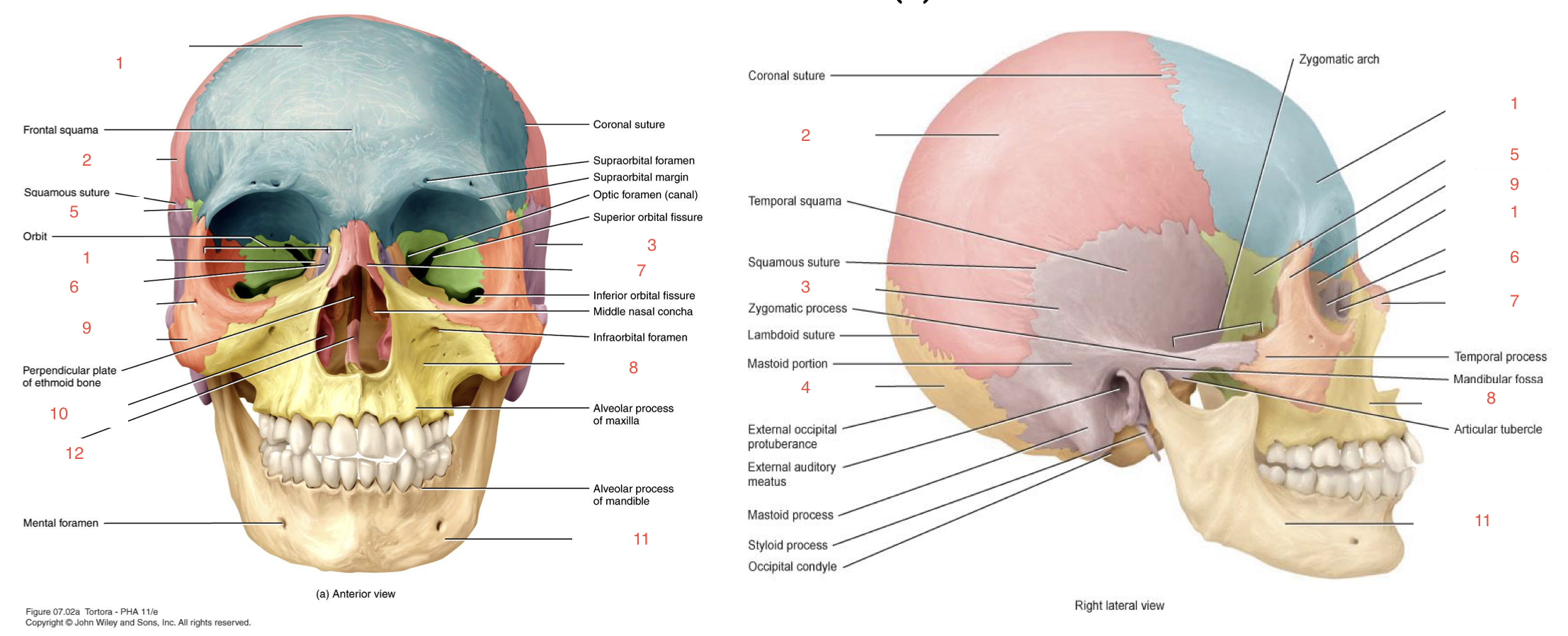
5
sphenoid bone

6
lacrimal bone

1
ethmoid bone

7
nasal bone
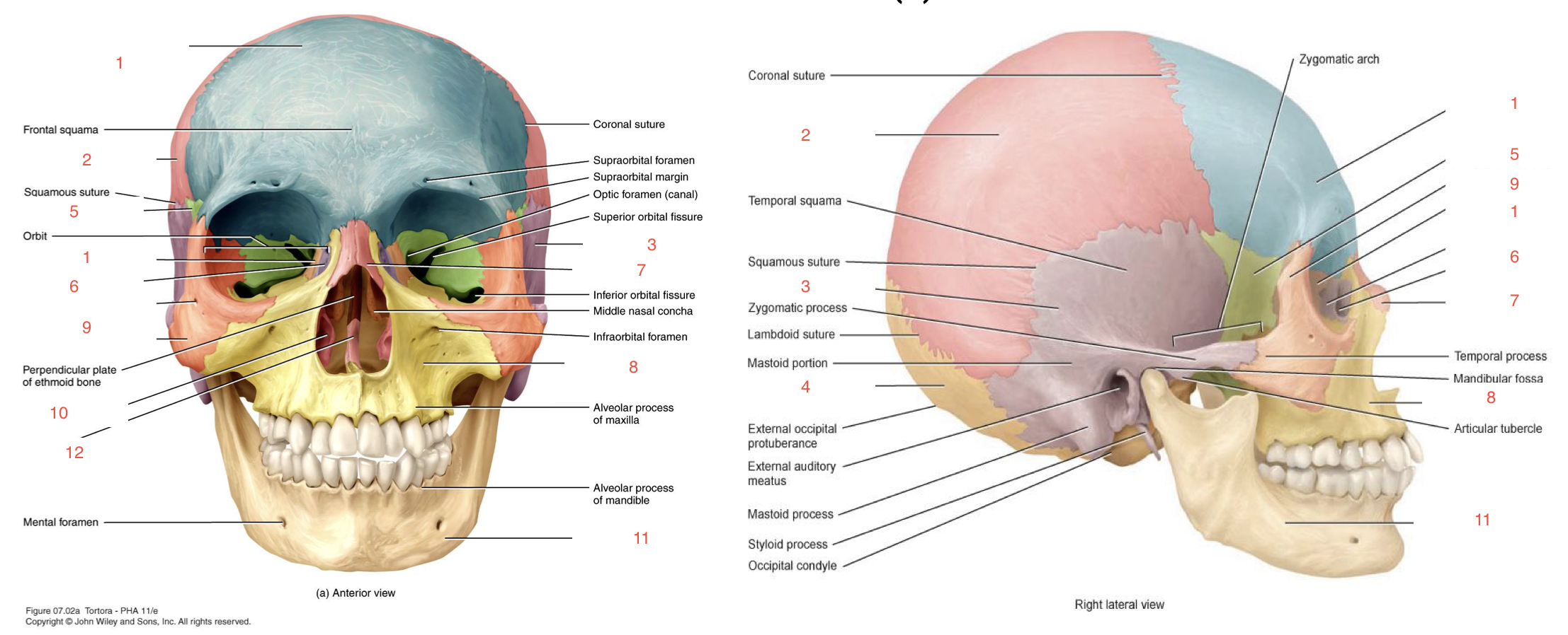
8
maxilla

9
zygomatic bone

10
inferior nasal concha

11
mandible

12
vomer
auditory ossicles
within the middle ear (petrous temporal)
transmit sound impulses
malleus
hammer
bone in the middle ear

incus
anvil
stapes
stirrup
hyoid bone
not attached directly to any other bone
in the neck inferior to mandible, suspended from the temporal bone by the muscles and ligaments
supports the tongue and larynx
vertebral column order and number
cervical 7
thoracic 12
lumbar 5
sacrum 5 merged to 1
coccyx 4 merged to 1
vertebral column primary curve
concave
thoracic and sacral
vertebral column secondary curve
convex
cervical and lumbar
thoracic cage
bony and cartilaginous structures
12 pairs of ribs, costal cartilage, sternum, 12 thoracic vertevrae
pectoral girdle
clavicle
scapula
lateral forearm bone
radius
medial forearm bone
ulna
hand bones
8 carpal bones
5 metacarpals
14 phalanges
hip bone parts
illium
ischium
pubis

pelvis
hip bone (os coxae)
sacrum and coccyx
lateral leg bone
fibula
medial leg bone
tibia
foot bones
7 tarsals
5 metatarsals
14 phalanges
joint
point of contact between two bones, bone and cartilage, or bone and tooth
hold the bones together allowing/restricting movement and flexibility
more mobility means less stability and vice versa
joint function classifications
no movement (synarthrosis)
slight movement (amphiarthrosis)
free movement (diarthrosis)
flexion
decrease angle of the joint
extension
increase angle of the joint
abduction
move away from the midline
“abduct”
adduction
move towards the midline
“add”
neck joint
atlanto-axial joint does rotation
shoulder joint rotation
circumduction
also in the hip
temporomandiubular joint movements
open is depression
close is elevation
anterior is protraction
posterior is retraction
foot movements
lift up ball is inversion
lift up outside is eversion
point toes is plantar flexion
bring up is dorsiflexion
radioulnar joint movement
palm posterior is pronation
palm anterior is supination
joint structural classifications
fibrous
cartilaginous
synovial
fibrous joints
no synovial cavity
bones held together by dense irregular connective tissue
sutures
syndesmoses
cartilaginous joints
no synovial cavity
bones held together by cartilage
synchondroses
symphyses
synovial joint examples
synovial cavity present
bones connected by dense irregular connective tissue (articular capsule), accessory ligaments
planar
hinge
pivot
condyloid
saddle
ball and socket
suture joints
fibrous
made of thin layer of dense irregular tissue
between bones of the skull
bones have irregular interlocking edges
synostosis: bony joint (fusion of two bones)
syndesmoses
fibrous
greater distance between the articulating bones more dense irregular connective tissue compared to sutures (ligaments)
interosseous membranes (radius/ulna, tibia/fibula) and distal tibiofibular joint
gomphosis (dentoalveolar joint) - periodontal ligament
synchondroses
cartilaginous (primary)
bones connected by hyaline cartilage
epiphyseal plates
1st rib and sternum
symphyses
secondary cartilaginous
articular surfaces covered with hyaline cartilage and united by fibrocartilage
pubic symphysis, between the manubrium and body of the sternum, intervertebral joints between the vertebral bodies
synovial joints
contain space between articular surfaces (covered by hyaline cartilage)
contains synovial fluid
articular (joint) capsule:
fibrous membrane (outer) - dense irregular connective tissue attached to the periosteum
synovial membrane (inner) - areolar connective tissue with elastic fibers, secretes synovial fluid
synovial joint parts
ligaments: stabilize the joints, intrinsic and extrinsic
articular discs and menisci of fibrocartilage between articular surfaces present in some joints
labrum: ring of fibrocartilage which depends the socket
bursa, tendon sheath: friction reduction
planar joints
articular surfaces: flat or slightly curved
movement: gliding or sliding in the plane of articular surfaces
intertarsal, interjarpal, acromioclavicular
hinge joines
articular surfaces: one convex the other concave
movement: flexion and extension (uniaxial)
knee (modified), elbow, interphalangeal, ankle
condyloid (ellipsoidal) joints
articular surfaces: one convex, oval shaped the other concave oval shaped
movement: flexion/extension, abduction/adduction, circumdaction (biaxial)
carpal and metacarpophalangeal joints
saddle joints
articular surfaces: saddle (modified condyloid)
movements: flexion/extension, abduction/adduction, circumduction (biaxial)
carpometacarpal between trapezium and metacarpal of the thumb
pivot joints
articular surfaces: rounded/pointed and a ring (bone and ligament)
movement: rotation around the central axis (uniaxial)
medial atlantoaxial, proximal radioulnar joint
ball and socket joints
articular surfaces: spheroidal surface fitting into a cup-like depression (socket)
movement: flexion/extension, adduction/abduction, rotation, (triaxial)
shoulder, hip joints
tissue types
epithelial
connective
nervous
muscle
epithelium
locations
external body surface
respiratory system
digestive system
urinary system
reproductive system
circulatory system
glands
function
protection
diffusion
absorption
secretion
good at these because tightly packed and polarised
epithelium classifications
shape and number of layers
shape: columnar, cuboidal, squamous
layers: simple, stratified, pseudo stratified
basal attachment
all epithelium lies on basal lamina followed by reticular lamina which makes up the basement membrane
apical side is outside world or inside body cavity
simple columnar epithelium
1 layer of tall, uniform cells
function: secretion and absorption
locations: lining of intestine, uterine tubes, bronchioles
specializations: goblet cells (cup like and produce/secrete mucus), microvilli and cilia
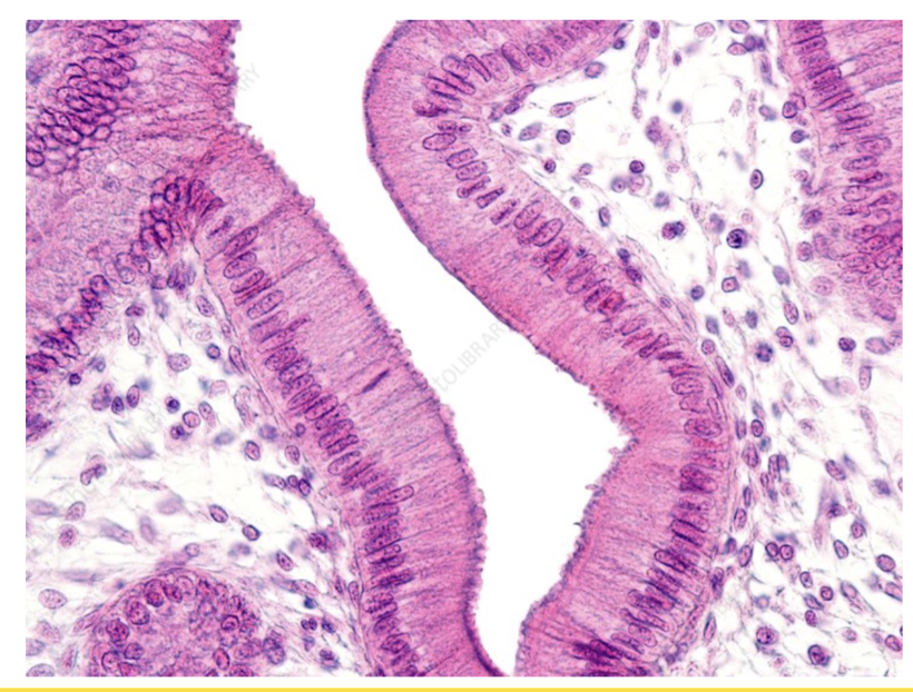
cilia vs microvilli
cilia: motile and help move contents like mucus, can maybe see individual cilia on microscope
microvilli: immotile and just to increase surface area for absorption, short and stubby so just looks like a solid sheath around
simple squamous epithelium
1 layer of wide, flat cells
function: diffusion and filtration
locations: alveoli and kidneys
specialized types: endothelium - lining blood vessels, mesothelium - lining the outside of organs
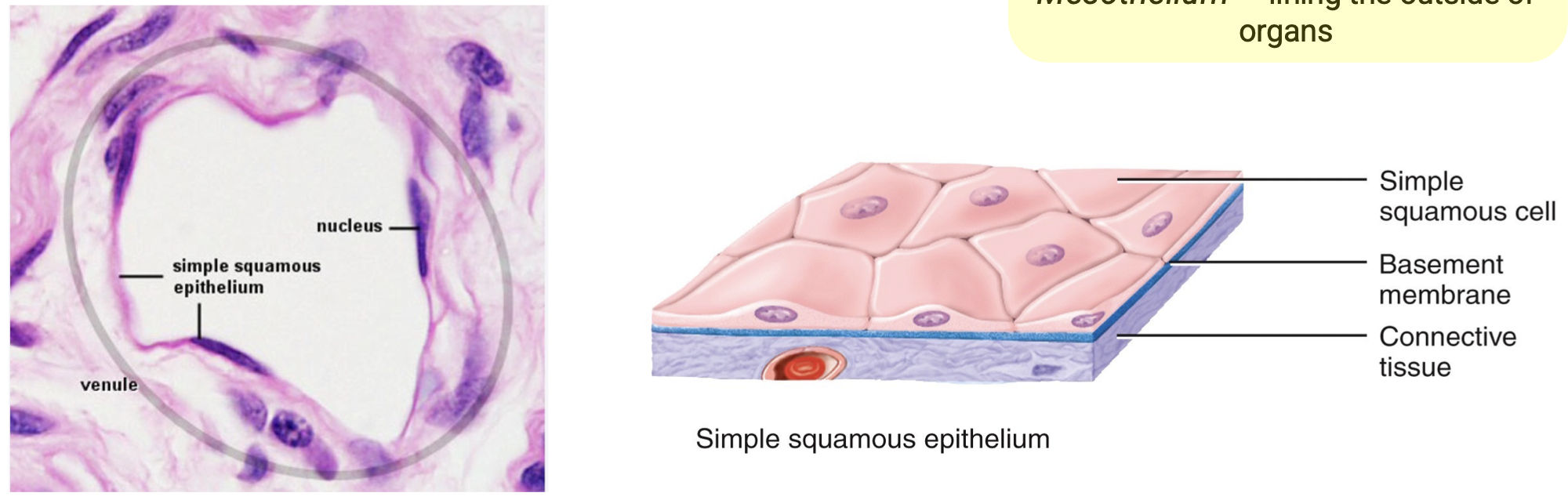
endothelium
simple squamous epithelium that lines blood vessels
mesothelium
simple squamous epithelium lining the outside of organs
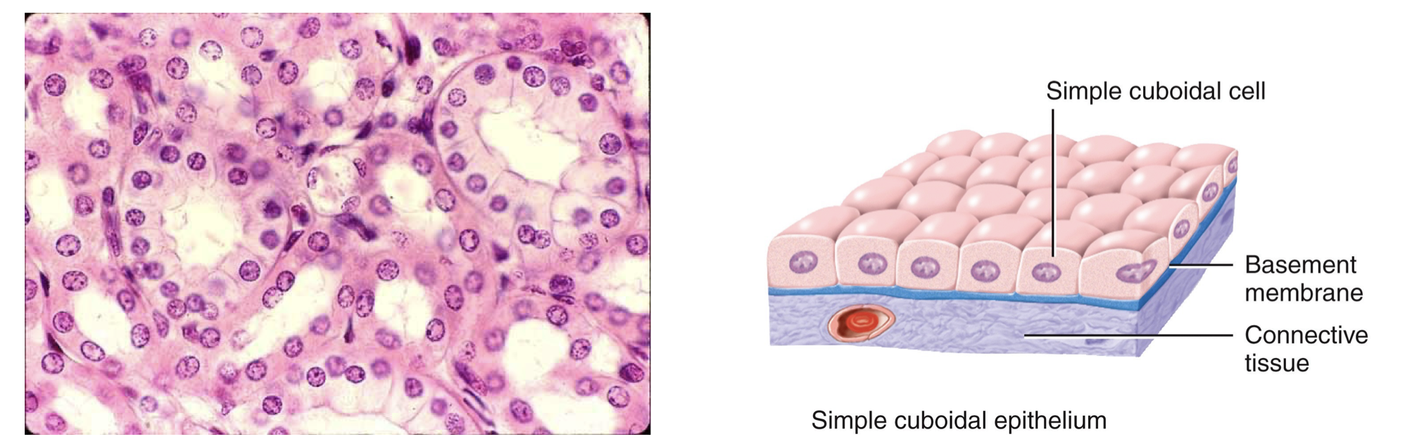
simple cuboidal epithelium
1 layer of cube-shaped cells
function: secretion, filtration and absorption
locations: glands and ducts, salivary and mammary
specializations: microvilli
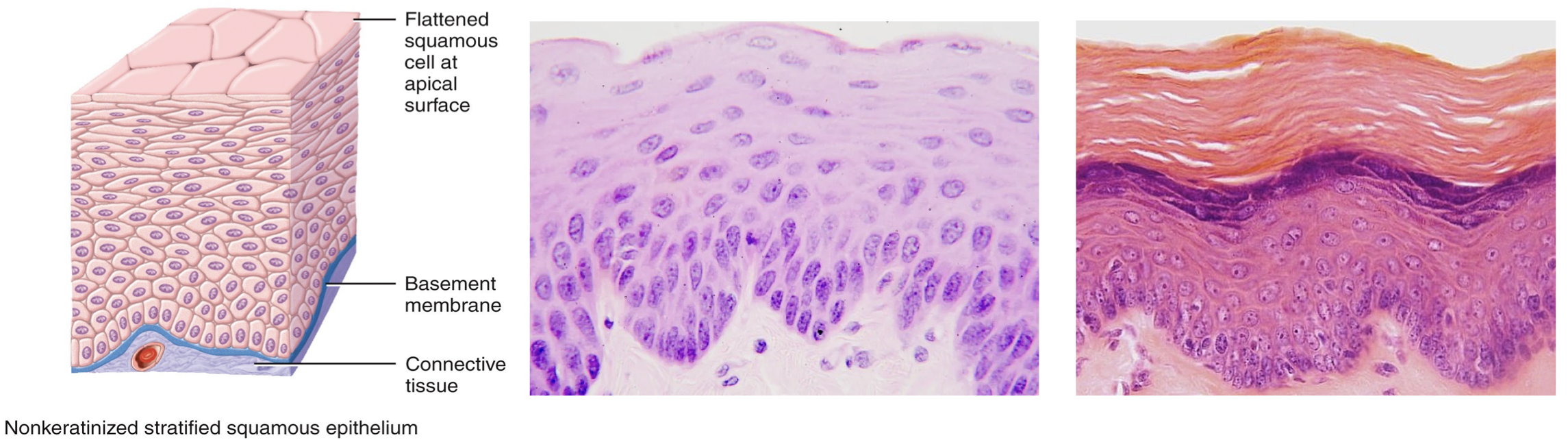
stratified squamous epithelium
multiple layers of cell - topmost are wide and flat
function: protection
locations: skin, oesophagus, and anal canal
specialization: keratinized
pseudostratified columnar epithelium
1 layer of cells of different heights
function: secretion and movement of luminal contents
locations: trachea, bronchi, nasal cavity
specializations: goblet cells and cilia

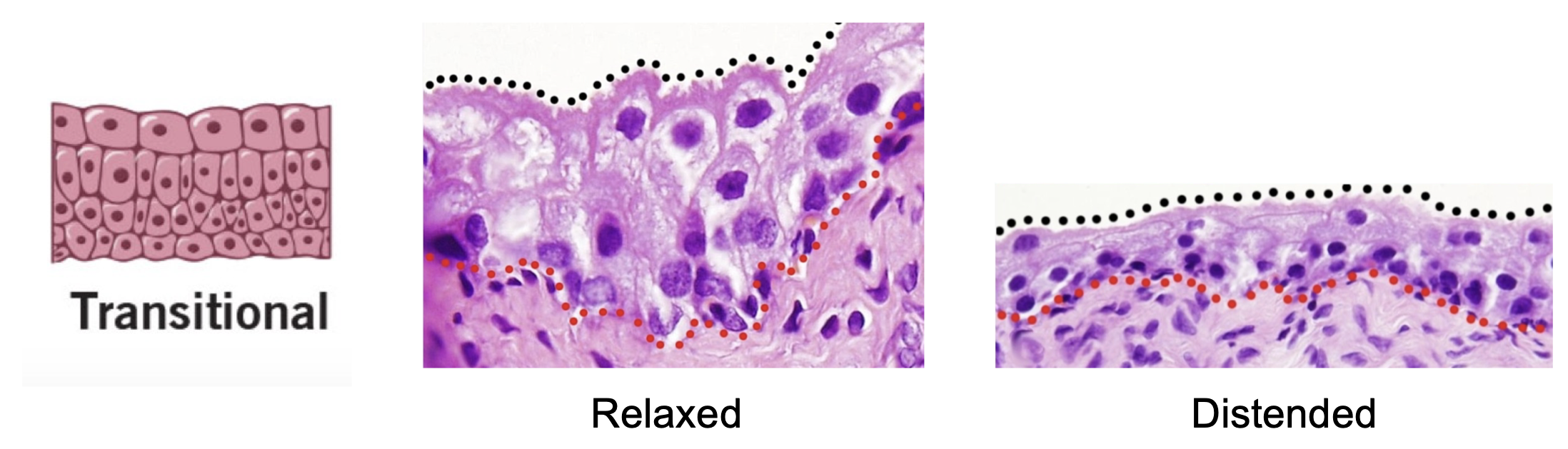
transitional epithelium
changing cell shape depending on distention of organ
function: stretch and protection
locations: bladder
specializations: apical hardening
connective tissue types
dense: regular, irregular, elastic
loose: areolar, adipose, reticular
dense irregular connective tissue
function: 3D stress resistance
locations: skin and around organs
ECM: collagen + few elastin

elastic connective tissue
dense
function: stretch
locations: heart, blood vessels, vocal cords
ECM: elastin + collagen

areolar connective tissue
loose
function: cushion and attachment, holds us together
locations: around organs and vessels
ECM: elastin + collagen + reticular + ground substance
