BIO-375 Exam #4
 Call Kai
 Learn
Learn Practice Test
 Spaced Repetition
 Match
Match Flashcards
 Knowt Play
1/75

Earn XP
Description and Tags
Cognition
Name | Mastery | Learn | Test | Matching | Spaced | Call with Kai | Chat |
|---|
No analytics yet
Send a link to your students to track their progress
76 Terms
Cognition
Mental processes relating to the acquisition, storage, manipulation, and retrieval of information.
Processing information
Association Cortex
A critical part of the cerebral cortex that is responsible for integrating sensory information and facilitating higher-order cognitive functions.
Neocortex that isn’t primary motor or primary somatosensory cortex
Parietal Lobe
Which lobe of association cortex is this?
Important for attending to stimuli and responding
Motivation leads to more attention
Multitasking = switching between what you are attending to
Contralateral Neglect Syndrome
A condition where patients fail to attend to, respond to, or perceive stimuli on the side of space opposite a brain lesion
Damage in right parietal lobe mainly
Deny presence of stimuli in left visual field
Right parietal lobe = attending to left and right visual fields
Left parietal lobe = attending to right visual field only
Temporal Lobe
Which lobe of association cortex is this?
Recognize specific objects such as faces
Present images of faces to monkeys and neurons become active
Angle and direction-specific neurons
Due to activity of populations of associated neurons
Agnosias
Difficulty recognizing, identifying, and naming (able to acknowledge) stimuli
Prosopagnosia
The inability to recognize faces
Inferior temporal lobe (the case of L.H.)
Frontal Lobe
Which lobe of association cortex is this?
Planning and decision making
Short-range and long-range planning
Delayed response task
Put food in one tray with another tray empty, cover trays, cause delay, monkey chooses tray and if they choose one with food they get the food
Cue → Delay → Response
Personality
James Watts & Walker Freeman- used technique of frontal lobotomy for mental illness treatment- disconnect personality/separate connections from frontal lobe to other parts of brain
Memories
Biological process by which the brain encodes, stores, and retrieves information through changes in neural circuits and synaptic connections
Synaptic plasticity
Synaptic Plasticity
Process of altering the strength of the synapse and making new connections with different neurons.
Short-Term
Long-Term
Short-Term Synaptic Plasticity
Synaptic plasticity that occurs within milliseconds to several minutes
Increase the [Ca2+] inside presynaptic neurons alters the amount of neurotransmitter released (increase NT)
Retrograde messengers- postsynaptic neuron sends short messages back to presynaptic neuron to alter amount of NT released
Long-Term Synaptic Plasticity
Synaptic plasticity that involves the changes in responsiveness occurring in the postsynaptic neuron
Long-term Potentiation (LTP)- increases responsiveness
Long-term Depression (LTD)- decrease responsiveness
Changes are not permanent
Long-Term Potentiation (LTP)
Type of long-term synaptic plasticity that increases the responsiveness occurring in the postsynaptic neuron
Add receptors to postsynaptic membrane
Requires high-frequency AP
Early Phase
Activation of NMDA receptors
Increase [Ca2+] intracellular
Increase number of AMPA
Late Phase- stabilization
Increase gene expression - increase synaptic proteins
Cytoskeletal changes → physically larger synapse
Astrocytic changes occur - synapse expands → astrocyte expands
Associativity
In reference to long-term potentiation
A second (low-frequency) neuron sends signal to add more receptors at same time as high frequency AP
Cooperativity
In reference to long-term potentiation
A bunch of low frequency AP neurons work together to change post-synaptic neuron
Long-Term Depression (LTD)
Type of long-term synaptic plasticity that decreases the responsiveness occurring in the postsynaptic neuron
Decrease gene expression
Decrease synapse size
Decrease number of receptors
Memory Process
Encoding
Acquisition
Consolidation (cellular level)
Storage (system consolidation)
Involves a lot of neurons
Integration with existing related memories
Retrieval
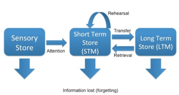
Atkinson-Shiffrin Memory Model (Tempooral)
Sensory information (immediate)
Iconic- visual sensory memories
Echoic- auditory sensory memories
Haptic- somatosensory memories
Short-term (working) Memory
Can rehearse to keep it here
Information can be retrieved from long-term memory
Long-term Memory
Storage of information from short-term memory
Declarative Memory (Explicit)
Memories that can be described
Episodic- recollection of events that were experienced
Semantic- naming or defining somethings
Nondeclarative Memory (Implicit/Procedural)
Memories that involve skills and habits
Motor skills - ex. riding a bike
Puzzle solving - ex. solving rubiks cube
Habituation/priming - superstitions
Memory Model
The working memory is separate from the short-term memory
Working memory- active processing system that manipulates information
Storage and retrieval to/from long-term memory
Short-term memory - temporary holding of new information
The Story of H.M.
This person had debilitating, untreatable seizures, and they removed his inferior-medial temporal lobe
Got rid of the seizures
Had severe anterograde amnesia with some retrograde amnesia
He couldn’t form new declarative/explicit long-term memories, but he could form new non-declarative implicit/procedural long-term memories
Inferior-medial temporal lobe important for creating new long-term memories - two different pathways for declarative vs. non-declarative memories
Learning Strategies
Help strengthen long-term memories
Repetition
Distributed practice
Retrieval-based learning
Motivation/attention
Rote (dry memorization) vs. meaningful learning
Association
Give meaningless items a meaningful context
Ex. create a story using the words
Increases learning and memory
Motivation
Increases learning and memory
Ex. Hungry individuals remember images of food better than non-hungry people
Leads to better attentiveness
More attention=better memory
Aficionados
An expert in something
Ex. Chess master
Remembering the movement of chess pieces better during an actual game than if the pieces were randomly moved
Repetition and experience
Cortical States
Different states of consciousness
Consciousness- a state of awareness of ‘self’ and the environment while retaining the ability to respond appropriately to stimuli
Sleep
A suspension of consciousness
EEG measures brainwaves in different locations of the brain the determine levels of cortical activity
Four stages and REM
Stage 1 (Sleep)
Becoming drowsy and nodding off, decreasing frequency, and increasing the amplitude of brainwaves
Stage 2 (Sleep)
Decreasing the frequency and increasing the amplitude of the brainwaves, sleep spindles (burst in activity of brain waves) occurs
Stage 3 & 4 (sleep)
Slow-wave sleep, delta waves occurring (giant waves in stage 4), deep/restorative sleep/most restful sleep
Sleep Stages 1-4
Slow rolling eyes
Decrease HR, BP, respiration, and metabolism
Decreased muscle tone and movement
REM Sleep
Increase in HR, BP, metabolism, and respiration
Rapid eye movements
Paralysis of large muscle groups
Twitching of smaller muscles
Circadian Rhythms
The internal clock
Suprachiasmatic Nucleus - located in the hypothalamus; master of the sleep/wake cycle
Photoentrainment- organisms internal biological clock synchronizes with external 24-hour light-dark cycle
Retinohypothalamic tract
Detect light intensity
Pineal gland produces melatonin
Melanopsin ipRGCs (retinal ganglion cells)- respond directly to light intensity- increase light = increase APs
Reticular Activating System
Active = Awake
Stimulate a sleeping cat’s brainstem → induce awakening
Cholinergic Nuclei - increase activity during REM sleep and awake
Raphe Nuclei- increase activity when awake (produce serotonin)
Locus Coeruleus - increase activity when awake (produce norepinephrine)
TMN (hypothalamus) - increase activity when awake (produce histamine)
Cholinergic Nuclei
Increase activity during REM sleep and awake
Raphe Nuclei
Increase activity when awake
produce serotonin
Locus Coeruleus
Increase activity when awake
produce norepinephrine
TMN (hypothalamus)
Increase activity when awake
produce histamine
Thalamus
Structure involved in sleep where if you stimulate when awake → induce sleep state
VLPO
As we transition into sleep cells from the _____ inhibit the TMN and the brainstem nuclei
Orexin Neurons
When we are awake, ______ stimulate the TMN and the brainstem nuclei
Reasons for needing sleep
Replenish glycogen levels - ATP
Cellular growth, maintenance, and repair
Conservation of energy/metabolism
Decrease free radicals in brain
Removal of waste
Other
Wound healing
Improved immune system
Memories
Sleep and Memories
When sleeping the brain can
Consolidate memories
Stabilize memories- strengthen synapses
Integrate new memories w/old memories
Dissect memories- consolidate the important qualities
Remove minutiae of the day (unnecessary details)
Approx. 6 hours of sleep is required for some aspects
Different stages of sleep influence different memories
Deep sleep = increase memory
REM Sleep
When during sleep do our vivid and story like dreams mostly occur?
Hypnic Jerk
The rapid jerking with a sensation of falling that wakes you up.
Non-REM Parasomnias
Sleep walking, sleep talking, sleep-related eating disorders, night terrors (pavor nocturnus-deep sleep)
REM Parasomnias
Nightmare disorder, sleep paralysis, REM sleep behavior disorder
Insomnia
Sleep deprivation that decreases cognitive abilities and causes the accumulation of sleep debt
Decreased attention
Cognitive skills impaired
Reaction times slowed
Language skills decreased
Can be fatal - Fatal familial insomnia/sporadic fatal insomnia
Decrease
Sleep needs ___(increase or decrease)___ with age.
Emotions
A complex reaction pattern involving experiential, behavioral, and physiological elements.
Duchenne
This person studied facial expressions of different emotions using electrodes.
Facial expression is a large unconscious part of our emotions
Dimensional Theory of Emotions
Emotions are very complex
Arousal level/Activation
Valence/Positive vs. Negative

Volitional (Classic Motor)
Emotional Expression
What are the two descending pathways/systems of facial expressions?
Volitional (Classic Motor)
Which descending pathway of facial expression is this?
Motor cortex
Pyramidal tracts
Spinal cord/brainstem
Emotional Expression
Which descending pathway of facial expression is this?
Anterior Cingulate Gyrus
Extrapyramidal tracts (reticular formation)
Spinal cord/Brainstem
James-Lang Theory of Emotion
Theory of emotion that is very linear
Experience → Physiological response → Emotion
Stimulus causes a physiological response which illicit an emotion

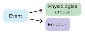
Cannon-Bard Theory of Emotion
Theory of emotion that separates the emotional response from the physiological response.
Stimulus → Thalamus → Emotional Response & Physiological Response
Philip Bard
Determined the notion of “sham rage”
Generated rage through electrical stimulation in cats
Removal of hypothalamus = no sham rage
When hypothalamus kept = sham rage
Walter Hess
Studied behavior in cats by stimulating the hypothalamus via electrodes to produce rage/behavioral changes in the cats.
James Papez
This scientist argued that there were other structures involved in emotion
Ex. Fornix, hippocampus, and mammillary bodies
Amygdala
Orbital and Medial prefrontal cortex
Cingulate Gyrus
Hippocampus
What are some of the other structures that were determined to be involved in emotion.
Kluver-Bucy Syndrome
The removal of medial temporal lobes (remove amygdala) leads to the loss of fear and aggression toward people (in animals typically fearful of people)
Lost control of their emotions
Hyperactivity and hypersexuality
John Downer
A scientist who severed the corpus callosum and optic chiasm
Eye without amygdala
With the right eye covered, information can’t get to other side of brain with amygdala
Respond with lack of fear
Amygdala
This structure is very important for producing fear and aggression.
Post-Traumatic Stress Disorder (PTSD)
A mental health condition triggered by experiencing or witnessing terrifying events like war, abuse, or disasters.
Very intense sensory experiences
Synaptic plasticity within amygdala at the heart
The Case of S.M.
This individual had amygdala degeneration and is missing her amygdala’s.
She has a difficult time recognizing or feeling fear
Her other emotions remain intact
Language
The symbolic representation involving
Syntax- symbolic order of words
Lexicon- vocabulary; meaning of symbols/words
Grammar- rules; whole structure of a language
Prosody- emotion or affect
Speech
Structures involved in production of ______
Lips, tongue, jaw, lungs, larynx
Dysarthria
Dysarthria
A motor speech disorder due to damage to CN X or associated structures in speech production.
Paralysis or paresis of muscles used in speech production
Arcuate Fasciculus
The tracts that connect Broca’s area and Wernicke’s area.
Broca’s Aphasia
Motor Aphasia/Expressive Aphasia
Can understand/comprehend language
Individuals know what they want to say but struggle to produce speech resulting in halting and short sentences
Wernicke’s Aphasia
Receptive Aphasia/Fluent Aphasia
Can produce speech but it doesn’t make any sense
Fluent but meaningless speech, poor comprehension, word salad
Conduction Aphasia
A language disorder caused by damage to the arcuate fasciculus and is characterized by fluent speech and good comprehension but difficulty repeating words or phrases.
Lateralization/Split-brain
Sever the connection between the two hemispheres of the brain
Certain cognitive processes and functions are more dominant in one hemisphere of the brain than the other
Info from RVF processed in left hemisphere- can be named and described- direct access to language center
Info from LVF processed in right hemisphere- can’t be named and described- no access to language center
If a split-brain patient had their language center in the left hemisphere, how would they respond to information in both the right and left visual fields?