Topic 9.1: Dental Anatomy
 Call Kai
 Learn
Learn Practice Test
 Spaced Repetition
 Match
Match Flashcards
 Knowt Play
1/47

There's no tags or description
Looks like no tags are added yet.
Name | Mastery | Learn | Test | Matching | Spaced | Call with Kai | Chat |
|---|
No analytics yet
Send a link to your students to track their progress
48 Terms


How does the Maxillary Central Incisor develope?
The initial calcification occurs at 3-4 months
The crown is completed at 4-5 years of age
The crown errupts in the age of 7-8
The root is completed with 10 years of age.


How is the facial anatomy of the Maxillary central incisor?
Flat facial surface
The incisal edge is relatively straight.
The distoincisal angle is curved while the mesioincisal edge is flat.
The root is very big and makes up 2/3 of the tooth.
The crown length (10.5mm) is greater than crown width (8.5mm).


How is the root shaped and how many root canals does the maxillary central incisor have?
Outline of the root is cone shaped and located slightly distal to center line of tooth.
It has one root canal


How is the palatal anatomy of the maxillary central incisor?
Cervical outline on palatal is more concave than on facial.
The cingulum occupies the cervical half of the crown. It has distal and mesial marignal ridges
The Lingual fossa in incisal ½ of crown surface
Mesial and distal marginal ridges


How is the palatal anatomy of the maxillary central incisor?
Cervical outline on palatal is more concave than on facial.
The cingulum occupies the cervical half of the crown. It has distal and mesial marignal ridges
The Lingual fossa in incisal ½ of crown surface
Mesial and distal marginal ridges


How is the incisal anatomy of the maxillary central incisor?
Mesiodistal width is greater than the faciolingual diameter.
Incisal ridge is perpendicular to mesiodistalbisecting plane.
Crest of cingulum is slightly distal to the bisecting plane of crown
Incisal ridge is straight


How does the Maxillary lateral incisor develope?
The initial calcification occurs at 1 year of Age.
The crown is completed after 4-5 years of age.
The crown erupts at 8-9 years of age.
The root is completed after 11 years of age.


How is the Facial Anatomy of the Maxillary Lateral Incisor?
Crown outline is trapezoidal
Convex in all directions
Incisal edge more rounded than central
Cervical line is elliptical


What are the differences of the Facial of the Lateral to the Central Maxillary incisor?
Both mesio- and distoincisal angles of the crown are rounded, but the distoincisal angle is more.
The cementoenamel junction is more rounded then the one of the central incisor.
The incisal edge is relatively more rounded and not straight.
It is smaller and narrower than the central incisor.
Has more distinct lingual anatomy


How is the lingual anatomy of the lateral maxillary incisor?
Cingulum limited to cervical third of crown
Mesial and distal marginal ridges form a V
The lingual fossa is relatively deep and has a triangular shape.
It has a Lingogingival groove at distal side of cingulum that may even extend across the cementoenamel junction onto the root (only in lateral)


How is the root of the Lateral maxillary incisor?
Root is conical and straight.
The root length is generally same or can be longer as the central.


How is the mesial and distal anatomy of the Maxillary lateral incisor?
Facial crown outline is convex and lingual is slightly concave.
Incisal ridge in line with mid point of faciolingual diameter.
Apex of root is facial to faciolingual long axis bisector.


Incisal Anatomy of Maxillary lateral incisor
Crown is wider mesiodistally by only 1mm and crown converges sharply toward the lingual surface. Incisal edge is converged.


How does the Maxillary Canine develop?
The initial calcification occurs with 4-5 months
The completion of the crown occurs from 6-7 years
The crown errupts in the ages of 11-12
The root is completed in the age of 13-15


How is the Facial Anatomy of the Maxillary Canine?
Has a asymmetrical anatomy in which the distal portion is bigger and has a bigger ridge
Mesial cuspal ridge shorter than distal cuspal ridge
Pentagonal crown shape
Canines have a cusp
Is the widest of all anterior teeth (8mm)


How is the Palatal/Lingual anatomy of the maxillary canine?
Cingulum is smooth and well developed and occupies the cervical third of lingual surface
It has 2 lingual fossas: mesiolingual and distolingual and 1 lingual ridge that separates both fossas and connects the cusp tip and the cingulum
The crest of the lingual ridge curves slightly to mesial on mesiodistal long axis bisector which makes the distolingual fossa larger than the mesiolingual.


How is the mesio/distal anatomy of the maxillary canine?
Triangular outline and smoothly convex
Cusp tip is never linugal and usually slightly facial to faciolingual axis bisector
convex cingulum and crest of lingual ridge slightly concave


How is the incisal anatomy of the maxillary canine?
Distal side is longer and mesial is shorter and rounder
Longer faciolingually than mesiodistally
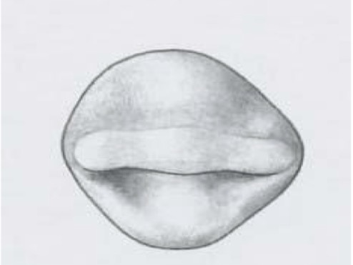

How is the chronology of the first maxillary premolar?
The initial calcification occurs after 18-21 months of age
The completion of the crown takes place after 5-6 years of age
The crown erupts after 10-11 years
The root is completed after 12-13 years
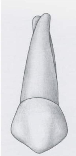

How is the facial anatomy of the first maxillary premolar?
Cusp tip is relatively pointed and slightly distal on mesiodistal long axis bisector
Mesial cuspal ridge of facial cusp longer than distal
Facial ridge running along axially from facial cusp


How is the lingual anatomy of the first maxillary premolar?
entire tooth converges towards the lingual
Lingual cusp shorter than the facial cusp
The lingual cusp tip is slightly mesial to the mesiodistal long axis bisector
Both lingual cusps of premolars swing to mesial aspect


How is the mesial/distal anatomy of the first maxillary premolar?
Mesial marginal development groove on mesial surface
Distal aspect does not have a groove


How is the occlusal anatomy of the maxillary first premolar?
Has two major cusps: facial and lingual
the linugal cusp is 1mm shorter
Faciolingually wider than mesiodistally (2mm)
Mesial cuspal ridge of facial cups islonger than distal
cusp tip of facial cusp is positioned distal to mesiodistal bisector
central groove connects mesial and distal triangular fossae and extends over mesial margina

How is the root of the maxillary first premolar?
2 roots: facial and lingual
At least 2 root canals and if there is a third, it will be in the facial root
Rarely single root with 2 canals


How is the chronology of the maxillary second premolar?
The initial calcification occurs after 24 months
Completion of crown 6-7 years of age
The tooth erupts after 10-12 years of age
The root is completed after 12-14 years


How is the facial anatomy of the maxillary second premolar?
Facial anaromy
Pentagonal shape
2 cusps: facial and lingual with nearly equal size
Distal cuspal ridge longer than distal
Facial ridge not as prominent as that of the maxillary 1st premolar


How is the Root of the maxillary second premolar?
Root length longer
Typically has one root with 2 pulp canals


How is the lingual anatomy of the maxillary second permolar?
Outline of both cusps visible
Both cusps are in line
Lingual cusp little rounder than facial


How is the occlusal anatomy of the maxillary second premolar?
Cusp tips in line with each other and slighty mesial to the mesiodistal bisector
Line angles more rounded and balanced look
mesial and distal irregular triangular fossae near the center
Several supplemental grooves
Central groove smaller than of the first premolar


How is the chronology of the maxillary first molar?
The initial calcification of the maxillary first molar occurs at birth
The crown is completed during the 3-4 years of age
The crown erupts in the of 6
The root is completed after 9-10 years
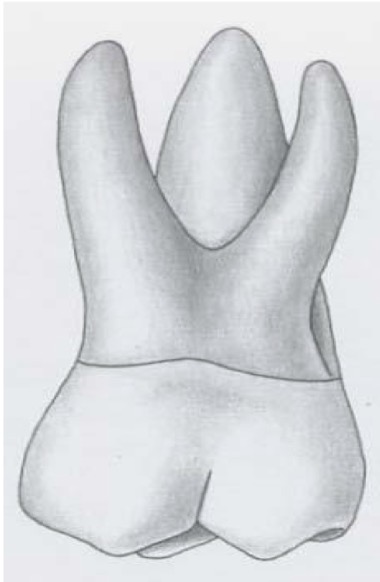
How is the facial anatomy of the maxillary first molar?
The crown has a trapezoidal outline
Facial groove that extends occlusocervically between the mesiofacial and distofacial cusps to middle of facial surface
Facial cusps more pointed and taller than lingual
Mesial cusps taller than distal


How is the root of the maxillary first molar?
Three roots with 3-4 root canals and root depressions
Lingual root longest then mesiofacial and then distofacial
Mesiofacial root has 50% probability of having 2 root canals


How is the lingual anatomy of the maxillary first molar?
Only lingual cusps visible
Mesiolingual cusp is the largest and the mesiodistal the widest
Lingual groove begins at lingual border as an extension of distal oblique groove
Fifth (carabellis) cusp is located on the lingual aspect of the mesiolingual cusp
cuspal edges parallel to and about 2mm cervical to the occlusal outline of mesiolingual cusp


How is the mesial anatomy of the maxillary first molar?
carabellis cusp is visible
mesiofacial root covers the distofacial root


How is the distal anatomy of the maxillary first molar?
Distofacial root is the smallest


How is the occlusal anatomy of the maxillary first molar?
4 well developed cusps and 1 carabellis cusp
Wider faciolingually than mesiodistally (1mm)
Crown wider mesially than distally and wider lingually than facially
Cusp sizes →: ML, MF, DF, DL,CB
Oblique ridge from ML to DF cusp that is produced by the meeting of the triangular ridge of distofacial cusp and distal cuspal ridge of mesiolingual cusp
Sometimes there is also a transverse groove of the oblique ridge that crosses it from central to distal fossae


How is the chronology of the maxillary second molar?
The initial calcification occurs at 3 years of age
The crown is completed with 7-8 years of age
The crown erupts in the age of 12 years
The root is completed after 14-16 years


How is the root of the maxillary second molar?
3 roots: L → MF → DF
3-4 Root canals
Less divergent than 1st molar
Limited depressions


How is the facial anatomy of the maxillary second molar?
Facial cusps are smaller in height than first molar
Distofacial cusp shorter than the mesiofaciak cusp
Facial groove is shorter
Mesiofacial and distofacial roots visible


How is the lingual anatomy of the maxillary second molar?
Mesiolingual cusp is more nearly centered and bigger and more developed than the distolingual cusp
Single lingual root visible
Very rarely 5th cusp


How is the distal anatomy of the maxillary second molar?
More of occlusal anatomy visible
Distofacial smaller than mesiofacial


How is the occlusal anatomy of the maxillary second molar?
similar occlusal anatomy to the maxillary 1st molar
Msdt diameter smaller (1 mm)
Distal cusps are smaller
Usually has more secondary grooves.
Wider faciolingually than mesiodistally.
no carabellis


