PARA L4 +L5 poultry endoparasites
1/68
There's no tags or description
Looks like no tags are added yet.
Name | Mastery | Learn | Test | Matching | Spaced | Call with Kai |
|---|
No analytics yet
Send a link to your students to track their progress
69 Terms
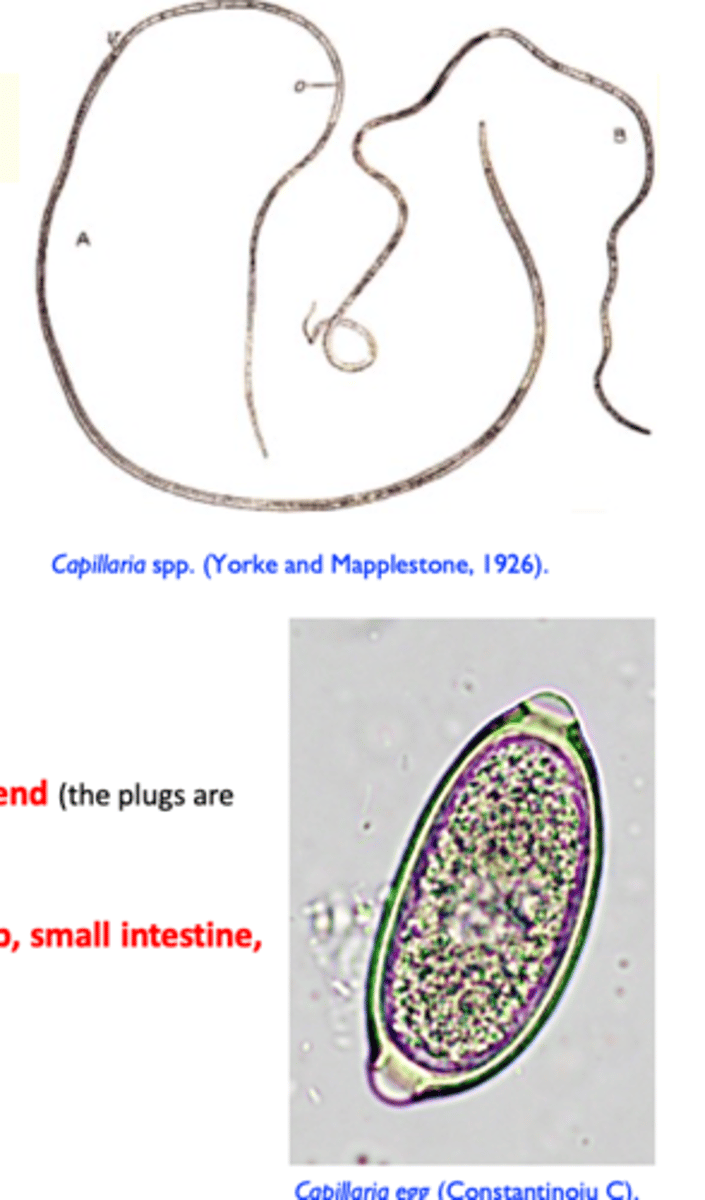
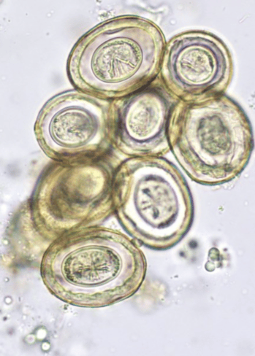
Capillaria spp. (hairworms) morphology
Small and slender parasites (capillary body difficult to see with the naked eye), similar to Trichuris;
Size: extremely thin worms (capillary);
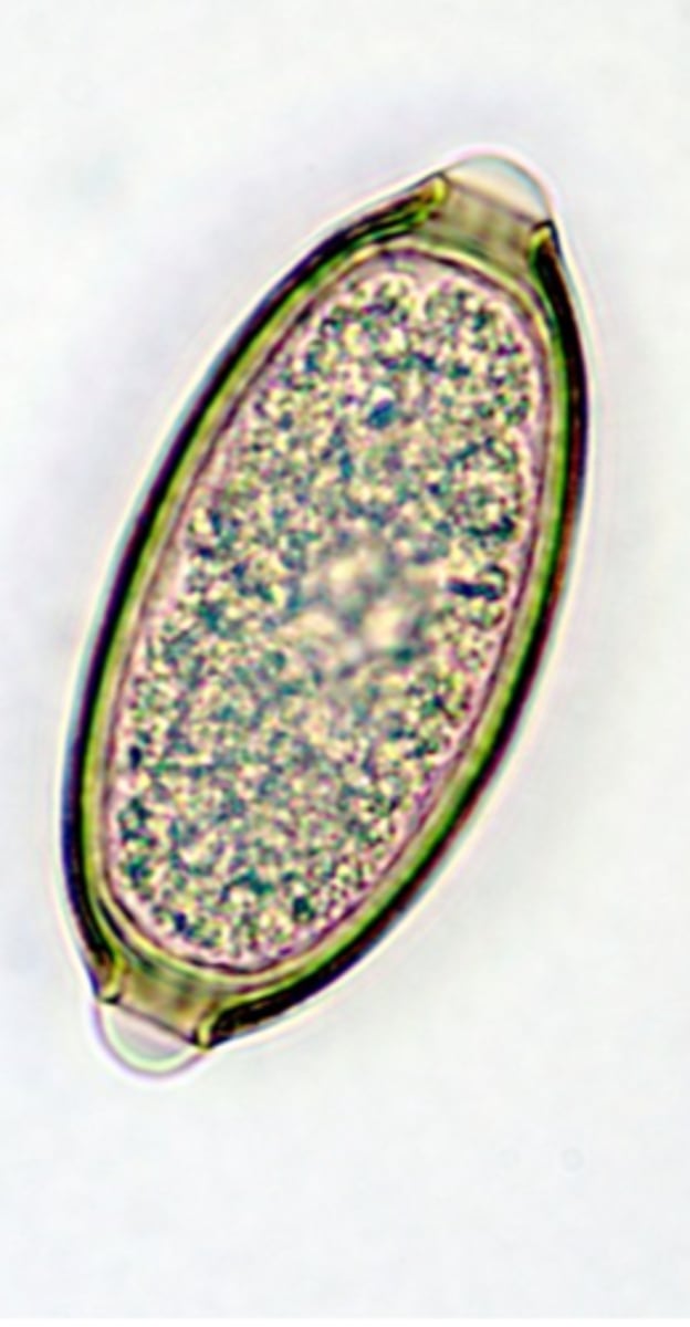
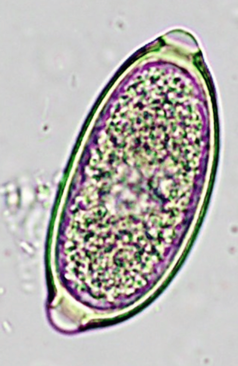
Eggs similar to those of Trichuris
• Thick shelled, barrel shape with a plug at each end
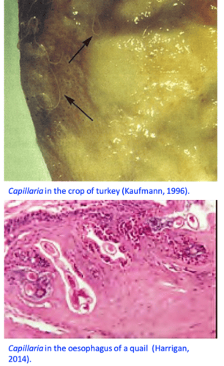
Capillaria spp. (hairworms) LOCATION
different regions of the digestive tract - mouth, oesophagus, crop, small intestine, cecum;
Capillaria spp. (hairworms) LIFECYCLE
a) Indirect
• IH represented by earthworms
->birds become infected after ingesting earthworms containing infective larvae;
b) Direct
•Infective larvae develop inside eggs in 1-4weeks
-> birds become infected after ingesting embrionated eggs;
Capillaria spp. (hairworms)
Epidemiology
Worldwide distributed parasites, present in outdoor systems (all species) and indoor systems (C. obsignata);
Outside pens of poultry may remain contaminated for long time
Young birds are more susceptible to infection, adult birds may serve as carriers;
Capillaria spp.
Clinical signs and pathology
Oesophagus and crop: Capillaria annulata, Capillaria contorta
Heavy infections: inappetence, weakness, emaciation, frequent swallowing attempts, death
Marked thickening of the mucosa, diphtheric membranes, sloughing of the mucosa, food may accumulate in the crop etc;
Capillaria spp. Diagnosis
Necropsy and careful examination of the mucosa of the digestive tract;
Microscopic examination of the mucosal scrapings -> worms/eggs;
• Patent infections: detection of eggs in the faeces:
Capillaria spp. Treatment
Imidazothiazoles
Levamisole
Benzimidazoles
Flubendazole
Capillaria spp. Control
Species with IH
Anthelmintic treatments -> house the birds or
Regular anthelmintic treatments -> move on fresh ground
Species without IH
Hygiene, heat treatments of surfaces
Regular anthelmintic treatments
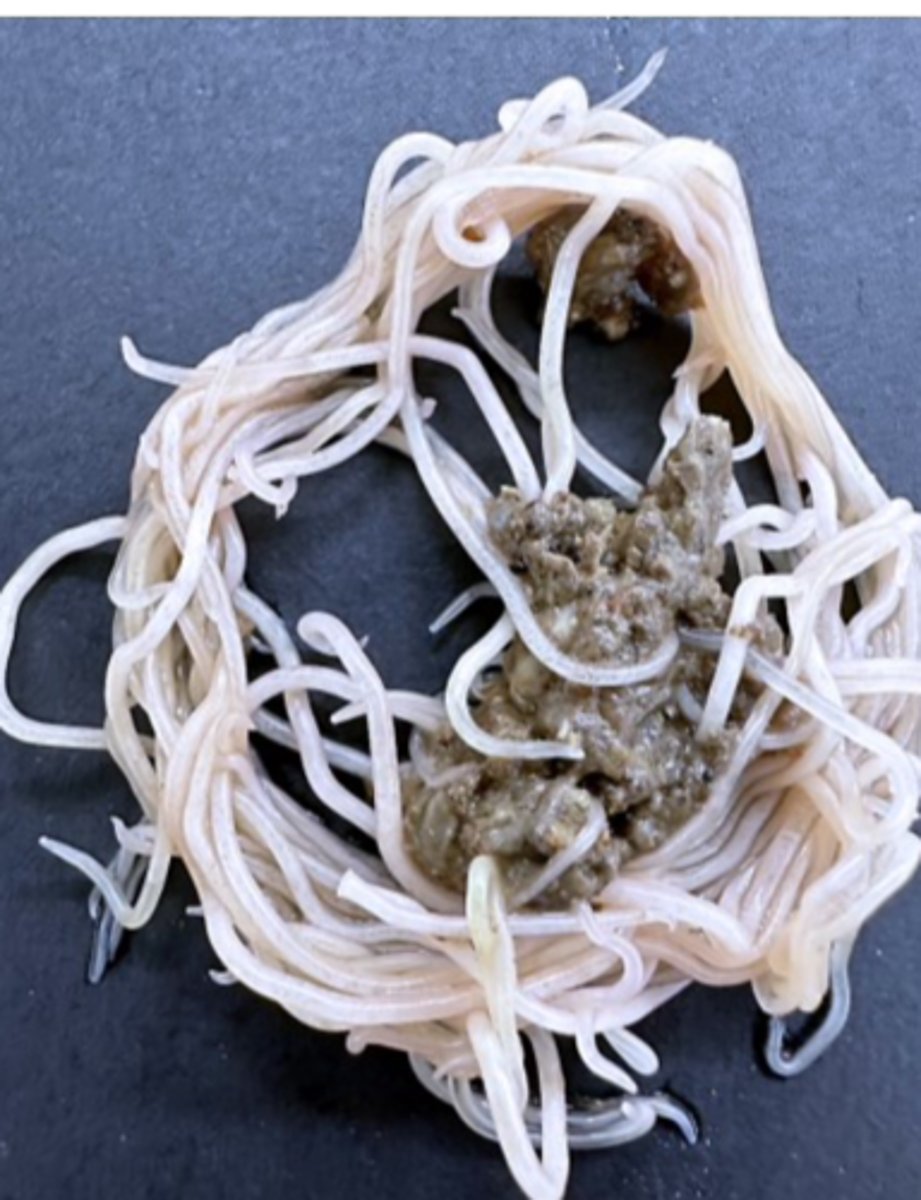
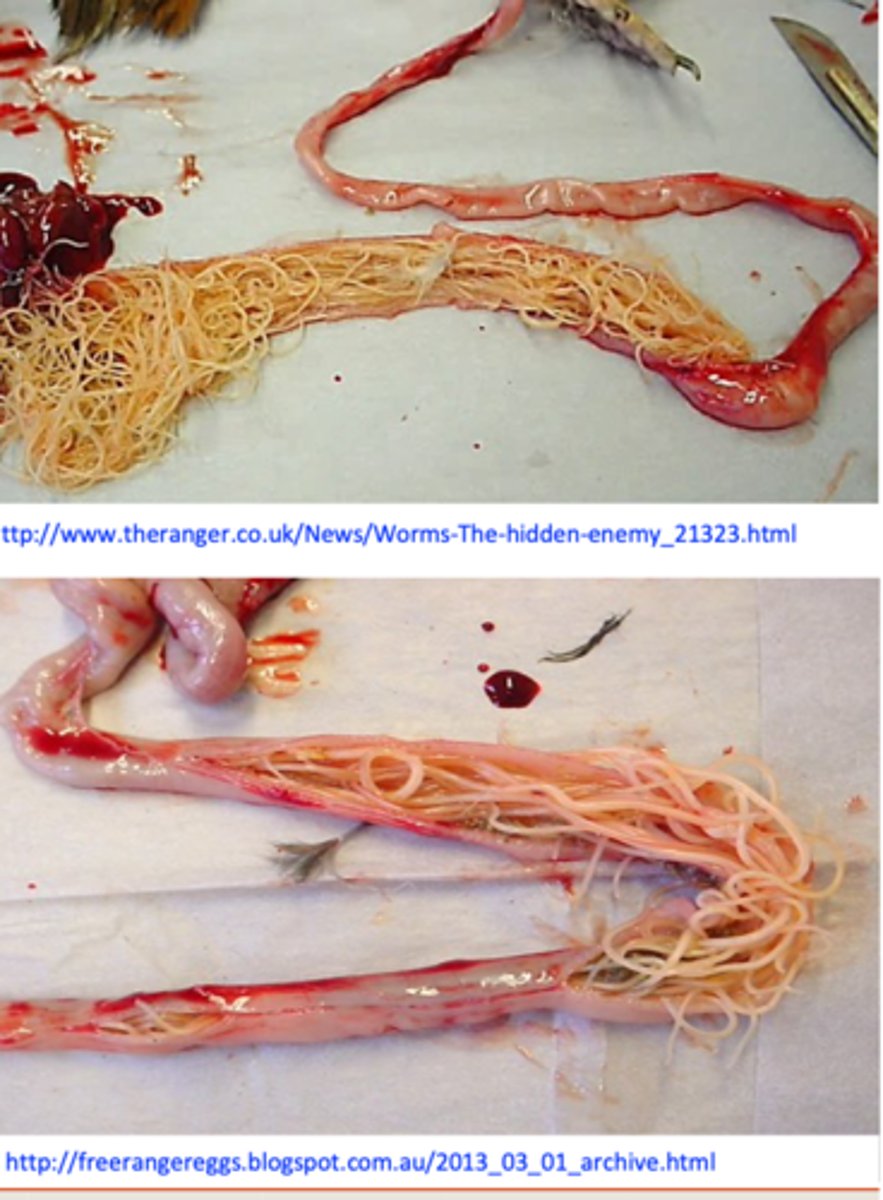
Ascaridia galli
Location
Small intestine of chicken, turkey, dove etc;
Feeding
• Intestinal content;
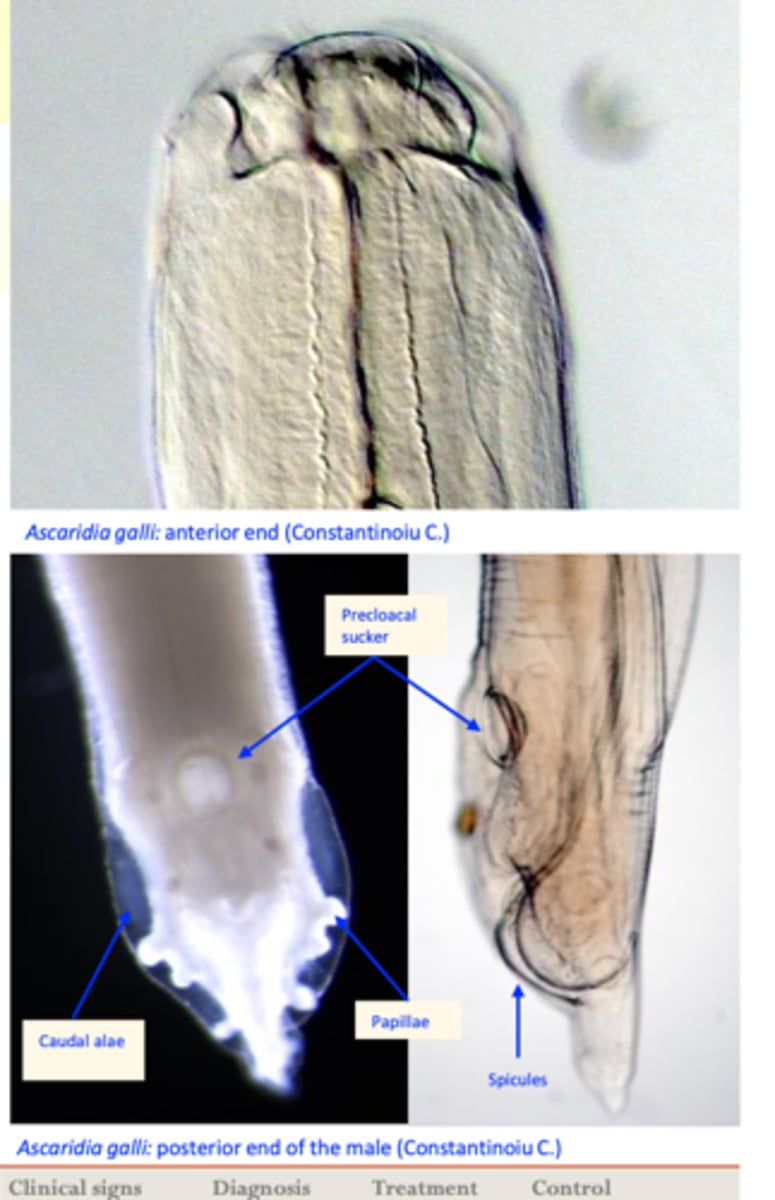
Ascaridia galli
Morphology
• White worms, up to 12 cm long;
Anterior end
• 3 lips around the mouth;
Posterior end
• Male: A circular precloacal sucker with a thick
cuticular rim;
Ascaridia galli
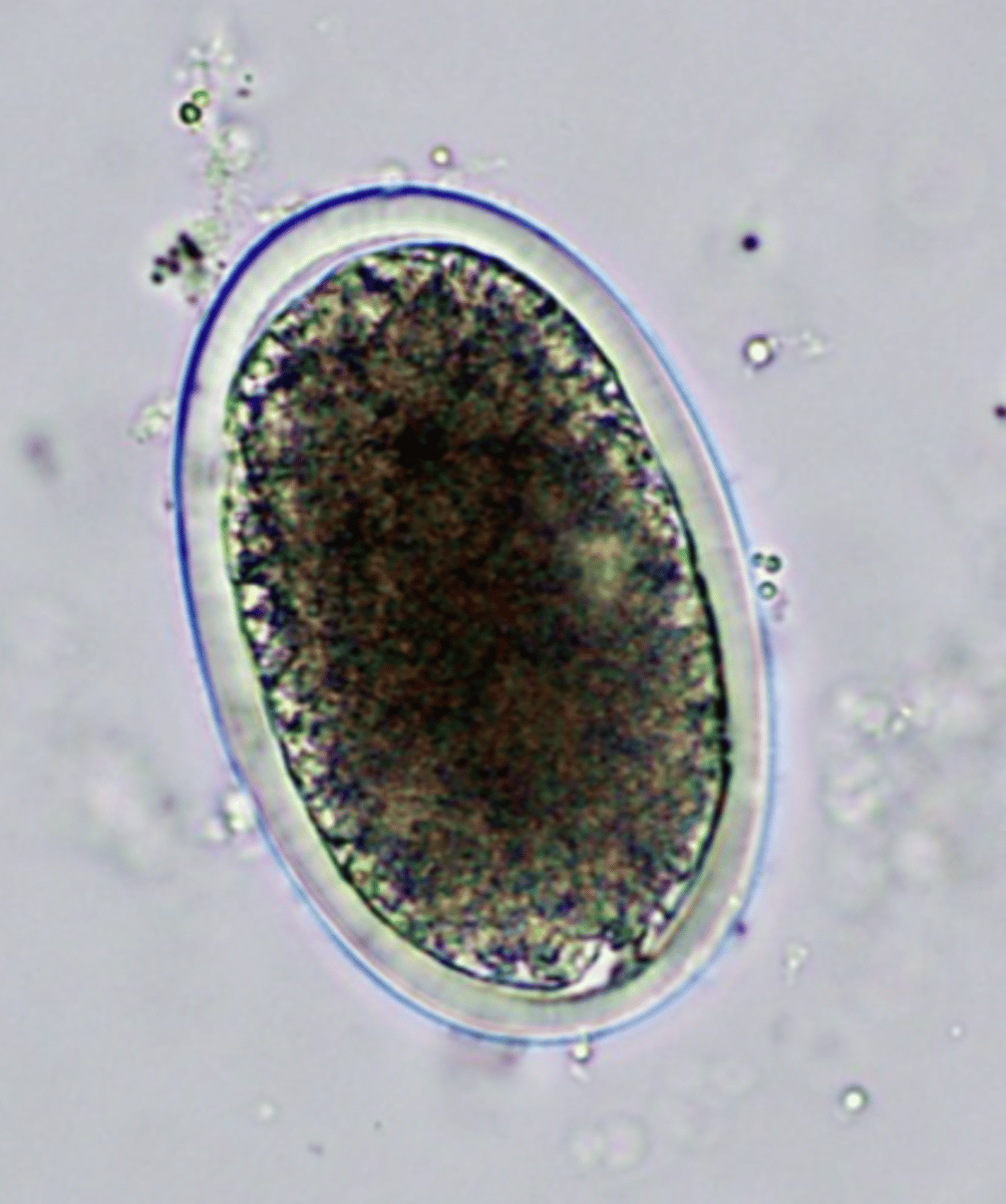
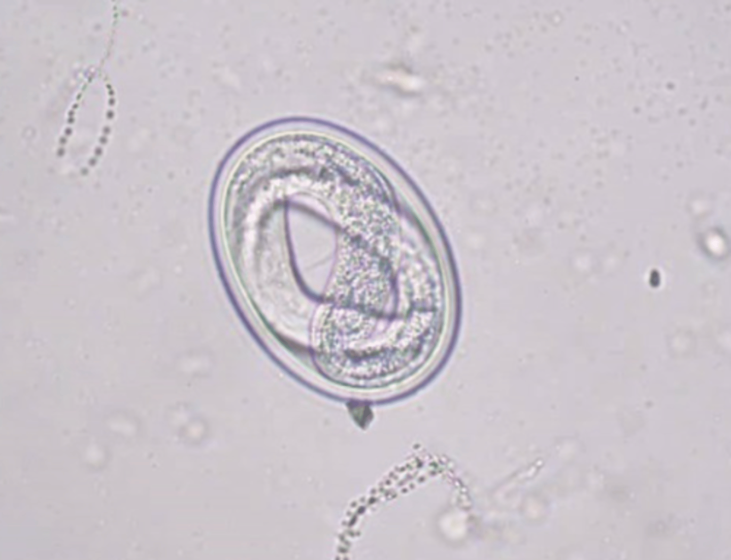
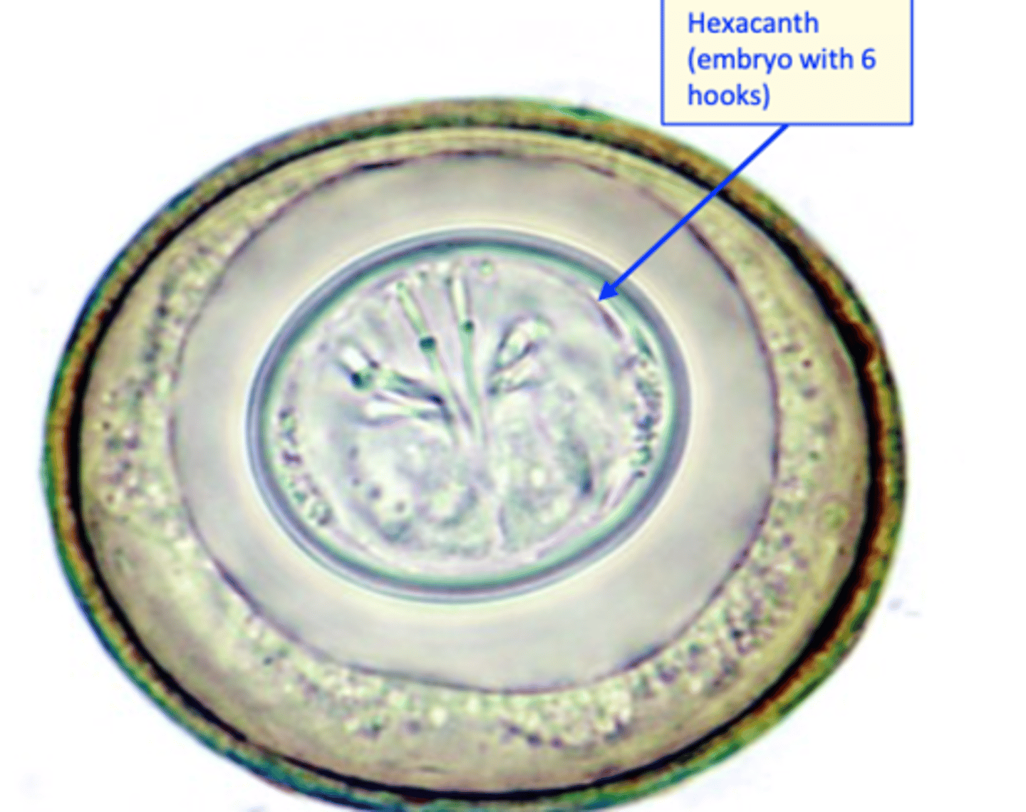
EGG Morphology
Shape: ellipsoidal, thick, smooth shell;
Colour: brown;
One cell inside.
Ascaridia galli
Life cycle
Infection of the host:
Ingestion of embrionated eggs;
After infection larvae develop in the wall of the
small intestine
PPT: 4-8 weeks
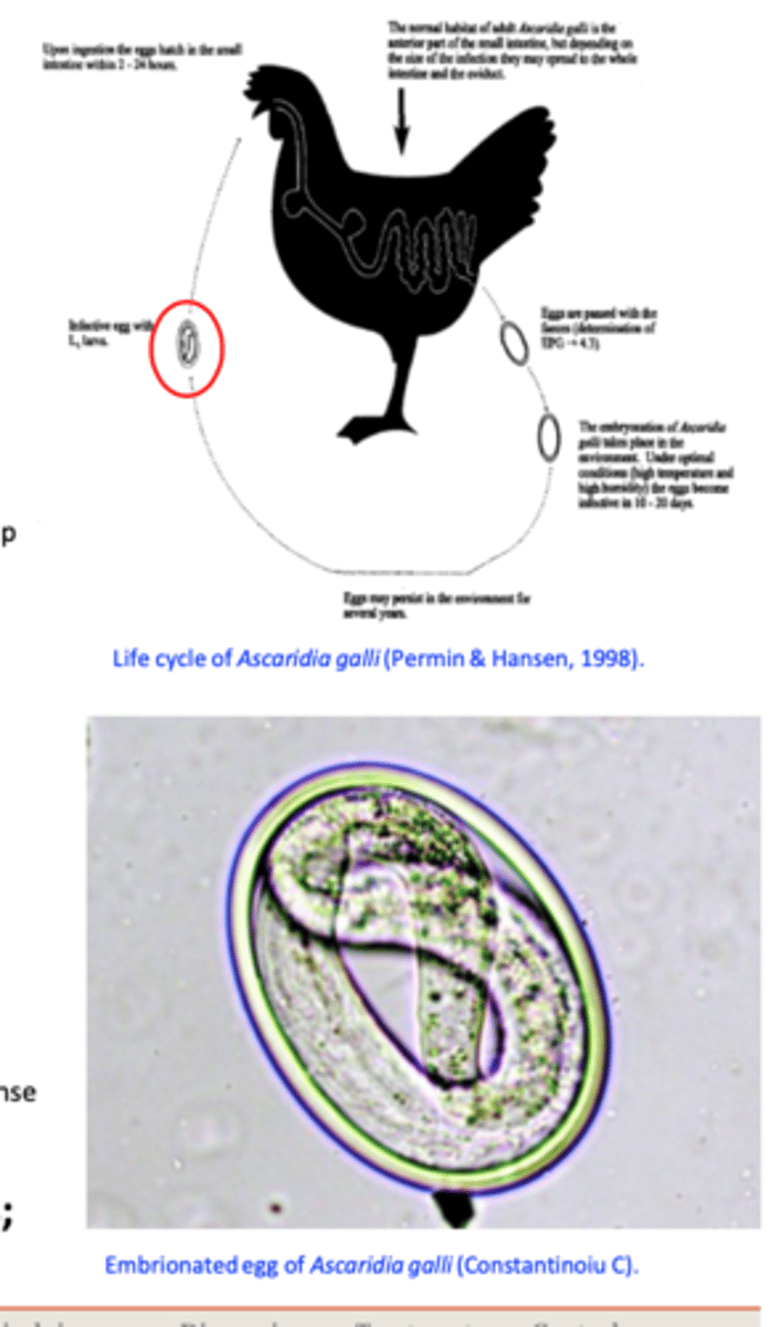
Ascaridia galli EGG
Ascaridia galli
EPIDEMIOLOGY
Spread all over the world;
Indoor and outdoor systems;
Sources of infection: birds that have unapparent infections (adults) and the ground - eggs can survive in the environment for long time
Common in chickens younger than 3 months
Ascaridia galli
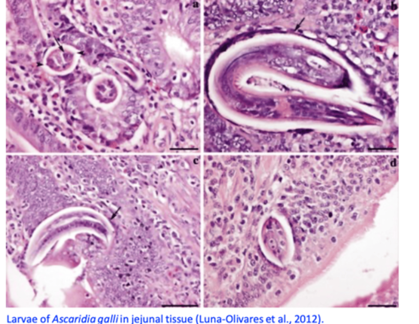
Pathogenesis/Pathology
I. Main effects caused during the larval histotrophic phase
Penetration of the mucosa of the intestine by large numbers of larvae -> enteritis, haemorrhagic enteritis;
During histotrophic phase: haemorrhage and loss of blood, destruction of the glandular epithelium;
II. Large numbers of adult worms:
Alter the intestinal functions reduced weight gain;
Block the intestine (usually lower half of the intestine) death;
Ascaridia galli
Pathogenesis -> HISTO
Ascaridia galli
Clinical signs
Anaemia, intermittent diarrhoea, anorexia, unthrifty, weight loss etc;
Ascaridia galli
Diagnosis
History (age), clinical signs;
Visualization of eggs in the faeces
- Elipsoidal,
- Thick, smooth shell;
- One cell inside.
Eggs of Ascaridia galli are difficult to differentiate from eggs of Heterakis galinarum!
Ascaridia galli: treatment
• Piperazine: food or water
• Imidazothiazoles
• Levamisole: water
• Benzimidazoles
• Flubendazole: food
Ascaridia galli: Control
Rear the young birds separated from adult birds
Rear the young birds on pastures/ground not used previously by adults;
Use feeders and water systems that prevent contamination of food/water by faeces;
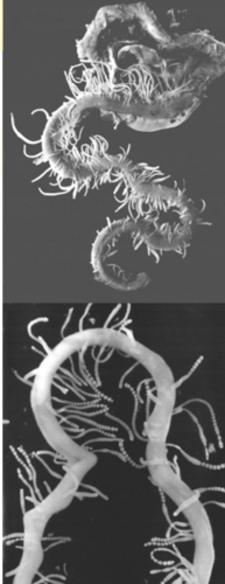
Tapeworms in poultry
Location: most species locate in the small intestine
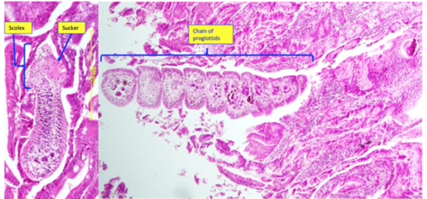
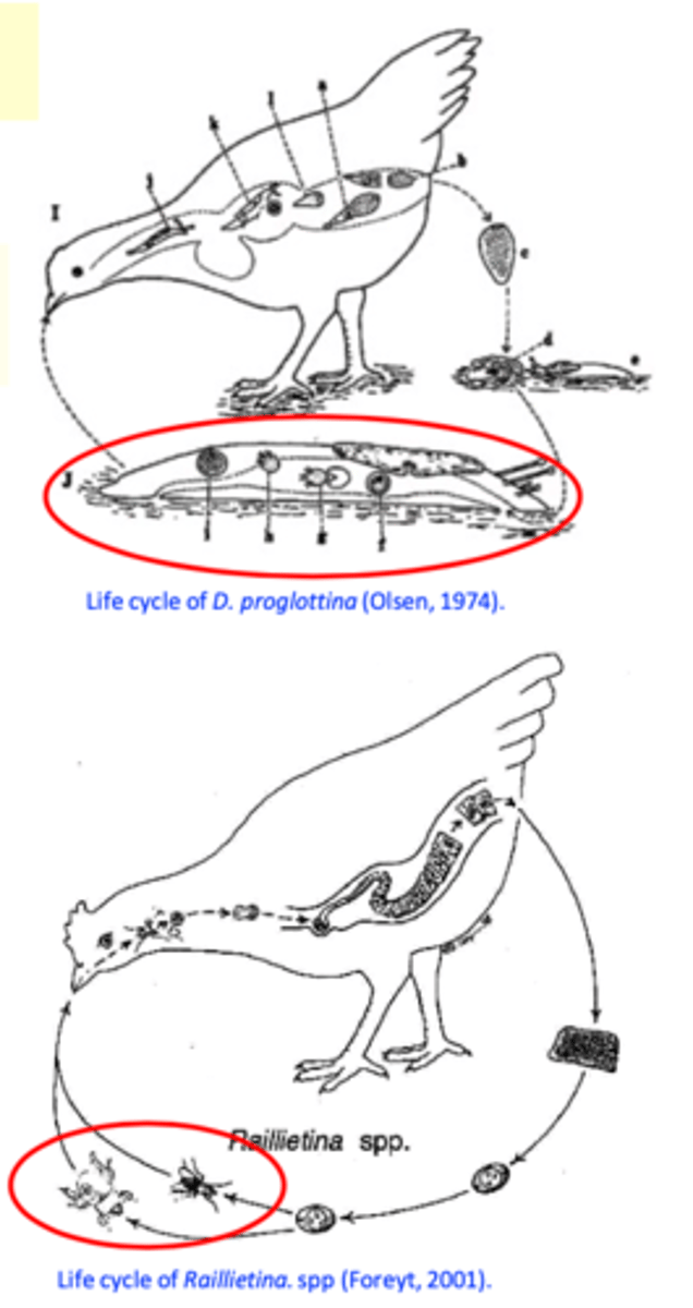
Tapeworms in poultry: Life cycle
• Various intermediate hosts (IH): houseflies, beetles, ants, snails, slugs etc;
• Gravid proglottids are passed in the faeces of birds -> proglottids/eggs ingested by IH;
• Infection of the birds: ingestion of IH;
PPT: 2-3 weeks
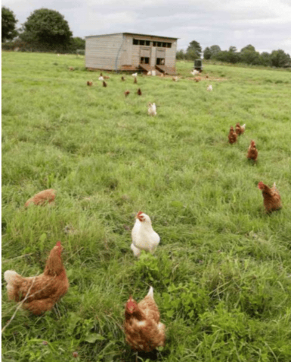
Tapeworms in poultry - Epidemiology
• Commonly occurs only in poultry reared in outdoor systems
In temperate areas infections are more common in warmer seasons
Birds younger than 3 months are more susceptible;
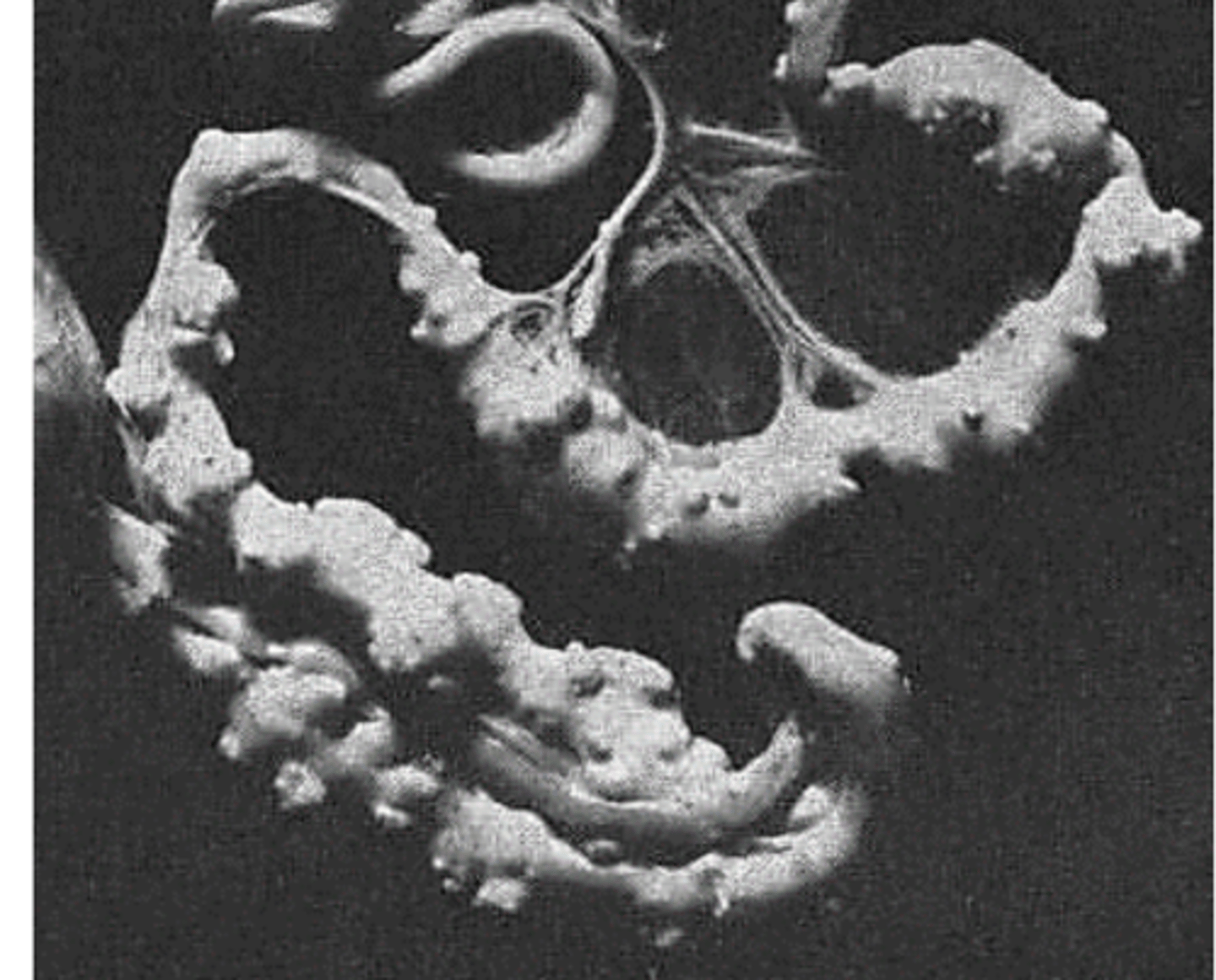
Tapeworms in poultry
Pathology/Clinical signs
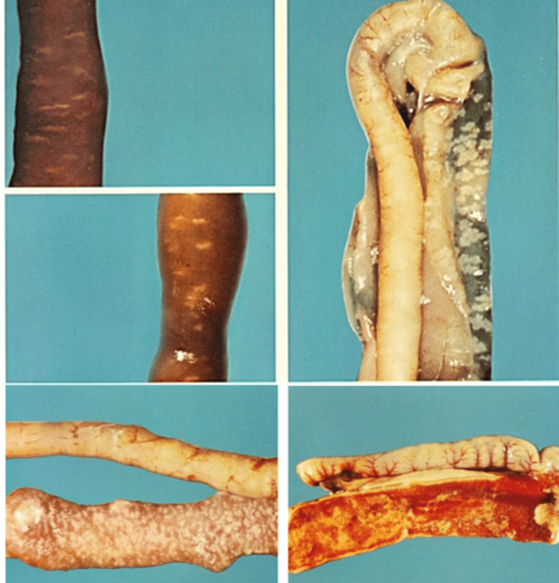
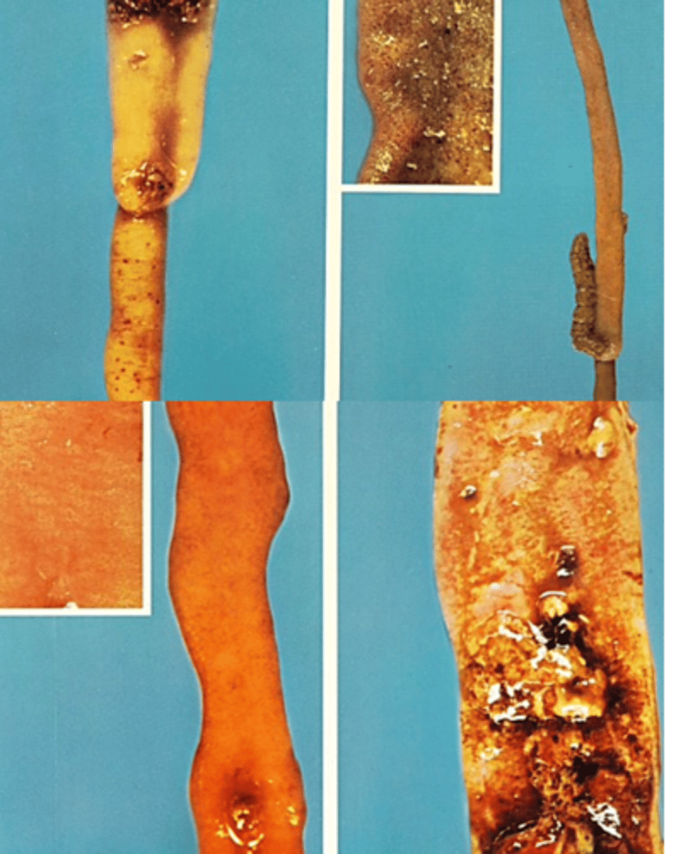
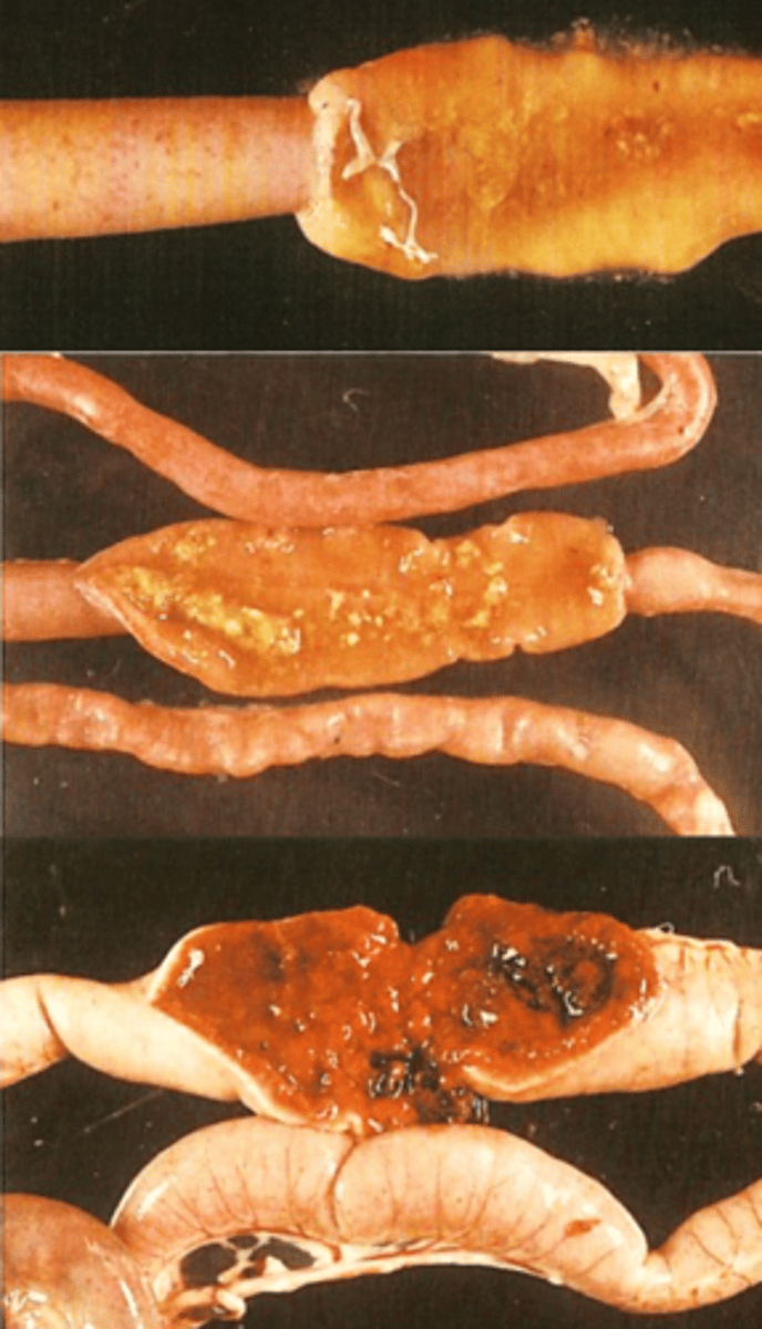
Raillietina echinobothrida is causing nodules at the attachment site;
Tapeworms in poultry
Diagnosis: live birds
• Clinical signs: not specific
• History
• Finding proglottids/eggs in the faeces
Tapeworms in poultry
Diagnosis: dead chickens
• Necropsy
• Histopathology
Tapeworms in poultry
Treatment and control
Flubendazol: food
Prevent contact with IH
Eliminate IH
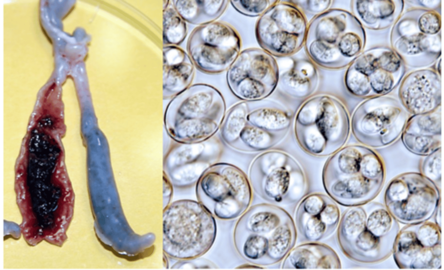
Poultry Coccidiosis/Eimeriosis
Poultry Coccidiosis/Eimeriosis DESCRIPTION
Coccidiosis is the most important protozoan diseases of chickens,
In chickens seven species of Eimeria have been described:
Develop at specific sites along the intestine;
Have different degrees of pathogenicity;
Short lifecycle and high reproductive potential -> severe outbreaks.
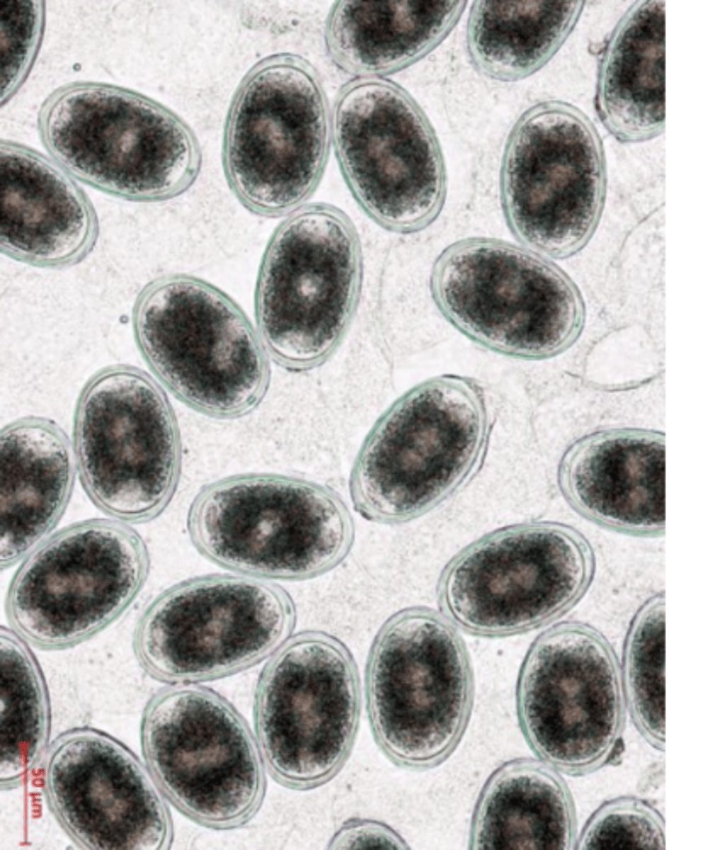
Poultry Coccidiosis/Eimeriosis
Morphology & life cycle
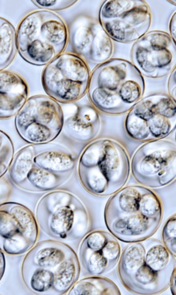
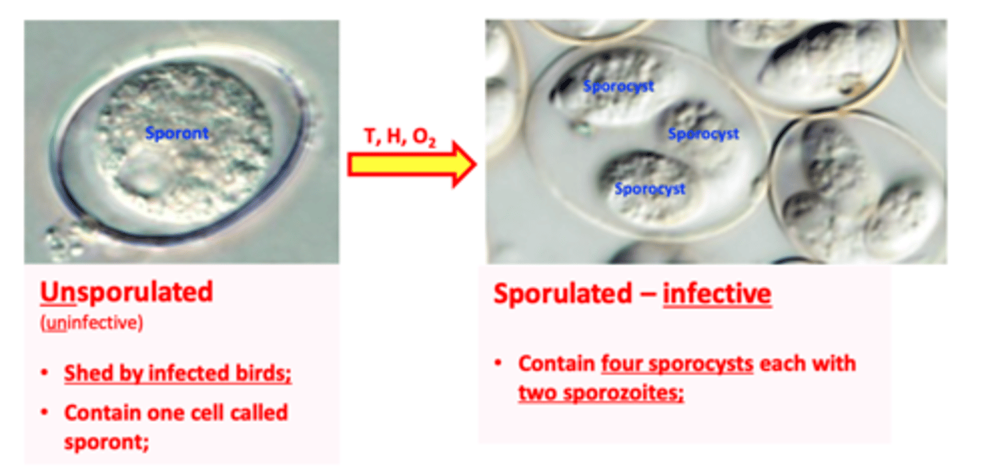
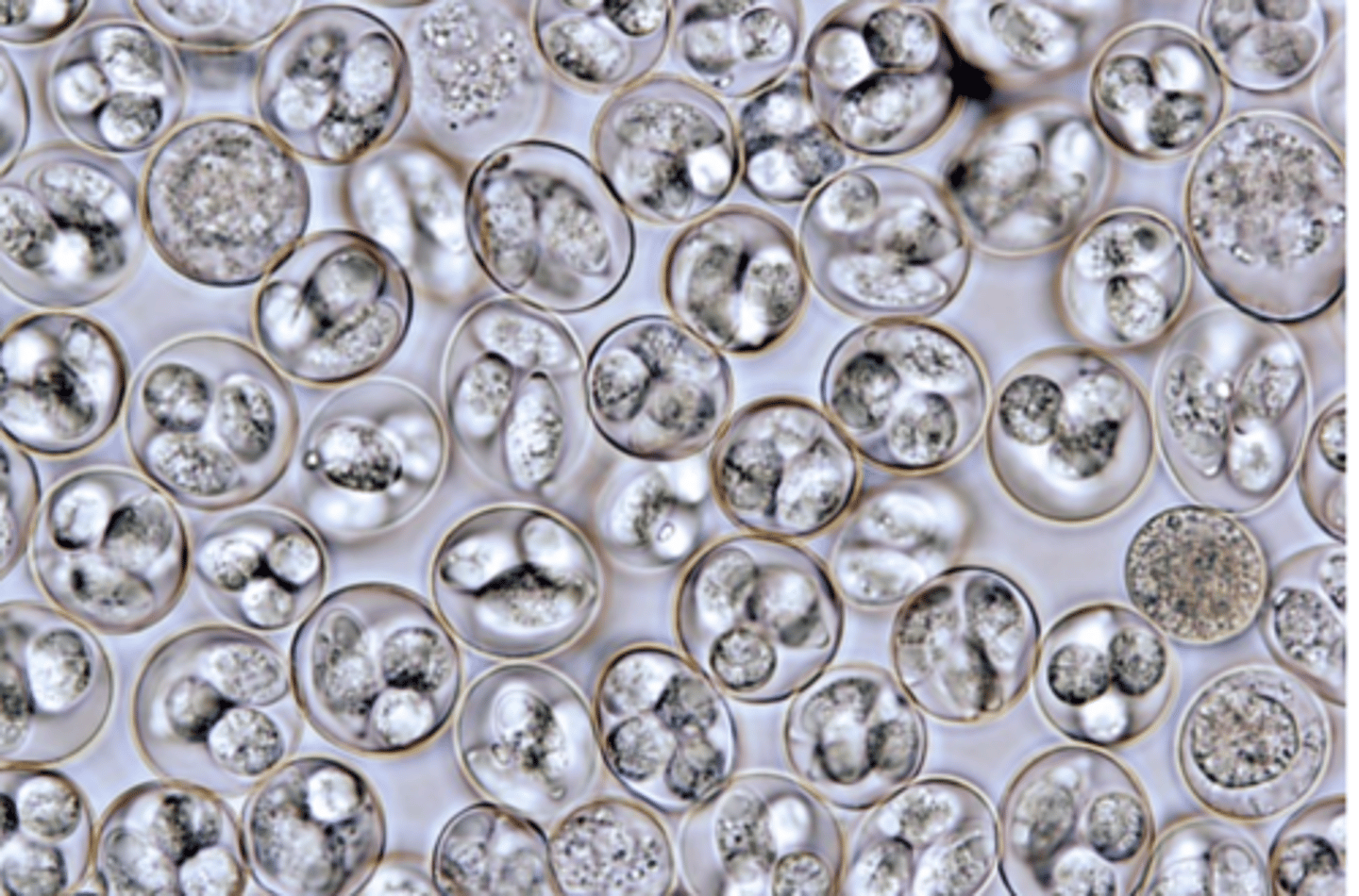
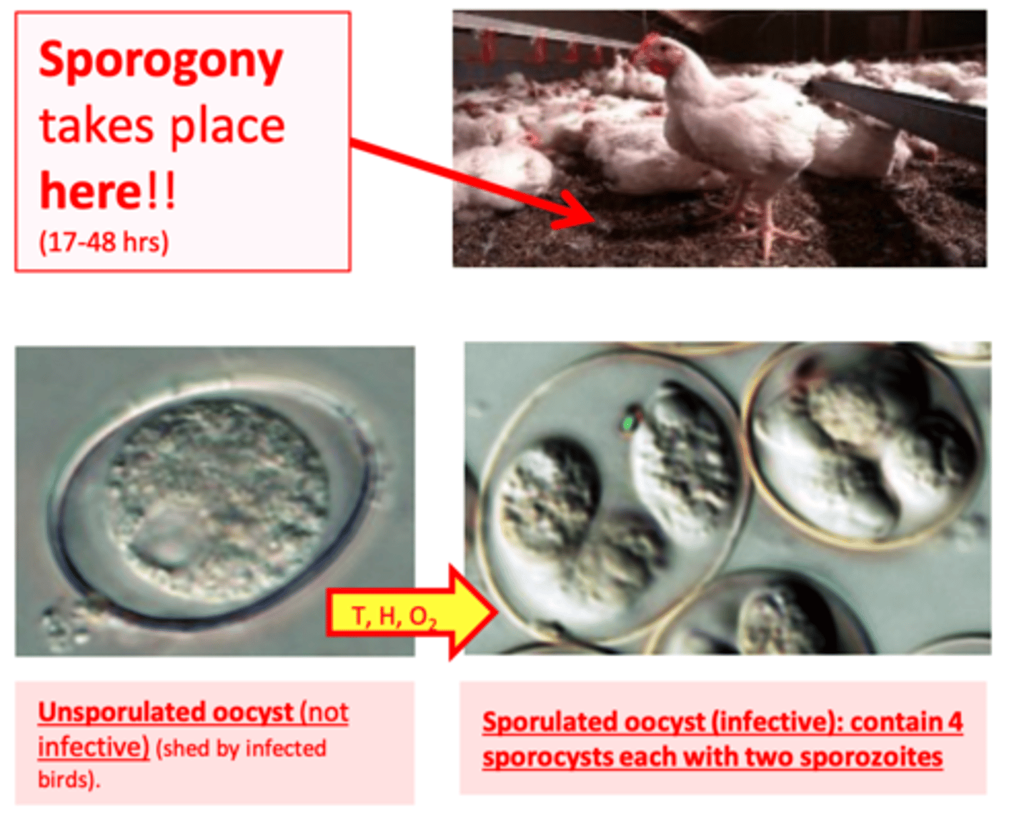
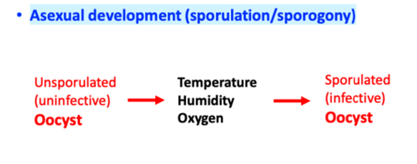
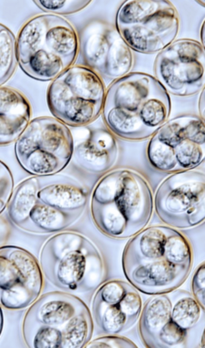
Oocyst: unsporulated -> sporulated
Poultry Coccidiosis/Eimeriosis
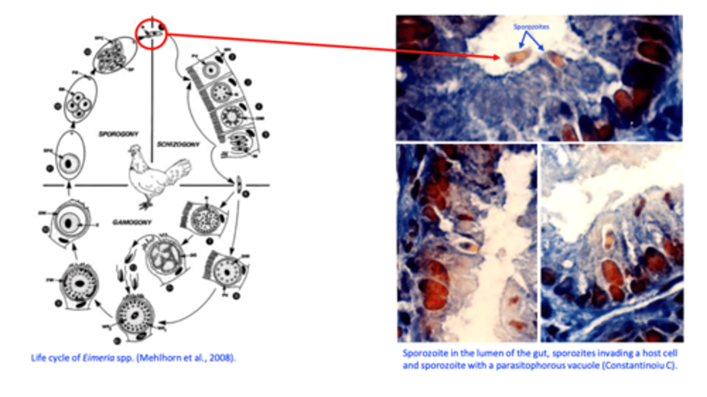
Sporulated oocysts: infective stage
Hosts become infected after ingestion of sporulated oocysts: infective stage
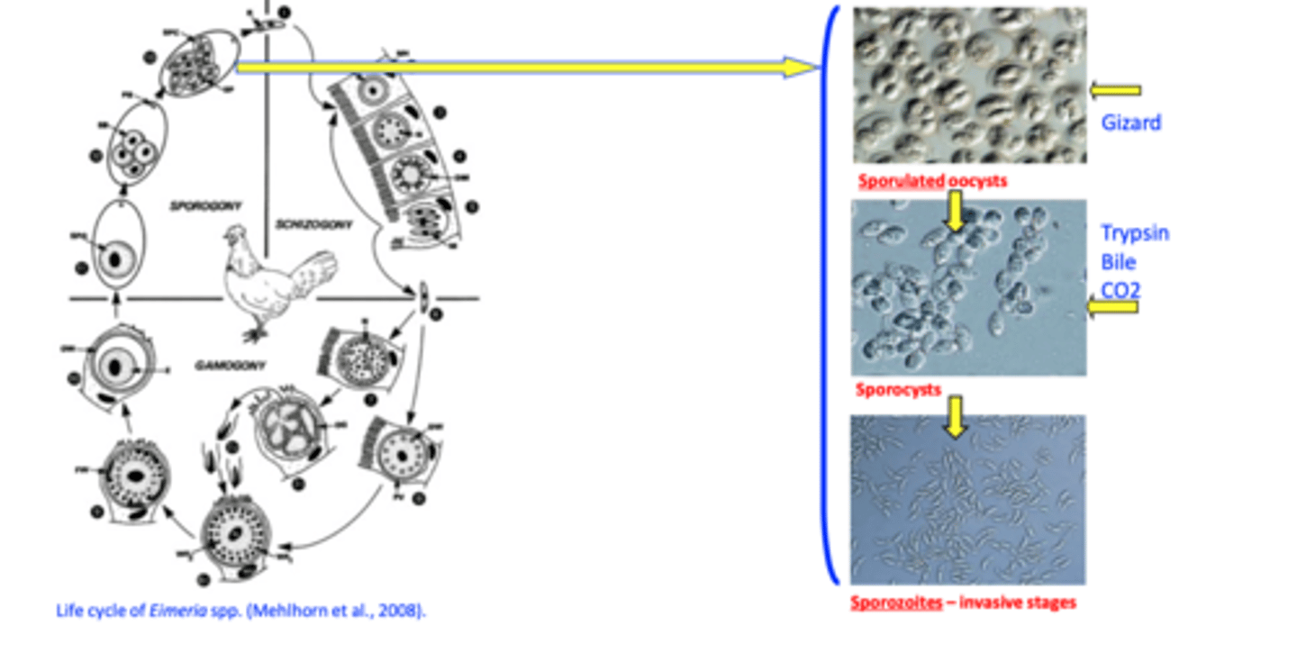
Sporozoites: invasive stage
Banana shaped cells
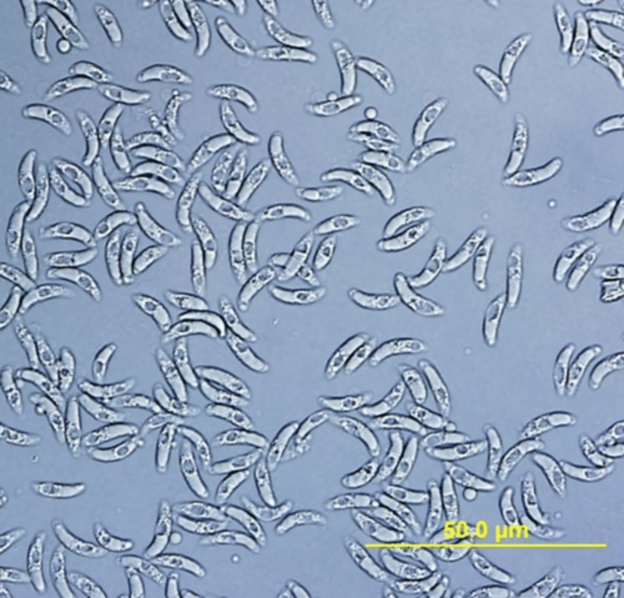
Sporozoites invade the host cells
Poultry Coccidiosis/Eimeriosis
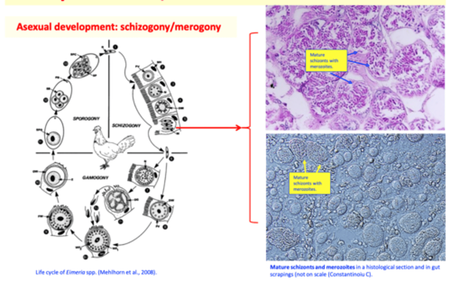
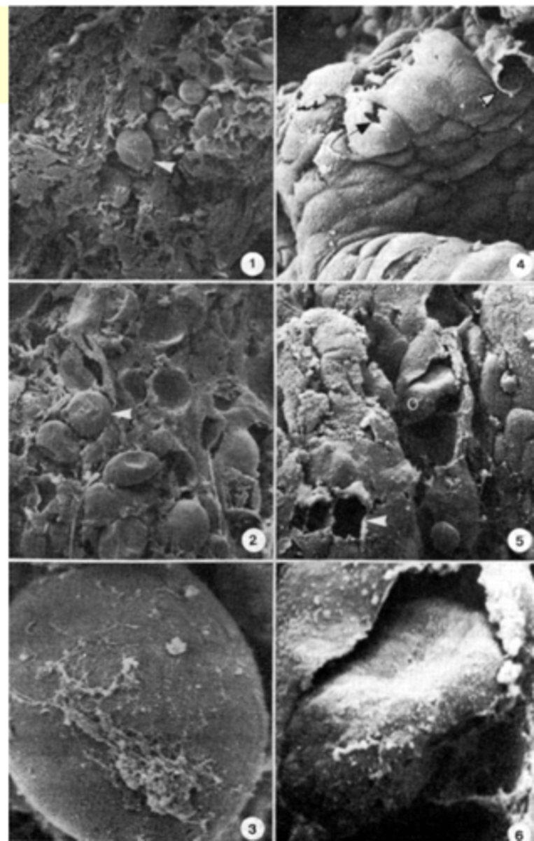
Asexual development: schizogony/merogony
Trophozoite: after invasion of the host cells the sporozite grows and turns into a trophozoite (rounded shape);
Immature schizont: the nucleus of the trophozoite divides -> immature schizont;
Mature schizont: the cytoplasm divides and merozoites are formed -> mature schizont;
Merozoites: similar structure to sporozoites;
• Will break out the schizonts and host cell;• Invade other host cells;• Resume the schizogonic development
Eimeriosis Merozoites: invasive stage
Poultry Coccidiosis/Eimeriosis
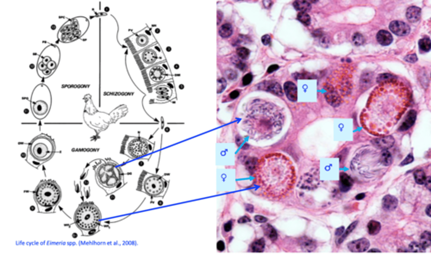
Sexual development: gametogony/gamogony
After ‘n’ schizogonic cycles the merozites invade other host cells and begin the sexual development
Merozoites will invade the host cells, grow and turn into either a:
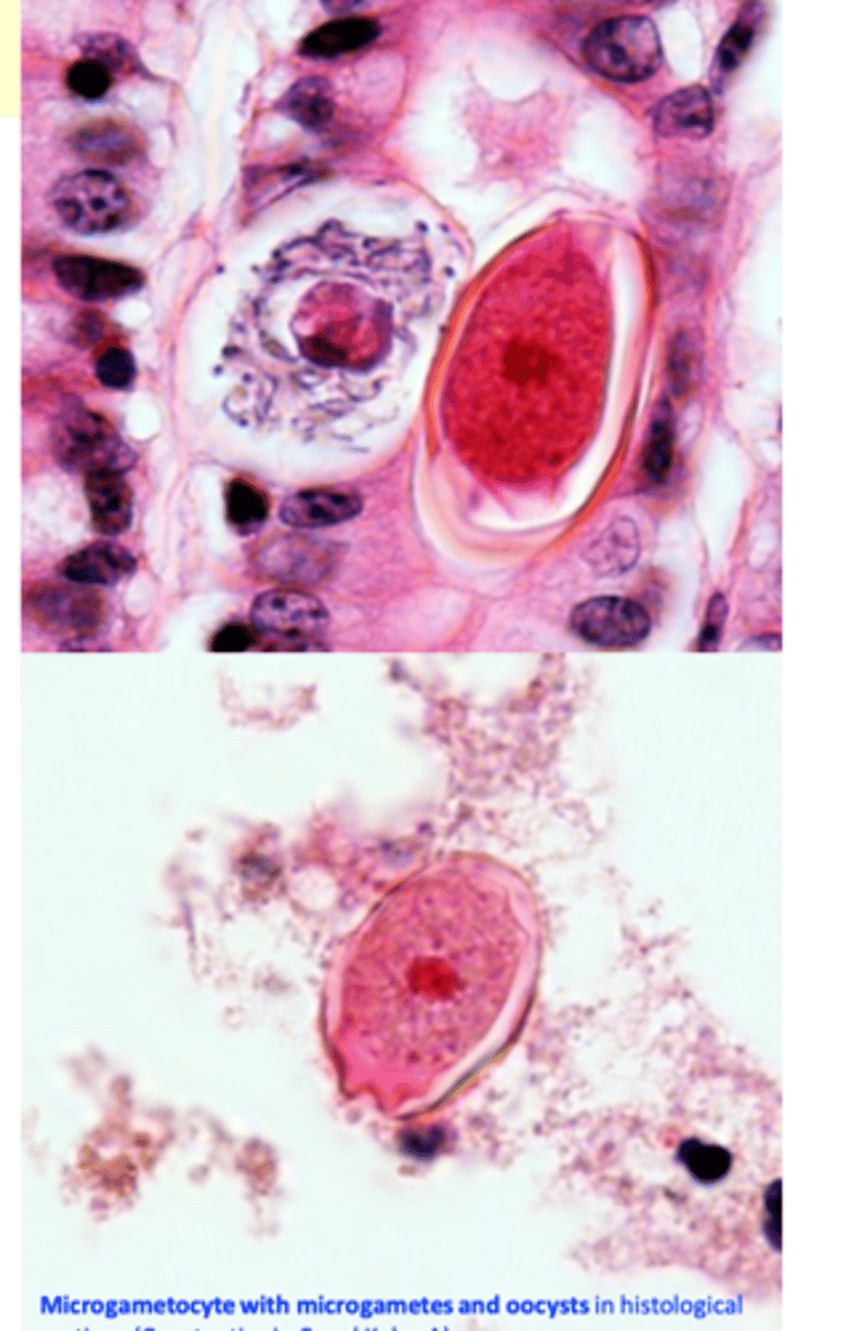
a) Microgametocyte (male) that will give rise to
many microgametes break the host cells and get into the lumen of the gut;
b) Macrogametocyte (female) that will give rise to a macrogamete;
Sexual development: EIMERIOSIS
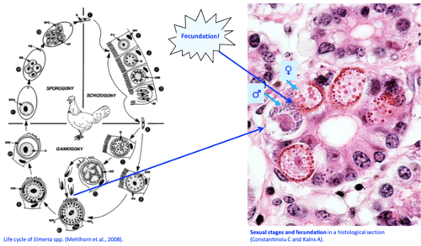
Poultry Coccidiosis/Eimeriosis
Fecundation
Poultry Coccidiosis/Eimeriosis
Macrogametes turn into oocysts
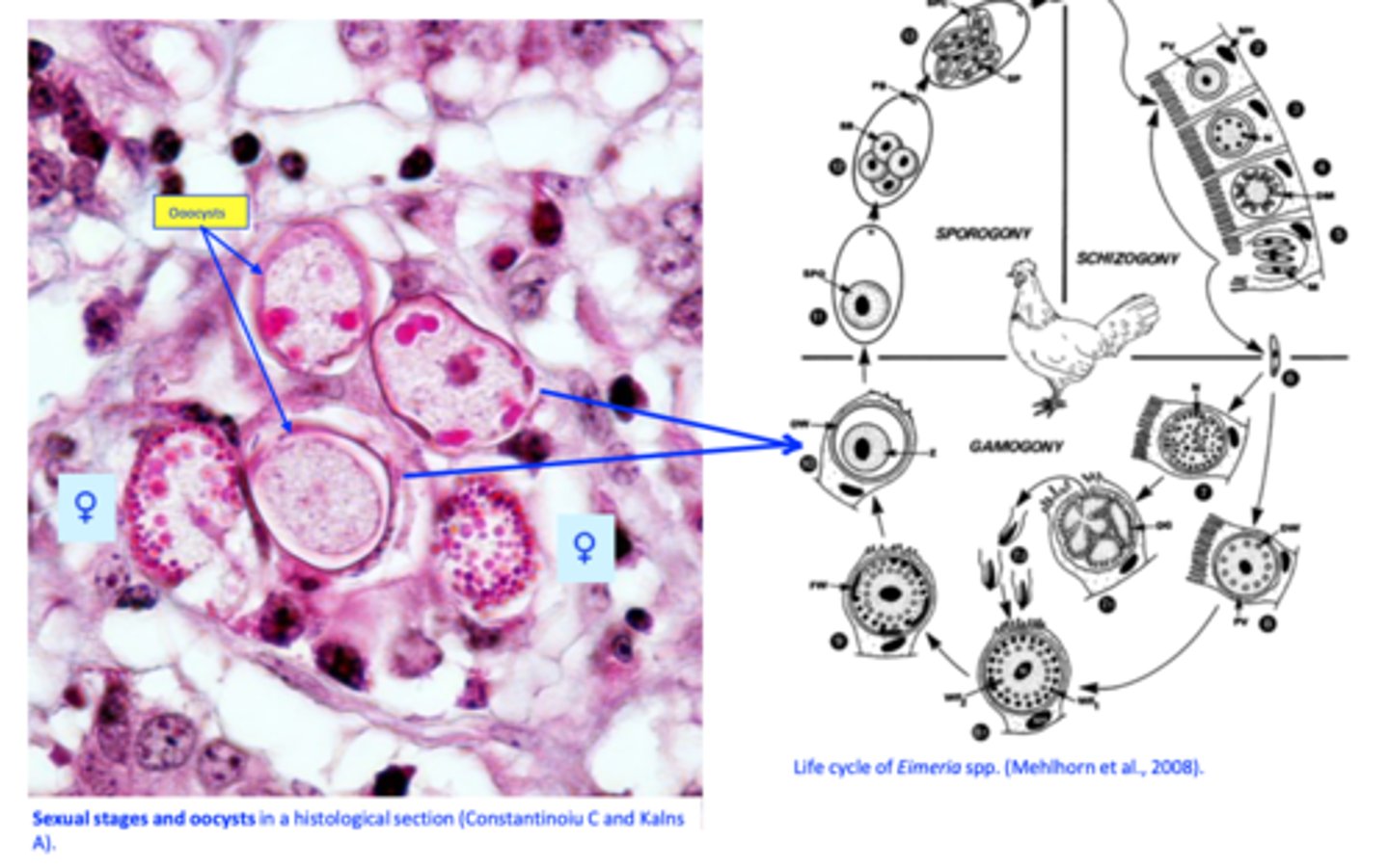
Poultry Coccidiosis/Eimeriosis
Oocysts are shed in the environment
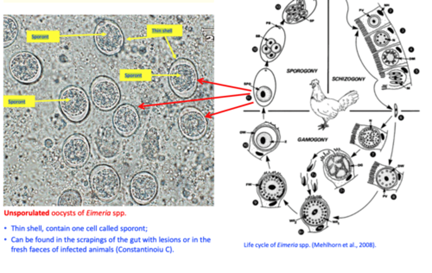
Poultry Coccidiosis/Eimeriosis
Sporulation/sporogony
The process by which the sporozoites are formed within oocysts and the oocysts become infective;
• Takes place in the environment;
• Depends on:
Temperature
Humidity
Oxygen
Eimeria life cycle: summary
Infection of the host: ingestion of sporulated oocysts.
1. Development within the host
2. Development in the environment
Poultry Coccidiosis/Eimeriosis
Epidemiology
Eimeria tenella
Eimeria necatrix
Locate in different segments of the intestine
Poultry Coccidiosis/Eimeriosis
Prevalence
• Chicken coccidiosis is spread all over the world
Common and causing significant losses in birds reared in farms on the floor but also in birds reared in outdoor systems
Outbreaks:
- Crowded of birds;
- Environment
Poultry Coccidiosis/Eimeriosis
Susceptibility
Severe cases common in chickens 3-6 weeks old
Poultry Coccidiosis/Eimeriosis
Sources of infection
Sick chickens with clinical coccidiosis that shed large numbers of oocysts/day
Chickens/adult birds with subclinical infections;
Sources of infection
Oocysts
Sources of infection
Oocysts
Resistant to the common disinfectants when used at usual concentrations but destroyed by ammonia and high temp
Might be transported by mice, rats, flies, boxes for eggs, tools etc.
Are introduced into new farms through contaminated equipment, vehicles, personnel etc.
Infection of the birds - eimeria
Ingestion of sporulated oocysts with water and food.
Pathogenesis - eimeria
• Species and strain
E. tenella and E. necatrix are the most pathogenic;
Dose and rate of infection
Infections with high numbers of oocysts in short time -> disease.
Pathogensis depends on:
Age, breed, immunologic experience of the host;
Pathogenesis
Eimeria and bacteria
Bacteria that normally reside in the digestive tract are required for production of the lesions specific to Coccidiosis;
Pathogenesis
Eimeria can cause:
Rupture the cells they develop -> villi atrophy, epithelial sloughing -> products from the gut can get into the blood;
Produce toxins that are released into the tissue, especially when schizonts break out;
Cause decreases in plasma carotenoids => depigmentation of carcasses => reduced value
Eimeria IMMUNITY
Protective immunity is species specific;
Size of inoculum and rate of infection:
In the farms: oocysts re-circulate in the floor => birds build up strong immunity;
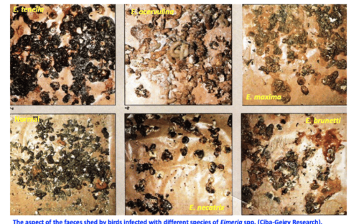
eimeria acervulina pathology
Eimeria acervulina
Locates in the duodenum
Scattered, white plaque-like lesions containing developing oocysts => in heavier infections lesions coalesce;
Intestinal wall is thickened;
Heavy infections => mucosa is bright red.
Eimeria maxima pathology
Locates in the jejunum;
Small red petechiae on the serosal surface;
Orange colored mucus or blood clots;
Eimeria necatrix PATHOLOGY
Locates in the jejunum and caeca;
2nd generation schizonts are the pathogenic stage;
White plaques or petechiae may be visible from the serosal surface;
Intestinal content streaked with blood;
Ballooning, hemorrhages;
On gut scrapings of the jejunum: large schizonts (60μm) that are diagnostic for this species;
No oocysts in the jejunum
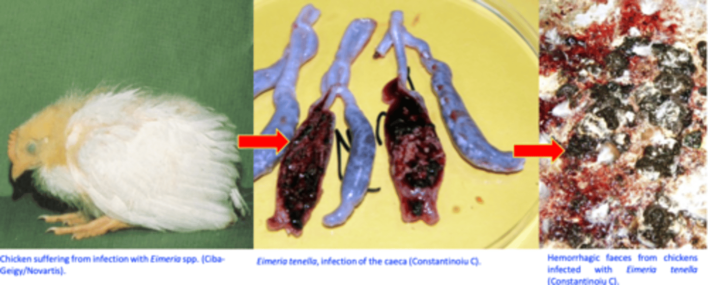
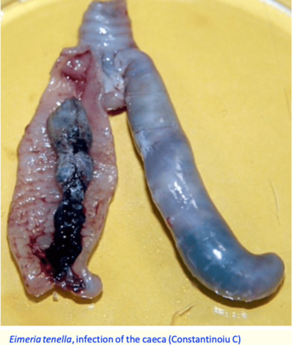
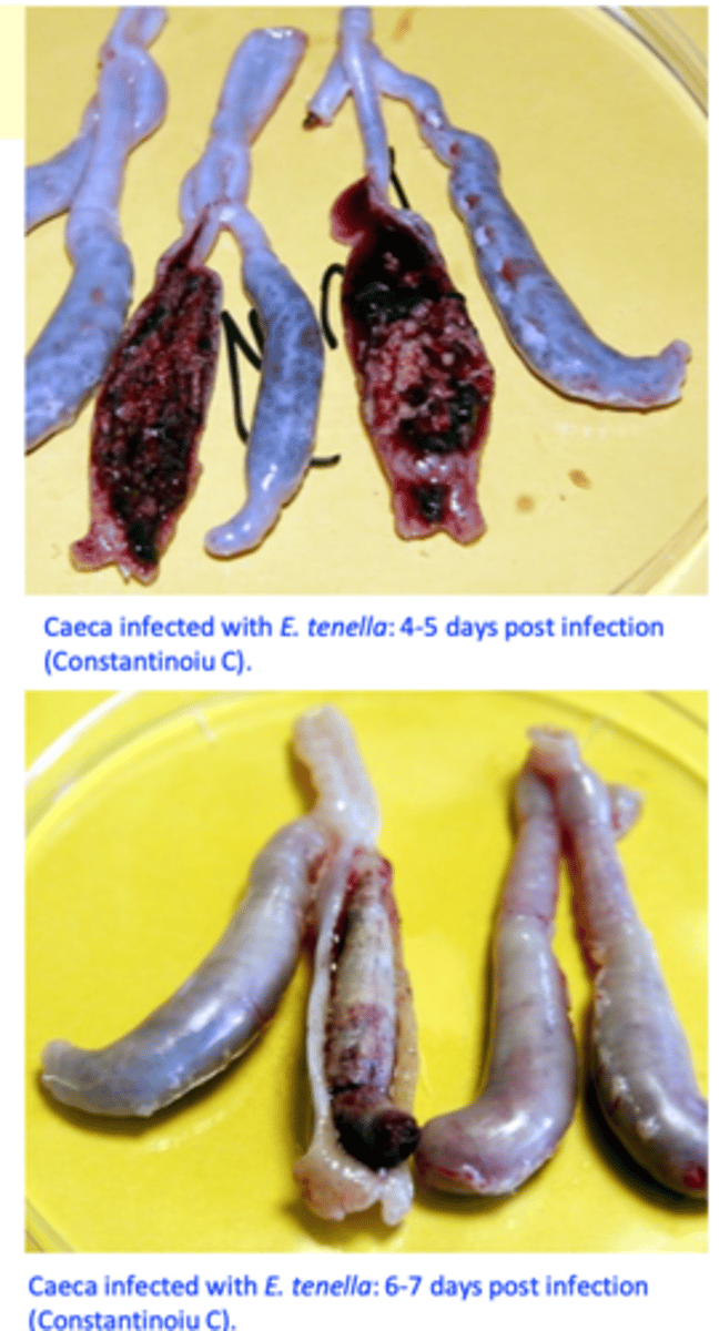
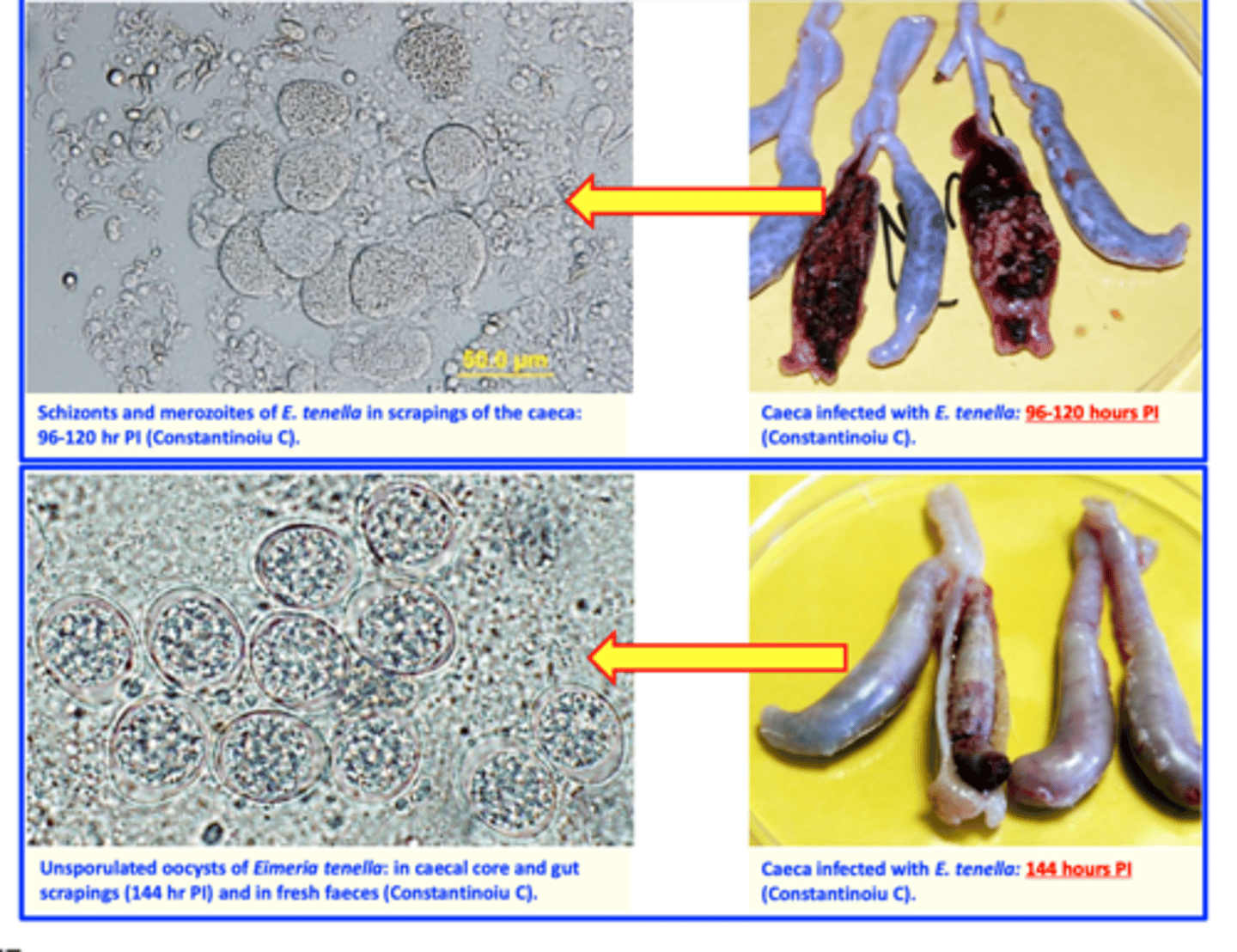
Eimeria tenella PATHOLOGY
Locates in the caeca;
2nd generation schizonts are the pathogenic stage;
Scattered pettechiae on the cecal wall;
Bleeding with clotting; caeca are much distended;
If the birds do not die the clot hardens as the mucosa cells join the blood to make a core;
Cecal wall is thick, wall greatly distended containing blood and/or large caseous cores;
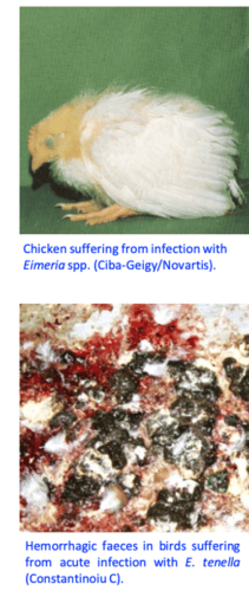
Clinical coccidiosis
Common in infections with E. tenella or E. necatrix;
Cease to feed and drink but later they show polydipsia;
The comb and gills are pale and atrophied;
Diarrhoea: with blood for E. tenella and E. necatrix;
Subclinical coccidiosis
Birds do not show obvious clinical signs but their performance is affected;
Difficult to diagnose
Clinical signs
Diagnosis
Live birds
- History
- clinical signs
- ID of parasites
-oocysts in faeces
Unsporulated oocysts
Diagnosis
dead birds
Pathology
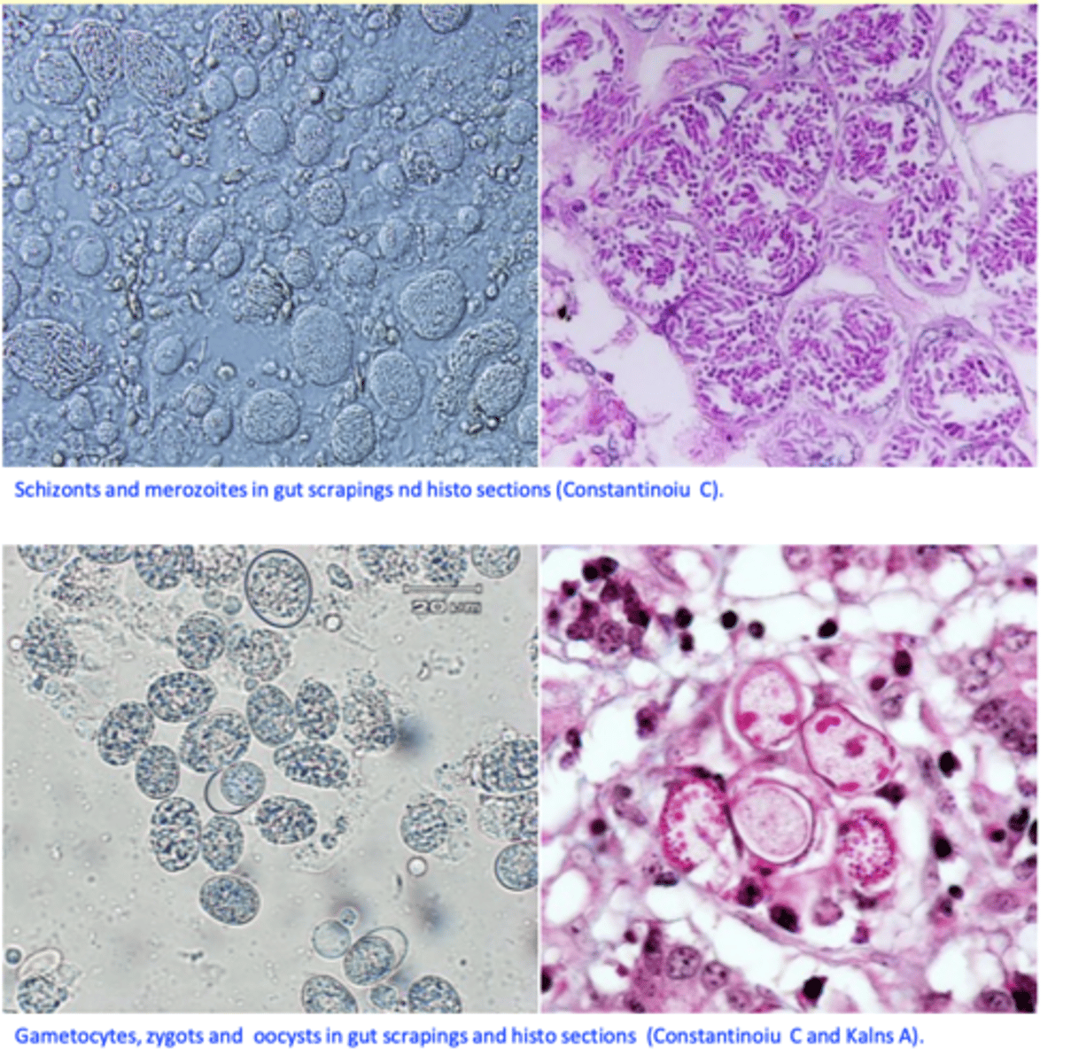
• Identification of the parasites:
• Schizonts, merozoites, gametocytes, oocysts in gut scrapings or histological sections;
Treatment & control
Curative treatment is often too late!
1) Sulfonamides
2) Amprolium
3) Toltrazuril - Plus vitamin A, Vitamin K and antibiotics?
Prevention
a) Hygiene and poultry house management
b) Chemoprevention:
c) Vaccination
• Live vaccines
Prevention
Broilers
A. Usually, anticoccidial drugs are given in the feed from one day of age until 5-7 days to market
• Two programs are common:
Shuttle programs: more common; two or more anticoccidials are used in successive stages
Straight programs: single anticoccidial is used throughout.
B. Live, attenuated vaccines
Anticoccidials used in the poultry industry
Ionophorous antibiotics
Amprolium
Nitrobenzamides
Clopidol and quinolines
Robenidine
Halofuginone
Triazines
Prevention
Breeder and replacement birds
Development of immunity is desirable;
Either vaccinate the flocks or allow development of the immunity under anticoccidial treatments;