L26: abdominal ultrasound
 Call Kai
 Learn
Learn Practice Test
 Spaced Repetition
 Match
Match Flashcards
 Knowt Play
1/83

There's no tags or description
Looks like no tags are added yet.
Name | Mastery | Learn | Test | Matching | Spaced | Call with Kai | Chat |
|---|
No analytics yet
Send a link to your students to track their progress
84 Terms

what structure is circled in pink?
liver
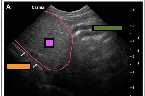
what structure is next to the green box?
duodenum
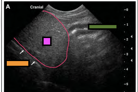
what structure are the white arrows pointing at near the orange box?
diaphragm
how is the echogenicity of the liver compared to the spleen?
uniformly hypoechoic with a coarser echotexture
how is the echogenicity of the liver compared to the renal cortices?
hyperechoic to isoechoic

what is the yellow arrow pointing to?
portal veins of the liver

what structure is the pink box in?
spleen

what structure is the orange box in?
liver
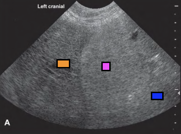
what structure is the blue box in?
fat
the liver should not cover more than ____ of the right kidney on ultrasound
one third

what structure is the blue box in?
liver

what structure is the green box in?
right kidney
how does the gallbladder appear on ultrasound?
anechoic teardrop structure with conical extension (cystic duct)

what structure is the yellow circling?
gallbladder

what is the purple circling?
biliary sludge in the gallbladder (normal in dogs)
which animal can the gallbladder occasionally be bilobed?
cats
how can you tell on ultrasound that there is hepatomegaly?
tips of liver lobes are ROUNDED
liver extends beyond the kidneys
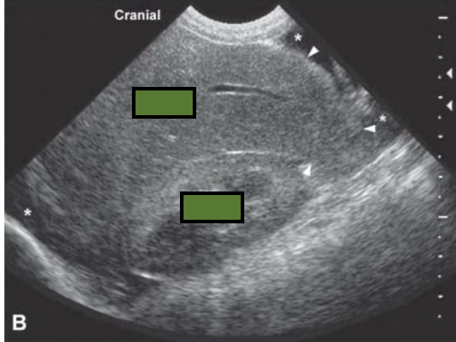
what is occurring in the following ultrasound?
hepatomegaly

what is occurring in the following ultrasound?
diffuse hypoechoic liver
what are the causes for a diffuse hyperechoic liver?
lipidosis
lymphoma
cirrhosis
what are the causes for a diffuse hypoechoic liver?
acute hepatitis
passive congestion
what are the causes for focal liver nodules?
nodular hyperplasia
extra-medullary hematopoiesis
metastatic disease
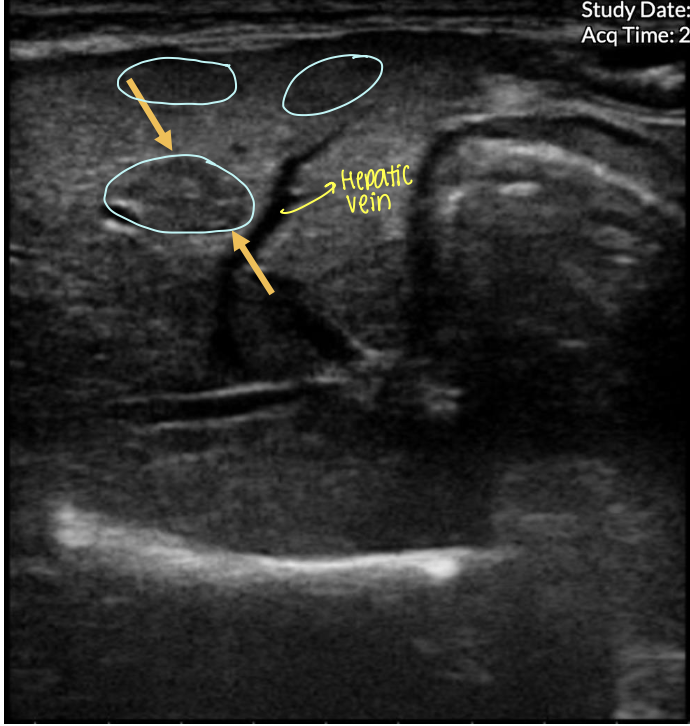
what is occurring in the following ultrasound?
focal liver nodules

This is a longitudinal scan of the liver from an 8-year-old MN MBD
diagnosed with lymphoma. What are the ultrasonographic findings?
diffused hypoechogenicity
where is the head of the spleen located?
left cranialdorsal abdomen often forming a hook between gastric fundus and left kidney
what drugs can effect the size and echogenicity of the spleen?
ketamine
diazepam
acepromazine
alpha-2 agonists
how are the branches of the splenic vein seen on ultrasound?
tubular anechoic structures

what structure is outlined in orange?
spleen
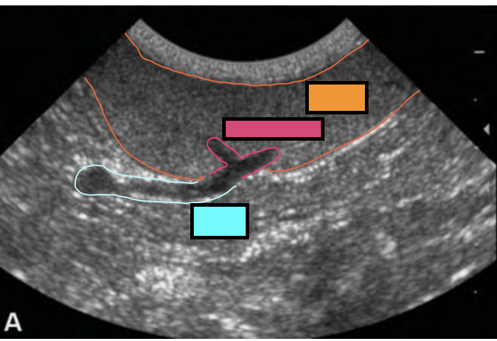
what structure is outlined in pink?
splenic hialus

what structure is outlined in blue?
splenic vein
how does the echogenicity of the spleen compare to the liver and renal corticies?
homogenous and fine echotexture covered in thin hyperechoic capsules
how does the echogenicity of the spleen compare to the renal cortex in cats?
isoechoic or mildly hyperechoic

what structure are the white arrows pointing to?
spleen

what is occurring in the following ultrasound?
splenomegaly

what is occurring in the following ultrasound?
multiple hypoechoic nodules on the spleen
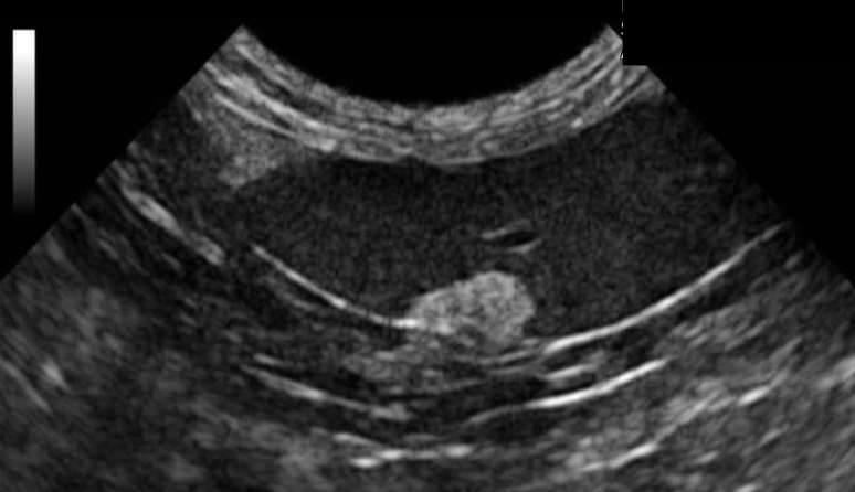
what is occurring in the following ultrasound?
hyperechoic nodules on the spleen
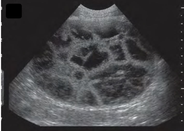
what is occurring in the following ultrasound?
heterogenous mass on spleen
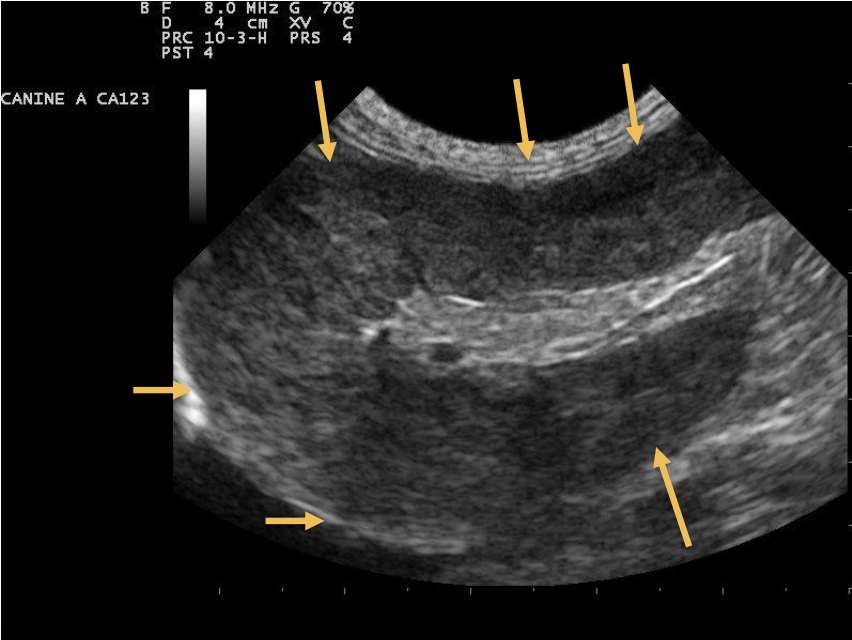
This is a long scan of the spleen of a 13-year-old FS Siamese cat presenting with
a history of abdominal distesion and anorexia. What are the ultrasonographic
findings?
diffused hypoechogenicity
which animal has a more oval shaped kidney?
cats
which animal has a more bean shaped kidney?
dog
what is the renal length in cats?
3-4.5 cm
what is the echogenicity of the renal medulla compared to the cortex?
hypoechoic
what is the echogenicity of the renal cortex compared to the liver?
hypoechoic or isoechoic
what is the echogenicity of the renal cortex compared to the spleen?
hypoechoic
what is the echogenicity of the renal pelvis?
anechoic
what is the height of the renal pelvis in the transverse plane?
2mm
what is the echogenicity of the renal sinus compared to the renal pelvis?
hyperechoic

blue box
renal pelvis

pink box
renal sinus
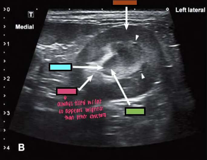
green box
renal crest

orange box
renal cortex

what is seen in the ultrasound of the kidney?
medullary rim sign
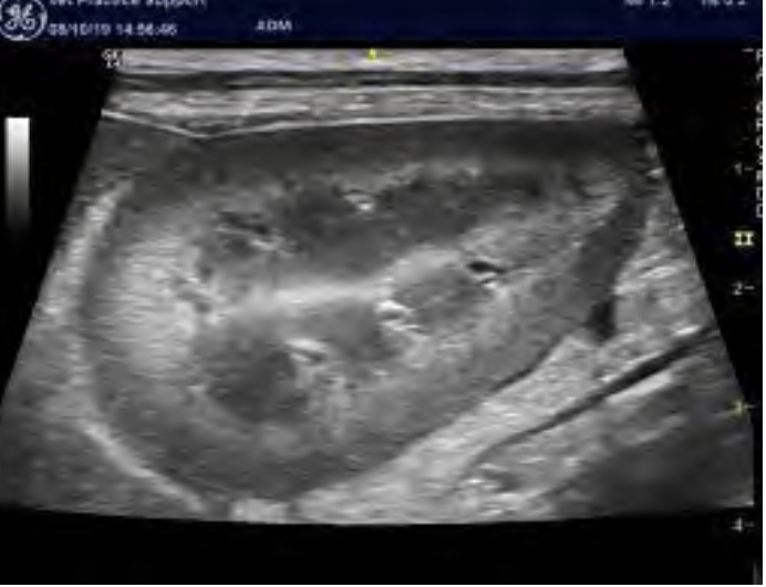
what is seen in the following ultrasound?
diffuse hyperechoic renomegaly
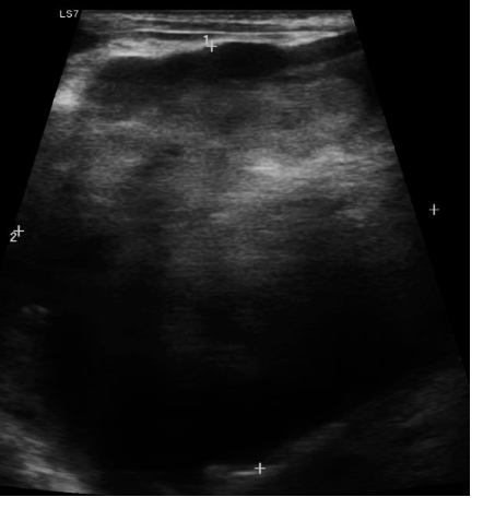
what is seen in the ultrasound of the kidney?
hypoechoic mass on cranial pole of kidney

what is seen in the ultrasound of the kidney?
perinephric pseudocyst

what is seen in the ultrasound of the kidney?
polycystic kidney disease

what is seen in the ultrasound of the kidney?
hydronephrosis

what is seen in the ultrasound of the kidney?
chronic kidney disease in cat

These are longs scans for the left and right kidney of a 7-year-
old DSH with a history of azotemia. What are the
ultrasonographic findings?
cystic structures within the renal cortices
what is the thickest portion of the small bowel in dogs?
duodenum
what are the layers of the GI starting from the lumen?
hyperechoic mucosal interface
anechoic mucosa
hyperechoic submucosa
anechoic muscular layer
hyperechoic serosa

what does the following ultrasound show?
transverse view of the stomach
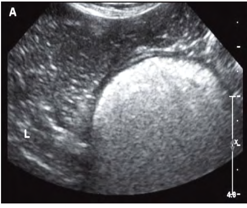
what does the following ultrasound show?
gas in stomach

what does the following ultrasound show?
food in the stomach

what does the following ultrasound show?
jejunum
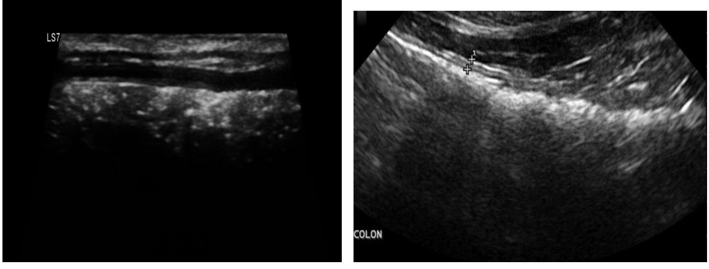
what does the following ultrasound show?
colon
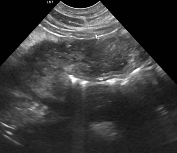
what does the following ultrasound show?
severe stomach wall thickening

what does the following ultrasound show?
severely thickened duodenum
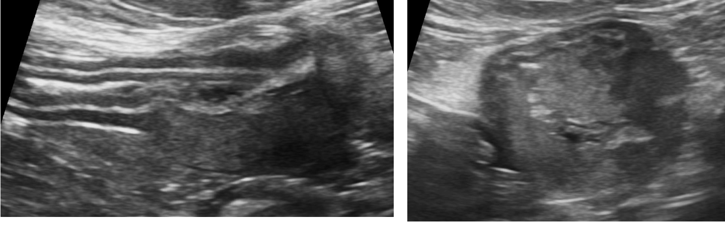
what does the following ultrasound show?
severely thickened jejunum → neoplasia

what does the following ultrasound show?
linear foreign body
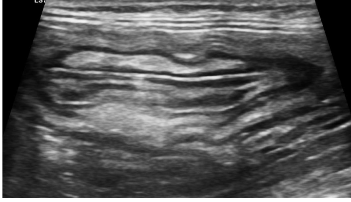
what does the following ultrasound show?
intussusception

what does the following ultrasound show?
intussusception

what does the following ultrasound show?
intussusception
what is the echogenicities of the layers of the bladder wall?
hypoechoic mucosa
hyperechoic submucosa
hypoechoic muscularis
hyperechoic serosa

what does the following ultrasound show?
bladder thickening with a luminal mass
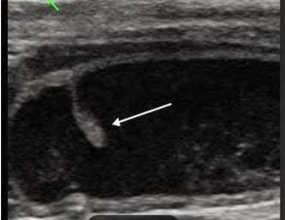
what does the following ultrasound show?
polypoid in bladder

what does the following ultrasound show?
cystitis
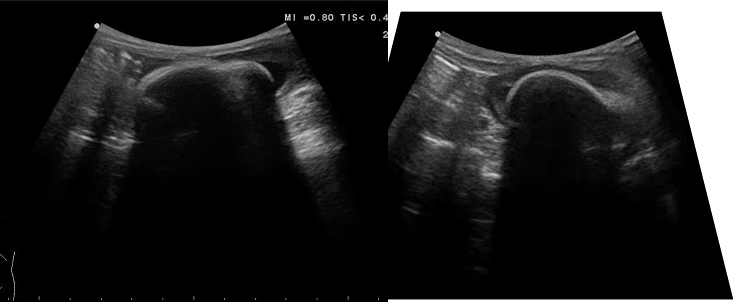
what does the following ultrasound show?
bladder calculi
what is the shape of the prostate in intact male dogs on sagittal images?
rounded to ovoid
what is the shape of the prostate in intact male dogs on tranverse images?
two prostatic lobes are symmetrical
how does the prostate appear in neutered dogs?
small hypoechoic and homogenous

what does the following ultrasound show?
benign prostatic hyperplasia

what does the following ultrasound show?
prostatic cyst
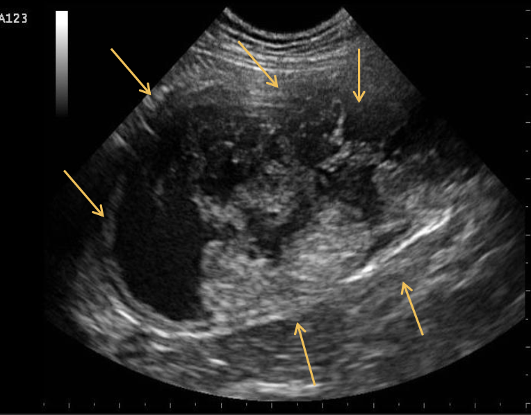
This is a 14-year-old, neutered male Malinois presenting with a history
of hemangiosarcoma of the tongue and scrotum. The patient also
underwent a splenectomy for the same reason a year ago. Currently
the patient is showing hematuria. What does the following longitudinal
scan of the urinary bladder show?
bladder mass