Looks like no one added any tags here yet for you.
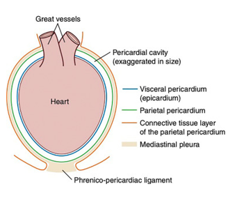
Pericardium Anatomy
Attached to?
Comprised of?
Contains?
Purpose?
Attached base of large vessels (aorta, PA)
Sac that covers heart
Comprised of 2 layers
Parietal (facing outwards)
Visceral (facing heart)
•Small amount of fluid in sac (0.3-1ml) – lubcricant
•Prevents over stretch of heart
Diseases of pericardium?
Pleural space disease
Fluid
Herniation
Restrictive Pericarditis (rare)
What is this?
Features?
Congenital pericardial diseases
Peritoneopericardial Diaphragmatic Hernia (PPDH)
•Communication between pericardium and peritoneum
•GI contents in pericardial space
PPDH - presenting signs
•Tachycardia, coughing, respiratory distress, exercise intolerance
•Vomiting
•Anorexia
•Some are asymptomatic
PPDH - CX
•Muffled heart sounds
•“empty abdomen”
•Thoracic borborygmus
•Pectus excavatum - chest wall deformity that causes a sunken or caved-in appearance of the chest
•Tachycardia
PPDH DX
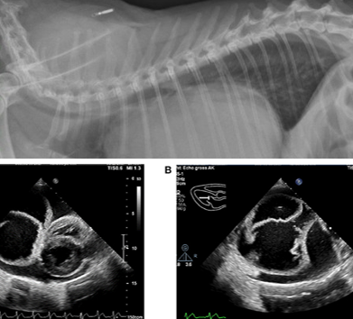
Radiographs
Might not be definitive if pericardial effusion - most cases dx with rads
Bowel loops in pericardial space
Cranial gastric axis displacement
Barium can confirm DX
Echocardiography
Pericardial effusion, bowels in pericardium
CT
DX but not usually necessary
PPDH - TX?
Cases with CX - surgery
Older cats - no clinical sign: may benefit from surveilance
Pericardial Effusion in dogs, what fluids we see?
•Blood
•Transudate
•Exudate
•Serosanguinous is most common
PE clinical consequences
Fluid accumulation in pericardium
Ventricles can’t fill → Diastolic failure → Cardiac tamponade and weak pulses
Venous return is reduced → venous distension
Congestion → Ascites / Liver distension
PE clinical presentation
Acute
•Collapse
•Tachycardia
•Pallour
Chronic
•Abdominal distension/ascites
•Exercise intolerance
PE physical exam signs
Beck’s triad
•Muffled heart sounds
•Jugular distension
•Weak pulses/BP
Tachycardia
Tachypnoea
Pale mucous membranes
Abdominal distension
Not all PE findings are present in all cases
PE - DDX
DCM
MVD
Liver disease (Ascites)
Sytemic illness
Respiratory disease
Heart sounds “muffled” in deep chested / overweight dogs
PE - dogs
Caused by?
Neoplasia
Haemangiosarcoma
Heart-base mass (chemodectoma)
Mesothelioma
Thyroid carcinoma
Idiopathic pericarditis (20-75% of cases)
Infective pericarditis (Rare)
PE TX?
•2 priorities
Emergency management
Oxygen
Anxiolytics
Emergency pericardiocentesis
Long term management
Idiopathic
Drainage
Partial pericardiectomy in recurrent cases
Neoplasia
Depends on type
PE TX - prognosis
•Depends on cause
•Idiopathic – good with a appropriate therapy
•Neoplasia – variable
PE in cats - Causes and tx?
•FIP - Antiviral drugs
•Lymphoma - Guarded prognosis
•Heart failure - Drain and rx heart failure