Ch 35 - Fetal Echocardiography
1/70
There's no tags or description
Looks like no tags are added yet.
Name | Mastery | Learn | Test | Matching | Spaced |
|---|
No study sessions yet.
71 Terms
When can fetal echo be performed transvaginally?
10-16 weeks
What is the optimum time for a complete anatomical fetal echo?
18-22 weeks
The most sensitive period in the first trimester for cardiac development is between _____
3.5 and 6.5 weeks
Cardiovascular system is the _____ organ system to reach functional state
first
By week ____, blood circulation begins
3
By week ____, heartbeat is seen
5
Primitive heart is a ______ structure that forms like a large blood vessel from _________ cells in cardiogenic area of embryo
tubular; mesenchymal
Paired _____ _____ _____ develop before the end of the _____ and begin to fuse, forming the primitive heart
endocardial heart tubes; 3rd week
Vascular system begins during the _____ week in the wall of the ____ _____, _____ _____, and ______
3rd; yolk sac; connecting stalk; chorion
The blood vessels begin to develop ____ after the vascular system
2 days
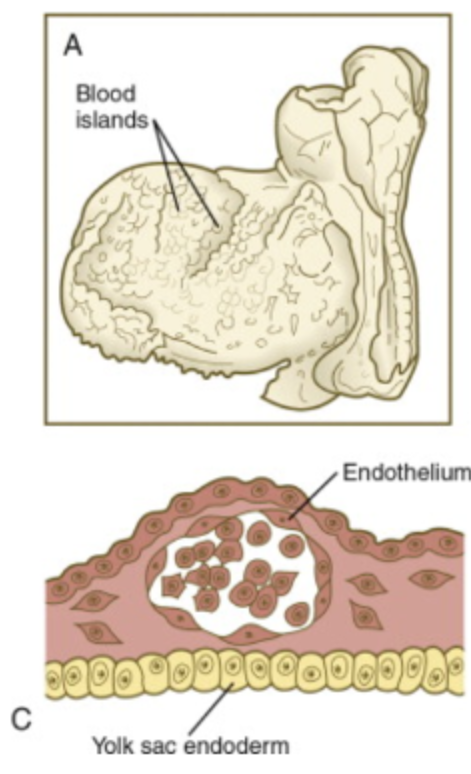
____ ____ are formed and cavities develop in the islands to form primitive ____ _____ and ______
Blood islands; blood cells; vessels
Primitive vessels form vascular networks in the wall of the ____ _____
yolk sac
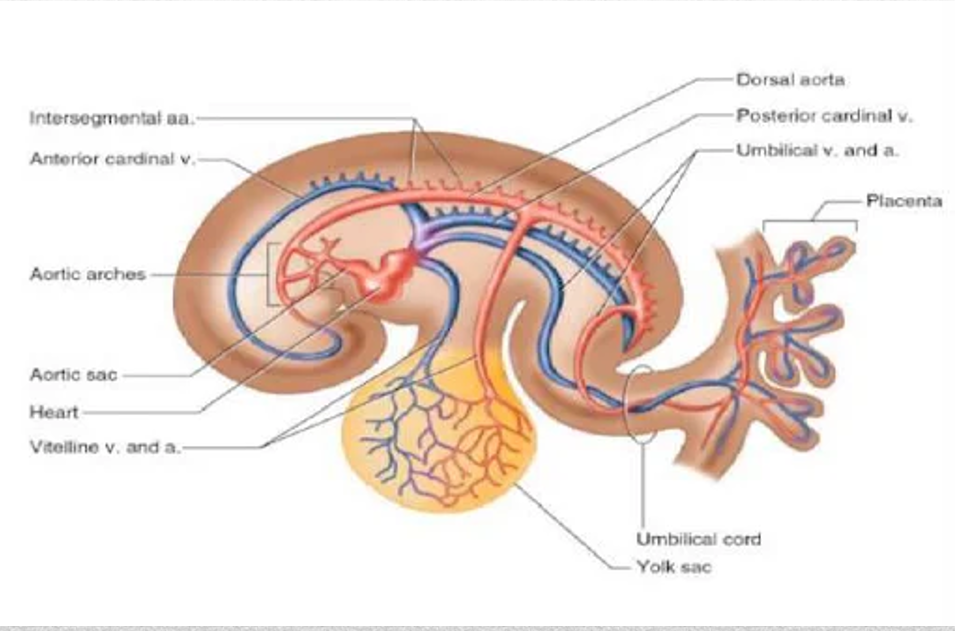
Cardinal veins return blood from the ______
embryo
Vitelline veins return blood from the _____ _____
yolk sac
Umbilical veins return ______ blood from ______; only ____ umbilical vein persists
oxygenated; placenta; one
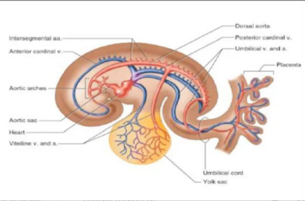
Two dorsal aortas fuse in the _____ half of the embryo to form ____ ____ _____
caudal; single dorsal aorta
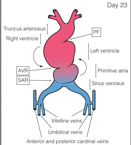
What is sinus venosus?
Caudal region of the primitive heart

The sinus venosus receives all blood returning to the heart from _____ _____ ____, ____ ____, ____ _____
common cardinal veins; vitelline veins; umbilical veins

The primitive atrium develops into the _______ and ______
right and left atria
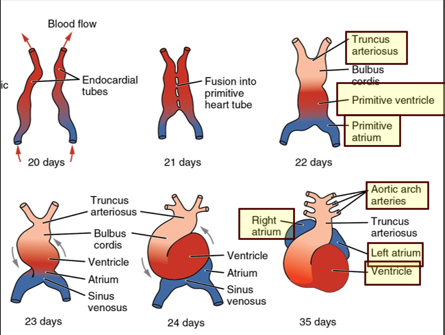
The primitive ventricle develops into the _____ ______
left ventricle

The bulbus cordis develops into the _____ _____
right ventricle

The truncus arteriosus dilates to form _____ ____ from which ____ ____ arise
aortic sac; aortic arches
When does division of the heart into four chambers occur?
During the 4th and 5th weeks
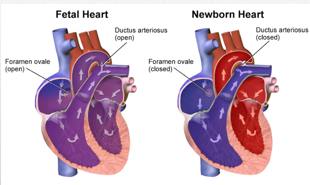
Communication is open between the right and left atrium of the fetal heart through the ____ _____
foramen ovale

Communication is also open between the aorta and the ______ _____ via the ____ _____
pulmonary artery; ductus arteriosus
Before birth, oxygenated blood is given to the fetus through the _____ _____ from the _____ to the heart
umbilical vein; placenta
Half of the oxygenated blood passes through, _____ _____, the rest bypasses the liver to through _______ into the _____
hepatic sinusoids; ductus venosus; IVC
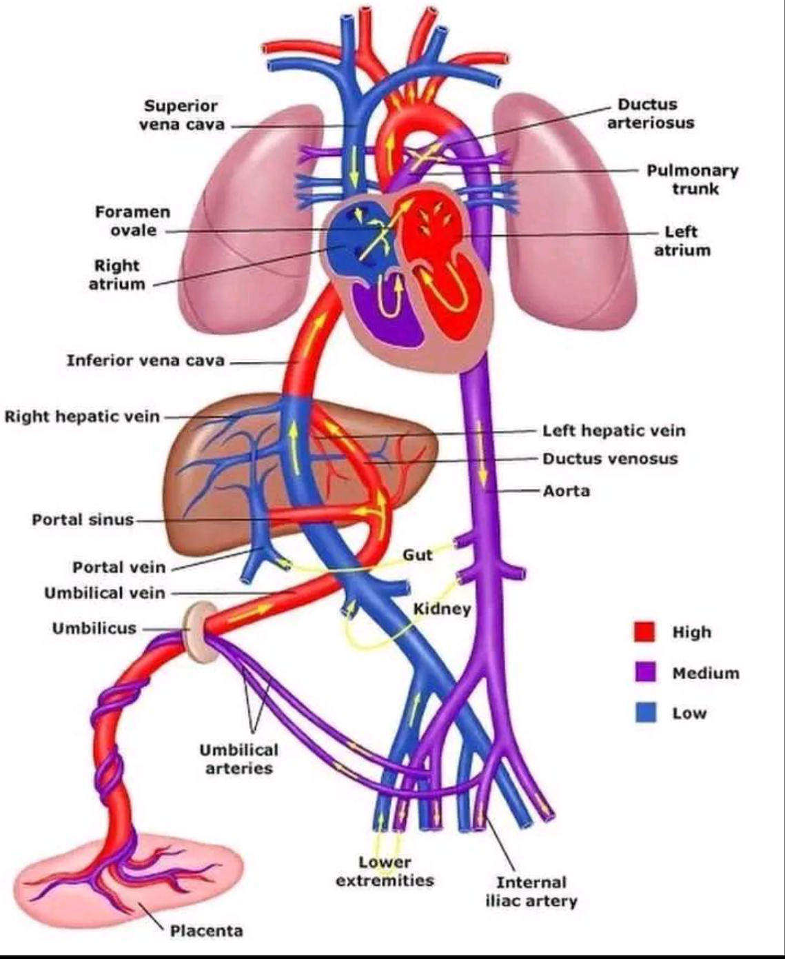
Blood flows from the IVC and superior vena cava and enters into the ____ _____
right atrium
Blood in the _____ atrium is ____ oxygenated than blood in the umbilical vein
right; less
Small amount of oxygenated blood from IVC is diverted by the ____ _____ and remains in the ____ ____ to mix with deoxygenated blood from the ____ and _____ _____
crista dividens; right atrium; SVC; coronary sinus
Most of the blood from IVC is directed by lower border of ____ ____ through the foramen ovale into ____ ____
septum secundum; left atrium
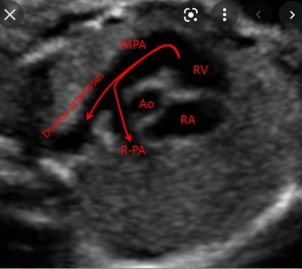
Blood in RA flows through the _____ valve into the _____ _____ and leaves through the ____ ____ ____
tricuspid; right ventricle; main pulmonary artery (MPA)
MPA bifurcates into right and left pulmonary artery branches that lead to their respective _____
lungs
Most of this blood passes through the connection of the ____ _____ into the ____ ____; only a very small amount goes to the lungs
ductus arteriosus; descending aorta
Blood mixes with small amount of _____ ____ as it returns from lungs via the four _____ _____ into the ____ ____
deoxygenated blood; pulmonary veins; left atrium
The pulmonary veins enter the posterior of the _____ _____
left atrium
The four pulmonary veins are named according to their locations:
Right upper
Left upper
Right lower
Left lower
Blood then flows from LA into LV through _____ _____ and leaves heart through _____ _____
mitral valve; ascending aorta
What are branches of fetal ascending aorta?
innominate artery
Left carotid artery
Left subclavian artery
The rest of the mixed blood in the descending aorta passes into the ____ ____ and is returned to the placenta for ________
umbilical arteries; reoxygenation
Circulation of fetal blood through placenta ceases at birth when the ___ ____ begin to function
neonatal lungs
What fetal cardiac structures are no longer necessary at birth?
Foramen ovale
Ductus arteriosus
Ductus venosus
Umbilical vessels
Omission of placental circulation causes immediate fall in blood pressure in newborn’s _____ and ____
IVC; RA
As lungs expand with air, there is a ____ in pulmonary vascular resistance. This causes an _____ in pulmonary blood flow and progressive _____ of the walls of the pulmonary arteries
fall; increase; thinning
Pressure in the left atrium becomes ______ than that in the right atrium. This causes the ____ ____ to close
higher; foramen ovale
With time, complete closure of foramen occurs from adhesion of the septum _______ to the left margin of the septum _____
primum; secundum
Septum primum forms the floor of the ____ ____
fossa ovalis
Ductus arteriosus constricts ___- __ after birth, once ___-sided pressures exceed the ____- sided pressures
24-48 hours; left; right
There is a small shunt of blood from aorta to _____ ____ until these pressures adjust to neonatal life
pulmonary artery (PA)
Once the ductus arteriosus closes, it turns into _____ ______ in the neonate
ligamentum arteriosum
If ductus arteriosus communication persists, it is called a _____ ____ _____
patent ductus arteriosus
Umbilical ______ also constrict after birth to prevent blood loss from the neonate
arteries
Umbilical _____ may remain patent for some time after birth
vein
What is normal fetal heart rate?
120-160 bpm
In first trimester, heart rate begins around ____ and increases to _____ before returning to normal rate and sinus rhythm
90 bpm; 170 bpm
Heart rate less than ____ is bradycardia
100 bpm (<60)
Heart rate greater than ____ is tachycardia
>200 bpm
What are fetal risk factors indicating fetal echo?
IUGR
Cardia arrhythmias - MOST COMMON
Abnormal amniocentesis
Abnormal amniotic fluid collections
Hydrops fetalis
Presence of extracardiac abnormalities in the fetus are associated with _____ _____ ____
congenital heart disease
What are extracardiac abnormalities that can cause congenital heart disease?
renal anomalies
gastrointestinal anomalies
Single umbilical artery
What are maternal diseases indicating fetal echo?
diabetes
lupus erythematosus
infections during pregnancy
If one parent has congenital heart defect recurrence risk ranges from ____-____
2.5-4%
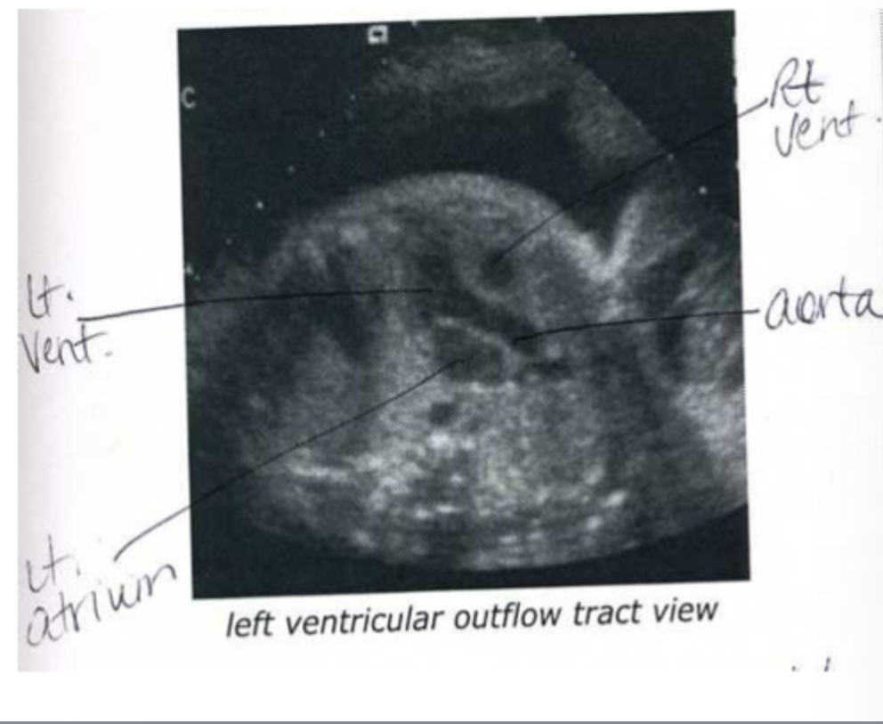
What is LVOT?
connects left ventricle to aorta
What is RVOT?
connects right ventricle to pulmonary artery
Chap 32
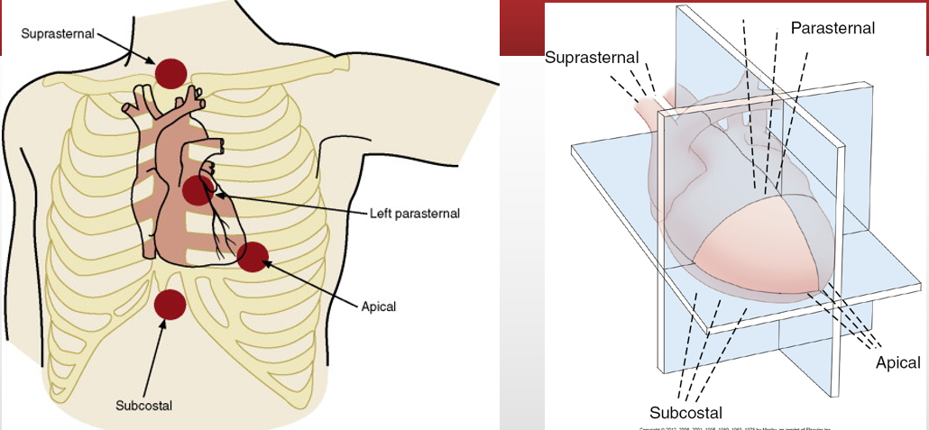
What is the transducer location when it is placed in the suprasternal notch?
Suprasternal

What is the transducer location when it is located near body midline and beneath the costal margin?
subcostal

What is the transducer location when it is located over the apex?
Apical

What is the transducer location when it is placed over the area bounded superiorly by the left clavicle, medially by the sternum, and inferiorly by the apical region?
Parasternal
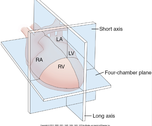
What is the imaging plane when it is perpendicular to dorsal and ventral surfaces and parallel with the long axis of the heart?
Long axis

What is the imaging plane when it is perpendicular to dorsal and ventral surfaces and perpendicular to the long axis of the heart?
short axis

What is the imaging plane when there is a apical view and it is parallel with dorsal and ventral surfaces?
4cH heart