Flashcards CCMA - Ch. 5 Integumentary System | Quizlet
 Call Kai
 Learn
Learn Practice Test
 Spaced Repetition
 Match
Match Flashcards
 Knowt Play
1/106

There's no tags or description
Looks like no tags are added yet.
Name | Mastery | Learn | Test | Matching | Spaced | Call with Kai | Chat |
|---|
No analytics yet
Send a link to your students to track their progress
107 Terms
The integumentary system includes...
the skin and accessory organs such as hair, nails, sweat glands, and subcutaneous tissue underneath the skin
What is the function of sweat glands?
cool the body
What is the function of Sebaceous glands
release sebum to lubricate the skin and hair follicles and help inhibit bacterial growth.
What is the function of Hair and hair follicles
assist in regulating body temperature.
What is the function of a network of blood vessels
permeates into other structures and glands to nourish them.
What is the function of nails?
protect the heavily used, exposed surfaces
The main functions of the skin include...
Protection, sensory reception, regulation of body temperature, and synthesis of vitamin D
Keratin
A protein that forms the outermost layer of the skin; it functions as a waterproof protective barrier that prevents fluid loss from the body; it also protects the body from absorbing excess water while swimming or bathing
What is the body's first line of defense against bacteria and other toxins?
Unbroken and intact skin
What do acidic oily secretions of sebaceous glands do?
Work to prevent bacteria and other organisms from entering the skin
Melanin
Pigment in skin cells that protects underlying tissue from damage due to ultraviolet exposure
Sensory reception of the skin
Skin contains numerous sensory receptors in the dermis that relay information to the brain to prevent or minimize injury or recognize textures; can detect heat, cold, pain, touch, and pressure
What is the normal internal temperature?
37°C or 98.6°F; but individuals may fluctuate and differ a few degrees from normal
What do fluctuations in the internal temperature influence?
The speed by which chemical reactions are conducted in the body
Through what two mechanisms does the skin aid in thermoregulation?
Both represent negative feedback in maintaining homeostasis; blood vessels and sweat glands; vasodilation to release heat when body temp increases (also the sweat glands become active) and vasoconstriction to conserve heat (also sweat glands become inactive)
Adipose tissue
In the subcutaneous layer of the skin; serves as an insulator
How does the skin help synthesize vitamin D?
UV light helps activate a precursor molecule in the skin
What does vitamin D do?
Helps the small intestine absorb calcium and phosphorus that are essential for normal bone metabolism and muscle function
What are the three layers of the skin?
epidermis, dermis, subcutaneous
Epidermis
Outermost layer of the skin; avascular; contains keratinocytes and melanocytes
Keratinocytes
Cells in the epidermis; as these cells move toward the surface, they lose nuclei and die; the dead cells contain keratin
Melanocytes
Cells in the epidermis; contain melanin which gives the skin its color and protects against UV radiation
Dermis
Middle layer of the skin; made up of thick connective tissue; contains two types of fibers: collagen and elastin; includes blood vessels, lymph vessels, hair follicles, and exocrine glands such as sebaceous and sweat glands
Collagen
Fiber in the dermis; prevents the skin from tearing
Elastin
Fiber in the dermis; provides resilience to skin
Subcutaneous layer
Bottom layer of skin; also known as hypodermis; contains fat cells that provide insulation to the body; also stores reserve calories
Skin lesions
Physical changes in the skin that may be caused by exposure to irritants or localized infections
Primary lesions
Appear immediately on the skin
What details should an MA note about a lesion?
Any drainage, pain, pruritus, excoriation, anatomical location, color, elevation, and texture of skin
Macule
Primary lesion; flat, colored spot on the skin (freckle); non-palpable skin lesion

Papule
Primary lesion; Solid elevation of less than 1 cm in diameter and has distinct borders (ex: a small raised pimple)
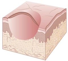
Nodule
Primary lesion; a firm and movable solid elevation 0.5-1 cm in diameter and extends deeper into the dermis than a papule.
A firm lesion extending into the dermis is an example of a nodule

Vesicle
Primary lesion; a small, raised blister that contains clear fluid inside or under the epidermis.
Herpes virus infection shows vesicles.
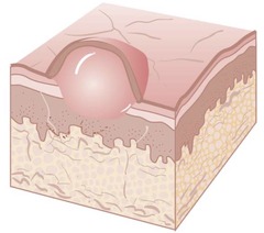
Bulla
Primary lesion; a large vesicle greater than 1 cm.
Burns exemplify bulla.

Pustule
Primary lesion; a vesicle with white cellular debris; it may be sterile or infected.
Acne is a form of pustule

Tumor
Primary lesion; a large solid mass that may be benign or malignant.
It is a large nodule and may be firm or soft.
Squamous cell carcinoma is an example of a tumor.

Plaque
Primary lesion; a solid, elevated, flat-top lesion with indistinct borders.
It is found on the skin or mucous membrane.
Psoriasis is an example of a plaque.
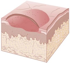
Wheal
Primary lesion; a transient, raised, edematous lesion caused by fluid accumulation in the dermis. It is a palpable, red, circumscribed swelling.
It results from transient edema in the dermis.
Intradermal skin tests produce wheals.

Secondary Lesion
may evolve from primary lesions. These lesions develop as a result of a complication of the primary lesion. For example, a rash that itches is a primary lesion, but an ulceration that occurs as a result of repeated scratching is the secondary, and possibly more serious, lesion.
Scales
Secondary lesion; thin, flaking layers of epidermis.
Psoriasis is an example of scales.
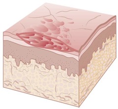
Crusts
Secondary lesion; a dried exudate on the skin.
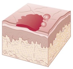
Fissures
Secondary lesion; a linear, wedge-shaped crack extending into the dermis
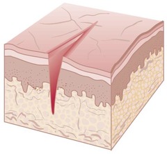
Ulceration
Secondary lesion; an area of tissue loss involving the epidermis and often the dermis
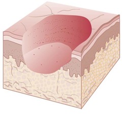
Scars
Secondary lesion; Scar tissue may form due to excess collagen production after injury.

Atrophy
Secondary lesion; a loss of some portion of the skin.

Impetigo
skin infection; caused by streptococcus or staphylococcus aureus. A common characteristic of impetigo is the formation of crusty lesions and small vesicles around the nose and mouth (may often be across the entire face).
Honey-colored exudate is excreted from areas of impetigo.
The treatment includes oral antibiotics and possibly topical antibiotic cream.
Impetigo is one of the highly contagious skin infections, most common in children.
Acne
skin infection; occurs when hair follicles are blocked with dead skin and oil from the skin. Acne vulgaris is common during puberty. Its causes include an inherited predisposition, hormonal fluctuations, exposure to heat and humidity, and the use of oily creams.
The treatment includes using benzoyl peroxide-based face wash twice daily and topical applications. In more severe cases of acne, an oral antibiotic may be prescribed for several weeks or months.
Acne involves the formation of blackheads, pimples, pustules, or larger abscesses. It also affects the sebaceous glands.
Rosacea
skin infection; characterized by inflammation, pustule formation, and visible dilated blood vessels (telangiectasia) and facial redness.
Treatment includes topical antibiotics and, in severe conditions, oral antibiotics.
Rosacea presents as frequently flushed skin on the face, particularly the nose, forehead, cheeks, and chin.
Cellulitis
skin infection; an acute bacterial infection that involves the subcutaneous tissue and manifests as redness, tenderness, swelling, and warmth at the site.
Treatment includes oral antibiotics. In some cases, patients are hospitalized and may receive IV antibiotics.
Cellulitis can progress from a small cut or any kind of skin injury. It can be serious and require hospitalization.
Fungal Infections
haracterized by pruritic lesions with a distinct border. Examples include tinea pedis (athlete's foot), tinea cruris (jock itch), and tinea corporis (ringworm).
Treatment includes topical antifungal agents, such as clotrimazole (Lotrimin), ketoconazole (Nizoral), econazole, or nystatin (Mycostatin).
Fungal infections are commonly caused by fungi that grow on keratinized tissues such as the skin, hair, and nails.
Viral Infections
Treatment includes analgesic and antipruritic medications. Vaccines can help prevent shingles.
The common viral skin infections include warts, herpes simplex (cold sores), and herpes zoster (shingles).
Warts
viral infection; small, raised skin growth caused by human papilloma virus
Cold Sores
viral infection; caused by herpes simplex virus type 1 and manifest as painful ulcers along the gumlines of the mouth or on the lips
Shingles
viral infection; an acute inflammatory disorder caused by herpes zoster virus and is characterized by a painful rash on the trunk of the body and occasionally on the face. Herpes zoster tends to follow the dermatomes
Scabies
Parasitic infection; caused by Sarcoptes scabiei and is characterized by intense itching, a body rash, and a sensation of something crawling on the skin. This condition is treated only through prescribed medication
Pediculosis
Parasitic infection; aused by lice. The Centers for Disease Control and Prevention (CDC) has posted recommendations on treatment for lice, including use of lice-medicated shampoo and steps to prevent reinfestation.
Seborrheic Dermatitis
inflammatory skin disorder; involves dry or moist, greasy-appearing scales and yellowish crusts on the scalp, eyebrows, eyelids, etc.
Seborrheic dermatitis affects the sebaceous glands by altering the amount and quality of sebum.
Symptoms include dry or moist, greasy-appearing scales, and yellowish crusts on the scalp, eyebrows, eyelids, and sides of the nose, behind the ears, and in the middle of the chest.
In adults, seborrheic dermatitis on the scalp refers to dandruff; in infants, it is referred to as cradle cap.
Seborrheic keratosis (age spots) occurs with aging and is characterized by benign, slightly raised, tan to black lesions.
Treatments for Seborrheic Dermatitis
Tar- or sulfur-based shampoos are used to treat seborrheic dermatitis of the scalp. Topical corticosteroids, such as triamcinolone diacetate, are used to treat inflammations of the skin.
Contact dermatitis
Contact dermatitis is an acute inflammatory skin disorder. It can be due to irritants or allergens found in lotions, latex, and poison ivy.
Symptoms include redness (erythema), edema, pruritus, and vesicles.
Oral corticosteroid medications, such as prednisone and methyl prednisolone, are used to treat contact dermatitis.
Eczema
an inflammation of the skin with unknown pathogenesis but may be associated with dry skin.
Symptoms include vesicular rash on the face, neck, and elbows and behind the knees and ears.
In young children, it may be due to food allergies. In older children and adults, stress or extreme temperatures may trigger flare-ups.
Primary inflammation of eczema is treated with topical corticosteroids and antihistamines such as diphenhydramine, cetirizine, and fexofenadine.
Treatment of inflammatory skin disorders
Secondary infections of inflammatory skin lesions are treated with antibiotics. Mild infections can be treated with topical antibiotics; severe infections may need oral or intravenous antibiotics. The treatment also includes medications to prevent itching and to reduce the inflammation.
Psoriasis
A chronic autoimmune disease characterized by discrete pink or red lesions covered with silvery scales.
Results from an autoimmune reaction that leads to rapid cell maturation and stimulates a build-up of scaly patches on the skin.
Treatment is palliative and includes exposure to UV light to slow down the cell proliferation, as well as coal tar application to relieve irritation.
Other treatment methods include methotrexate, retinoids, immunosuppressants, low-dose antihistamines, and biologic medications
systemic lupus erythematosus (SLE)
a chronic autoimmune inflammatory skin disorder. It may affect any connective tissue in the body, resulting in inflammatory changes in the skin, joints, muscles, and kidneys. Body systems affected by this condition include the urinary, skeletal, and muscular systems.
Symptoms include erythematous patches and plaques, alopecia, and photosensitivity. A "butterfly" rash extending from one cheek across the nose to the other cheek may also occur.
Treatment includes the use of nonsteroidal anti-inflammatory drugs (NSAIDs), low doses of corticosteroids, and cytotoxic drugs.
Cold Injuries
Severe or prolonged exposure to cold temperatures may lead to tissue damage that can progress to gangrene and may require amputation. In severe cases, prolonged exposure to cold can be fatal.
Frostbite
a cold injury caused by exposure to subfreezing temperatures. Blood vessels constrict, reducing blood flow to the affected area, which can lead to tissue damage and cell death.
Superficial frostbite
signs and symptoms include burning, tingling, numbness, and a white or grayish color of the skin
Deep frostbite
signs and symptoms include formation of blisters, with affected areas becoming hard, mottled, edematous, and blue or gray after thawing
Burns
caused by exposure to hot temperatures.
Burn injuries are classified as superficial-thickness (first degree), partial-thickness (second degree), or full-thickness (third degree) burns, depending on the depth of the wound
Rule of Nines
an assessment tool that helps assess the total body surface area burned. The major sections of the body are assigned a numerical value of multiples of nine, and the total burned area is calculated as a percentage. This number helps guide the treatment decision in terms of fluid resuscitation and further course of action.
Superficial (First-degree) burn
Minor sunburn is a superficial (first-degree) burn in which only the epidermis is affected. Dry skin and erythema (redness without vesicles) characterize superficial burns.
The burn may be painful, hyperesthetic, and tingling. Cooling may help relieve the pain.
Discomfort usually lasts about 48 hours, and desquamation occurs in 3-7 days.
Examples of first-degree burns include sunburns and steam burns without blister formation.
Partial thickness (second-degree) burn
Large thick-walled blisters (vesiculation), subcutaneous edema, with a wet, shiny, weeping surface characterize partial-thickness burn. Epidermis and dermis are damaged in this burn.
The burn may be painful, hyperesthetic, and sensitive to cold air.
Superficial partial-thickness burn heals in 10-14 days, whereas deep partial-thickness burn heals in 21-28 days.
Splashes of boiling water or quick exposure to flames may cause a second-degree burn.
Full thickness (third-degree) burn
This type of burn may appear white, brown, or black and typically has a dry, leathery surface.
Tissue disruption occurs, involving the epidermis and dermis and potentially extending into subcutaneous tissue.
Third-degree burns may result from prolonged exposure to heat (e.g., clothing catching fire) or from contact with corrosive chemicals.
Neoplasm
an abnormal growth. It may be benign (noncancerous) or malignant (cancerous)
Benign tumors
noncancerous tumors that do not show asymmetry or border irregularity.
Characteristics of benign tumors (masses):
Benign tumors are encapsulated, which helps prevent spreading.
Though these tumors increase in size, they remain within the shell.
Malignant Tumors
characterized by asymmetry, border irregularity, and elevation of the mole.
Characteristics of malignant tumors:
Local invasion: Malignant cells break through the basement membrane and invade surrounding tissue.
Carcinoma in situ: Malignant tumor is confined to the site of origin without invasion.
Regional spread: Malignant cells invade surrounding tissue without lymph node involvement.
Distant spread or metastasis: Malignant cells spread to lymph nodes with the development of secondary tumors in other organs such as lungs, liver, or bones.
What is the ABCDE rule?
A dermatologist should follow the ABCDE rule (early warning signs of malignant melanoma) to examine malignant melanoma.
A-Asymmetry: Unequal halves of mole
B-Border: Edges or border irregularity
C-Color: Appearance of different shades such as tan, brown, black, or white throughout the mole
D-Diameter: Increase in the size of the mole to more than 6 mm
E-Elevation: Raised and elevated mole
Allergy Testing
will help identify the source of an allergic reaction. A small amount of antigen (or antigen groups) is either percutaneously applied or intradermally injected and later the test site is examined for visible reactions. A localized skin reaction indicates the patient's allergic response to the tested antigen. The larger the localized skin reaction (wheal), the more severe the patient's allergic response to the tested allergen
Percutaneous Test Description
It is also called a scratch test and is performed on the fore arm, upper arm, or back. In children, this test is performed on the back because it provides a larger surface area for the test
Percutaneous Test Method
The surface of the skin is either labeled or numbered in rows 11/2 to 2 inches apart, and a small amount of allergen is applied on the skin. The skin is scratched or pricked so that the allergen moves under the skin surface. A reaction occurs 10-30 minutes after allergen exposure. A wheal (hive) forms at the site of the scratch if there is a positive reaction to the allergen.
Percutaneous Test Uses
A scratch test is used for allergic rhinitis, asthma, and detection of food allergies.
Percutaneous Test- Patient considerations
The patient must remain in the medical office for 30 minutes after the test because a delayed systemic allergic reaction can occur. Patients must also avoid the use of antihistamines for at least a week or longer prior to testing.
Patch Test Description
This test involves application of an allergen onto a patch, which is then placed on the patient's skin
Patch Test Method
Patches are placed on the arms or back of the patient, who is required to remain in the same place for 48 hours. They are later removed during consecutive office visits.
Patch Test Uses
This test helps detect delayed allergic reactions due to contact dermatitis
Patch Test - patient considerations
The patient is instructed to avoid bathing and any activity that may increase sweating, as sweating can increase skin irritation and provide false positive results.
Intradermal (Intracutaneous) Test Description
This is a sensitivity test in which extracts are injected into the intradermal layers of the skin.
Intradermal (Intracutaneous) Test Method
The extracts are injected in doses of 0.1-0.2 mL. When intradermal injections are used for allergy testing, usually 10-15 allergens can be tested at a time on each arm
Intradermal (Intracutaneous) Test Uses
The intradermal test is useful for diagnosing allergies to: Penicillin, Insect venom such as bee stings, Tuberculin or purified protein derivative (PPD) test (Mantoux test), and Valley Fever coccidioidomycosis test
Radioallergo-sorbent Test (RAST) Description
It determines the level of antibodies that are created when a sample of the patient's blood is mixed with different allergens in the laboratory.
Radioallergo-sorbent Test (RAST) Method
The procedure is easy to perform and requires only a single venipuncture. The actual tests that are performed on the blood sample differ in different regions of the country due to the variety of allergens.
Radioallergo-sorbent Test (RAST) Uses
The procedure is usually done to identify food allergies. The RAST procedure is indicated in certain conditions; for example, when: A patient cannot discontinue antihistamine medications, Accurate interpretation of skin test results is difficult due to a skin disorder, or The patient's skin test results are negative but the patient's signs and symptoms indicate that further testing may be required.
Excision biopsy
Removal of the entire lesion for examination and analysis
punch biopsy
Removal of a small section (for example, the center) from the lesion for analysis
shave biopsy
Cutting or shaving off a lesion or growth above the base line using a scalpel
How should MAs assist providers during biopsies?
Gathering the necessary equipment and supplies required for the procedure
Ensuring that the patient understands the procedure
Preparing the patient for the procedure using appropriate gowning, draping, and positioning
Verifying if the provider has obtained the patient's informed consent
Preparing the biopsy site by implementing steps as per the office protocol
Using appropriate personal protective equipment according to Standard Precautions while assisting the provider
Labeling the container containing the sample and make it ready for transport to the lab
Including the laboratory request forms along with the sample
Cleaning the area where this procedure was conducted
Disposing of all waste materials in the manner required, and disinfect and sterilize all equipment that was used in the procedure
Sanitizing hands and documenting the entire procedure
Provide patient education on how to care for the biopsy site
Chemical Peel (Chemexfoliation) - Procedure Steps
Topical agents are used to remove minor skin features, such as acne scars, hyperpigmentation, and fine wrinkles.
Tretinoin cream of 0.05% to 0.1% concentration (Retin-A) are agents used in chemical peels.
These agents may be carefully applied to prevent the solution from entering into the patient's eyes. Usually, a pea sized amount is the maximum amount used at a time.
Chemical Peel (Chemexfoliation) - patient considerations
The patient must avoid prolonged sun exposure and must use sunscreen with minimum SPF of 15 as photophobia is a side effect of chemexfoliation.
Botox Injections - procedure steps
Botox is a neurotoxin obtained from Clostridium botulinum, which is a type of bacteria that causes food poisoning. Botox treatment involves injecting these neurotoxins around specific places in the body such as the eyes, mouth, and forehead.
These toxins paralyze the muscles of face and cause the skin to feel smoother and younger.
Botox Injections - patient considerations
The patient must undergo treatment every 3 to 4 months. Because the muscles that cause wrinkles to form are paralyzed because of this treatment, patients may experience difficulty in showing facial expressions.
Dermabrasion - description
Dermabrasion is a procedure that involves a dermabrader, a handheld device that mechanically evens the layers of the skin. Medical skin specialists usually perform microdermabrasion in the dermatologist office.