401: Dental Triangles of the Neck - Part 1&2
 Call Kai
 Learn
Learn Practice Test
 Spaced Repetition
 Match
Match Flashcards
 Knowt Play
1/305

There's no tags or description
Looks like no tags are added yet.
Name | Mastery | Learn | Test | Matching | Spaced | Call with Kai | Chat |
|---|
No analytics yet
Send a link to your students to track their progress
306 Terms
Hyoid bone (it attaches to the muscles)
What bone does not articulate with any other bone?
Hyoid bone
Identify the structure

Region from the inferior edge of mandible of the skull down to clavicles and scapula
What region is considered the "neck"?
C1-C7 spinal nerves pass above correspondingly numbered vertebrae. C8 passes below C7 vertebrae
C1-C7 spinal nerves pass ___________ correspondingly numbered vertebrae. C8 passes ___________ C7 vertebrae
Support the weight of the skull
Provide attachment of muscles that can move the head and skull around in space
What is the purpose of the 7 cervical vertebrae?
Cervical vertebrae are smaller and lighter because they do not support as much weight
How do the cervical vertebrae compare to the other vertebrae?
Bifid (split - all cervical split except for C7)
How would you describe the C4 vertebrae spinous process?

Vertebral foramen
Identify the bone marking at #1

Posterior arch
Identify the bone marking at #2

Spinous process
Identify the bone marking at #3
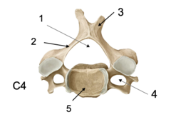
Transverse foramen
Identify the bone marking at #4

Vertebral body
Identify the bone marking at #5
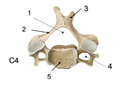
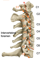
Vertebral arteries to provide blood supply to brain
What runs through the transverse foramen?
Vertebral arteries
What does the red line indicate?
Dens
Identify the bone marking

C1 (atlas)
What vertebrae supports the weight of the skull?
Transverse process
Identify the marking at #1

Anterior arch
Identify the marking at #2

Articular surface for occipital condyle
Identify the marking at #3
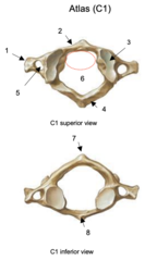
Posterior arch
Identify the marking at #4

Transverse foramen
Identify the marking at #5
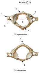
Vertebral foramen
Identify the marking at #6

Posterior tubercle
Identify the marking at #7

Anterior tubercle
Identify the marking at #8

C1 vertebrae does not have a vertebral body --> this is where the dens articulates
What does the dashed red line represent?

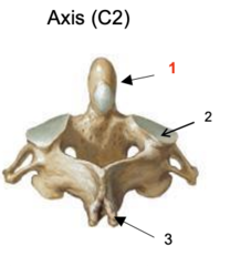
Dens
Identify the marking at #1
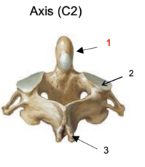
Articular surface for atlas
Identify the marking at #2
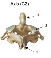
Spinous process
Identify the marking at #3
Atlas
What is the name of the C1 vertebrae?
Axis
What is the name of the C2 vertebrae?
Rotation point around which the atlas and skull can rotate as a unit
What is the purpose of the dens?
Primarily flexion/extension and lateral bending of skull of C1
What motion occurs at the atlanto-occipital joint?
Primarily rotation with some flexion/extension, lateral bending of C1 on C2
What motion occurs at the atlanto-axial joint?
Atlanto-occipital joint
What is the joint at #1?

Atlanto-axial joint
What is the joint at #2?

Foramen magnum
What is the red arrow pointing to?

Transverse ligament of atlas
What ligament is at #1?
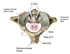
Alar ligaments
What ligament is at #2?

Posterior longitudinal ligament (runs from the base of skull around foramen magnum and posterior vertebral bodies)
Identify the structure at #1

Cruciate ligament
Identify the structure at #2
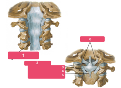
Superior longitudinal band
Identify the structure at #3

Transverse ligament
Identify the structure at #4
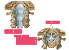
Inferior longitudinal ligament
Identify the structure at #5

Alar ligaments
Identify the structure at #6

Superior longitudinal band
Transverse ligament
Inferior longitudinal ligament
What bands and ligaments make up the cruciate ligament?
Prevents hyper flexion of cervical column
What is the function of the posterior longitudinal ligament?
Nuchal ligaments
Identify the structure at #1

Supraspinous ligament
Identify the structure at #2
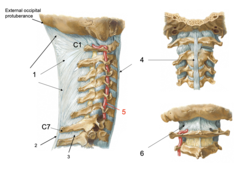
Interspinous ligament
Identify the structure at #3
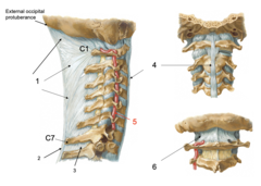
Anterior longitudinal ligament
Identify the structure at #4
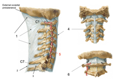
Vertebral artery
Identify the structure at #5

Posterior atlanto-occipital membrane
Identify the structure at #6
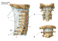
Forms septum in posterior neck and prevents hyperflexion of cervical vertebrae
What is the function of the nuchal ligament?
Prevent hyperflexion
What is the function of the supraspinous ligament?
Prevents hyperextension of cervical column
What is the function of the anterior longitudinal ligament?
Atlantoaxial joint
Identify the structure at #1
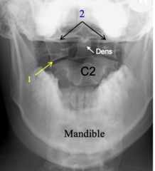
Lateral portion of C1 (atlas)
Identify the structure at #2

Equal in width and symmetry
As shown in red, how should the disc spaces be between cervical vertebrae?

Cervical spine injury (potentially whip lash)
- Vertebrae slip and stretch spinal cord
What clinical correlation is shown?

Mental process
Identify the structure at #1

Hyoid bone
Identify the structure at #2
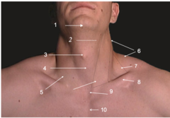
Thyroid cartilage
Identify the structure at #3
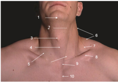
Cricoid cartilage
Identify the structure at #4
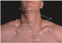
Sternocleidomastoid m.
Identify the structure at #5

Trapezius m.
Identify the structure at #6
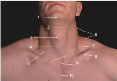
Supraclavicular fossa
Identify the structure at #7

Clavicle
Identify the structure at #8
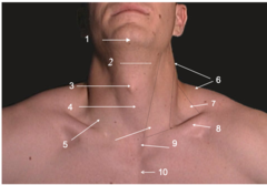
Jugular notch
Identify the structure at #9
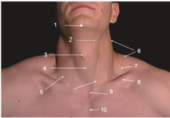
Sternum
Identify the structure at #10

1. Visceral compartment anteriorly
2. Bony-muscular compartment, posteriorly
3. 2 neurovascular compartments, laterally (cartoids, IJV, vagnus n.)
What are the three compartments of the neck?
Dens
What is shown at #1?

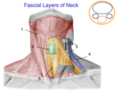
Investing fascia
Identify the fascia at #1

Superficial fascia
Identify the fascia at #2

Prevertebral fascia
Identify the fascia at #3
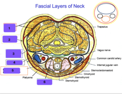
Carotid sheath
Identify the fascia at #4

Pretracheal (visceral) fascia
Identify the fascia at #5

Alar fascia
Identify the fascia at #6

Investing fascia
Identify the fascia at #1
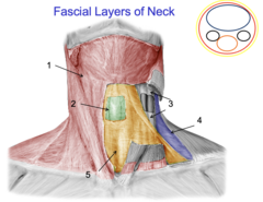
Alar fascia
Identify the fascia at #2

Carotid sheath
Identify the fascia at #3

Prevertebral fascia
Identify the fascia at #4

Pretracheal fascia
Identify the fascia at #5
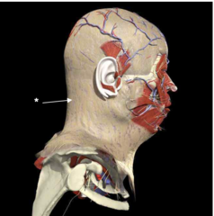
Superficial fascia
Identify the fascia at the star
Investing fascia
Identify the fascia at #1
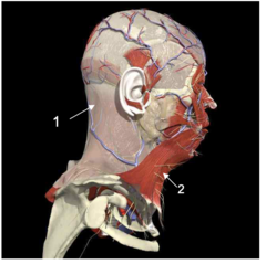
Platysma muscle
Identify the structure at #2

SCM
Identify the structure at #1

External jugular v.
Identify the structure at #2

Posterior auricular v.
Identify the structure at #1

Retromandibular v.
Identify the structure at #2

External jugular v.
Identify the structure at #3
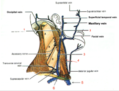
Internal jugular v. (deep to SCM)
Identify the structure at #4

Brachiocephalic v.
Identify the structure at #5

Subclavian v.
Identify the structure at #6
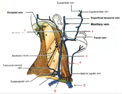
External jugular v.
What vein does the posterior auricular v. drain into?
External jugular v. (posterior division), Internal jugular v. (anterior division)
What vein does the retromandibular v. drain into?
Subclavian v.
What vein does the external jugular v. drain into?
Brachiocephalic v.
What vein does the subclavian v. drain into?
Brachiocephalic v.
What vein does the Internal jugular v. drain into?
External Jugular Vein Distention
What clinical correlate shows a markedly distended right external jugular vein (EJV) due to elevated central venous pressure (CVP)?