Histology Self Quiz
 Call Kai
 Learn
Learn Practice Test
 Spaced Repetition
 Match
Match Flashcards
 Knowt Play
1/100

There's no tags or description
Looks like no tags are added yet.
Name | Mastery | Learn | Test | Matching | Spaced | Call with Kai | Chat |
|---|
No analytics yet
Send a link to your students to track their progress
101 Terms
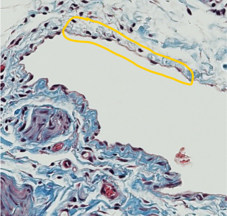
Which type of epithelium is encircled in yellow?
simple squamous
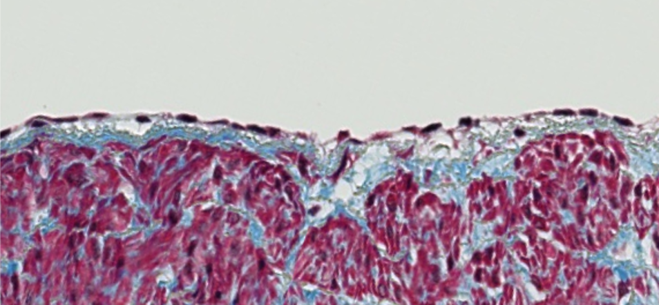
Which type of epithelium is lining the surface of this tissue?
simple squamous
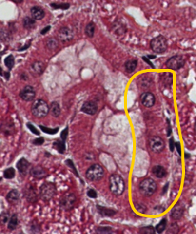
Which type of epithelium is encircled in yellow?
simple cuboidal
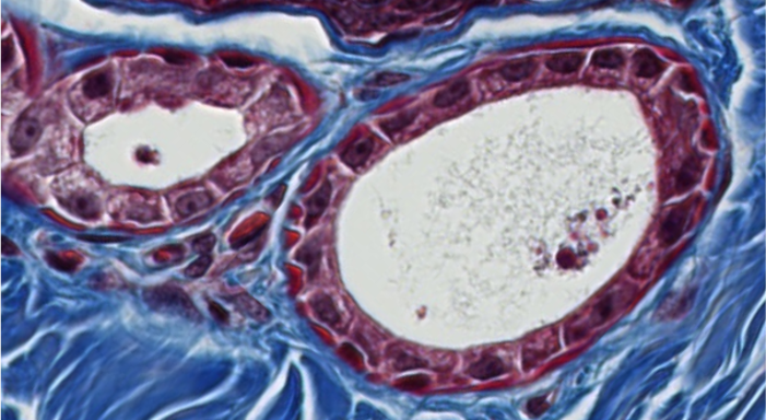
Which type of epithelium is lining the luminal surface of these two structures?
simple cuboidal
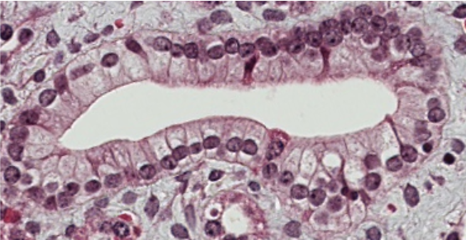
Which type of epithelium is lining the luminal surface of this structure?
simple columnar
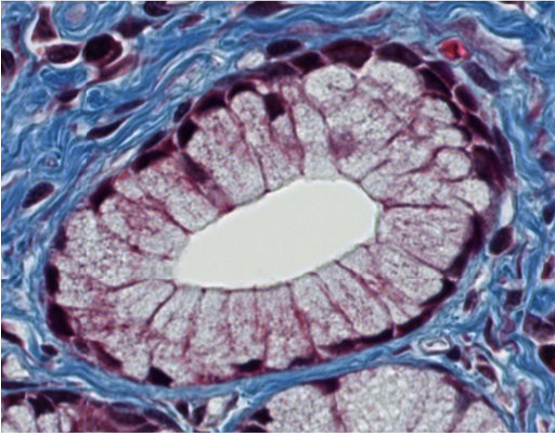
Which type of epithelium lines the luminal surface of this structure?
simple columnar
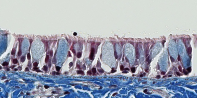
ID the type of epithelium lining the surface of this tissue.
pseudostratified columnar
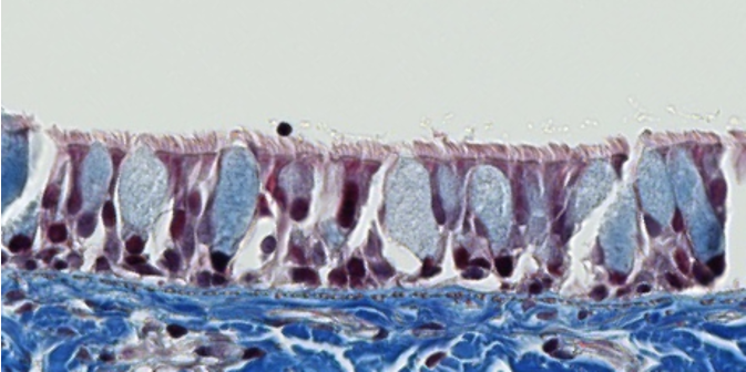
Which type of cell surface modification does this epithelium have?
cilia
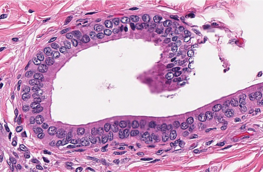
ID the type of epithelium lining the luminal surface of this duct.
stratified cuboidal
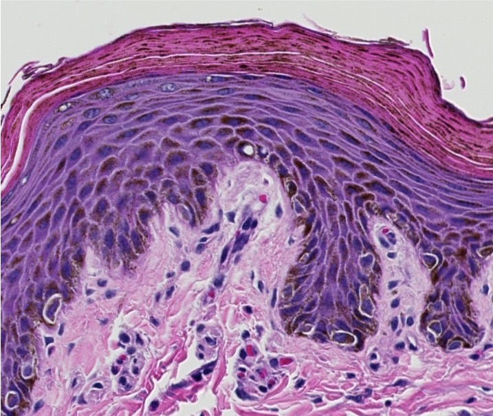
ID the type of epithelium lining the surface of this tissue.
stratified squamous
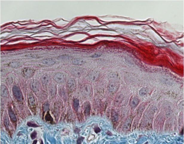
Is this stratified squamous epithelium keratinized or non-keratinized?
keratinized
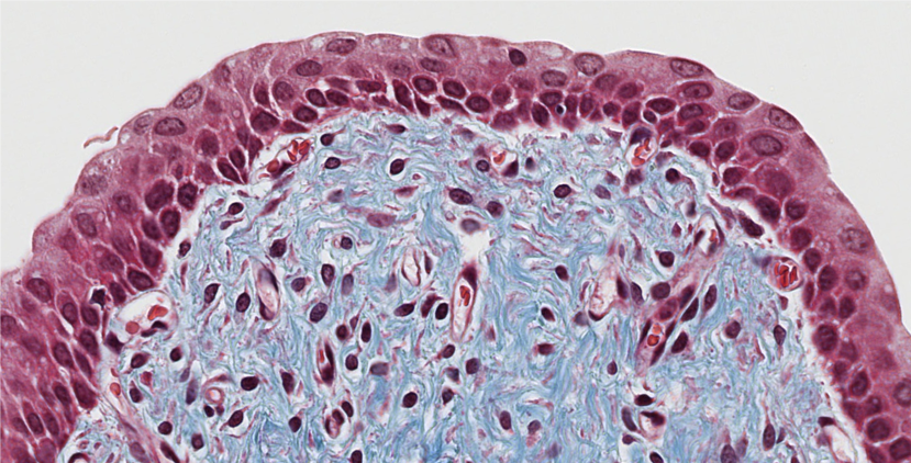
1. ID the epithelium lining the surface of this tissue.
2. Is this epithelium simple or stratified?
transitional
simple
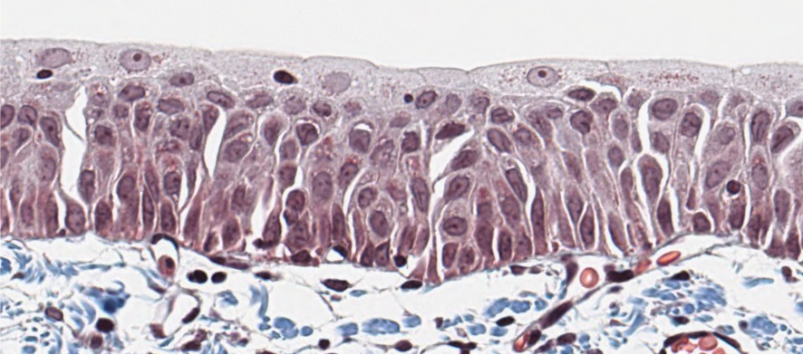
Is this transitional epithelium classified as pseudostratified or pseudostratified columnar?
Pseudostratified (respiratory epithelium is pseudostratified columnar)
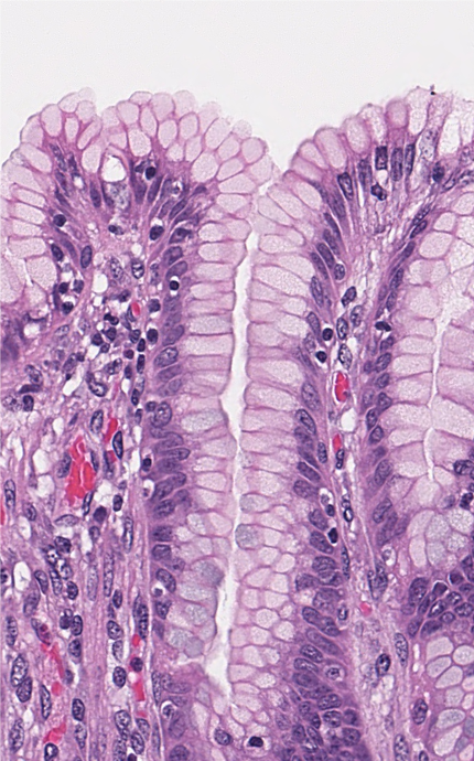
Which type of epithelium lines the surface of this tissue?
Simple columnar
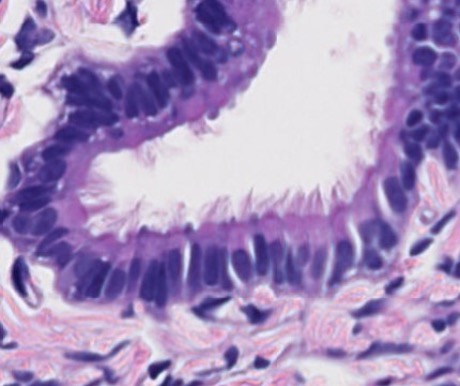
Do these cells have cilia or stereocilia?
cilia
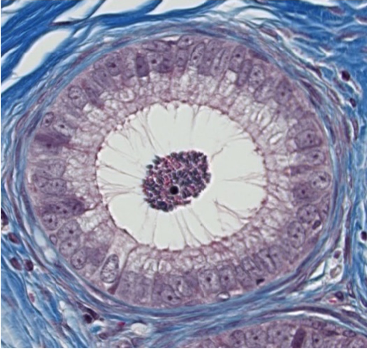
Do these cells have cilia or stereocilia?
stereocilia
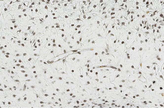
ID the embryonic CT.
mesenchyme
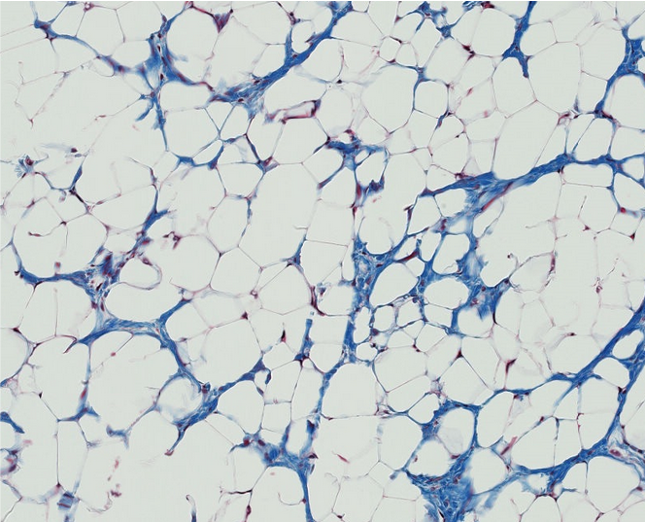
1. Which basic type of CT is this?
2. Can you be more specific?
Adipose tissue
White adipose tissue
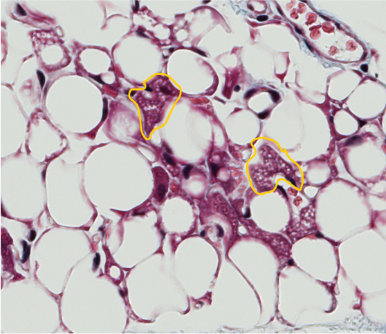
1. Brown adipose tissue cells are encircled.
2. White adipose cells are unilocular (one large lipid vacuole per cell). How do brown adipose cells differ?
They are multilocular (each cell has multiple small lipid vacuoles).
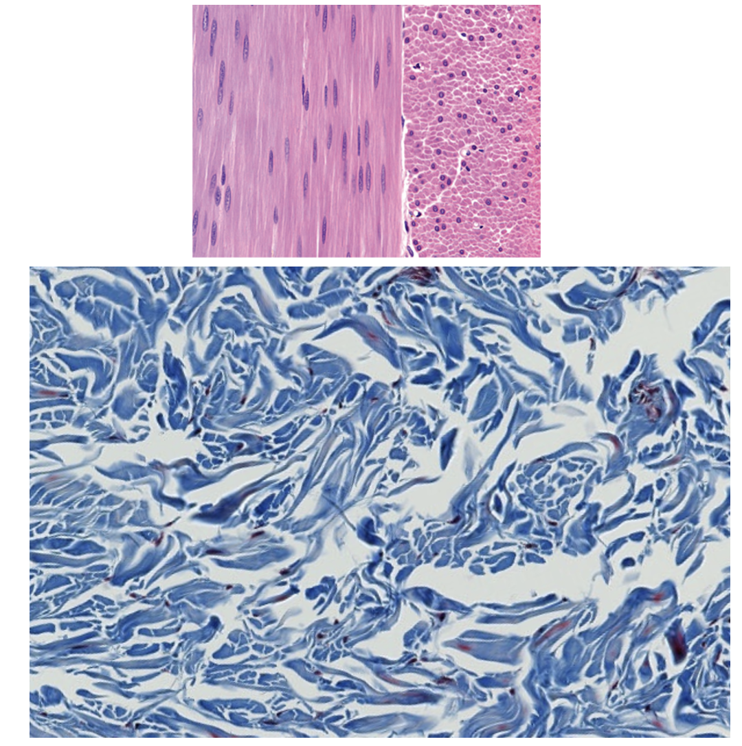
1. Which basic type of CT is this?
2. Can you be more specific?
dense CT
dense irregular CT
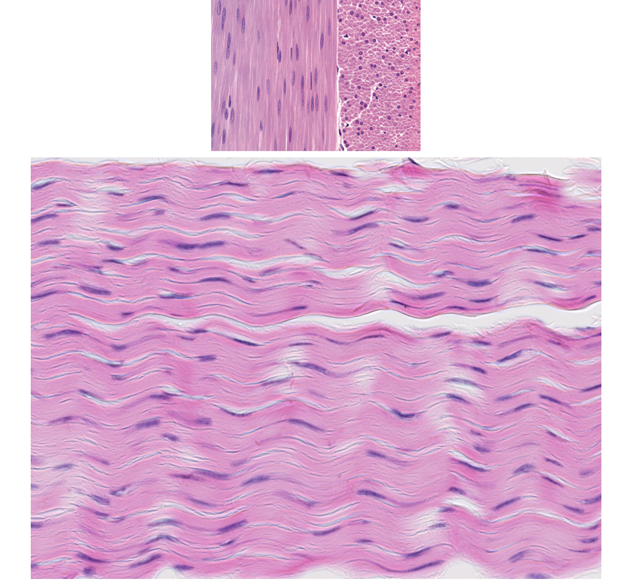
1. Which basic type of CT is this?
2. Can you be more specific?
Dense CT
Dense regular
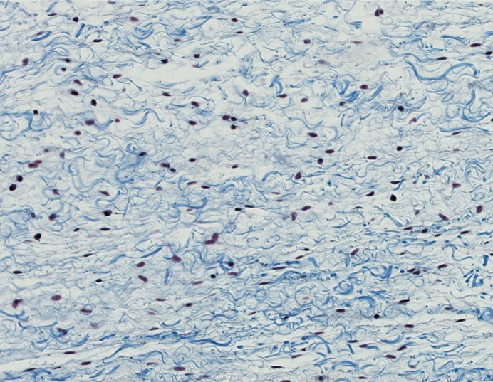
1. Which basic type of CT is this?
2. Can you be more specific?
3. What is another name for this CT?
1. Loose CT
2. Loose irregular
3. Areolar CT
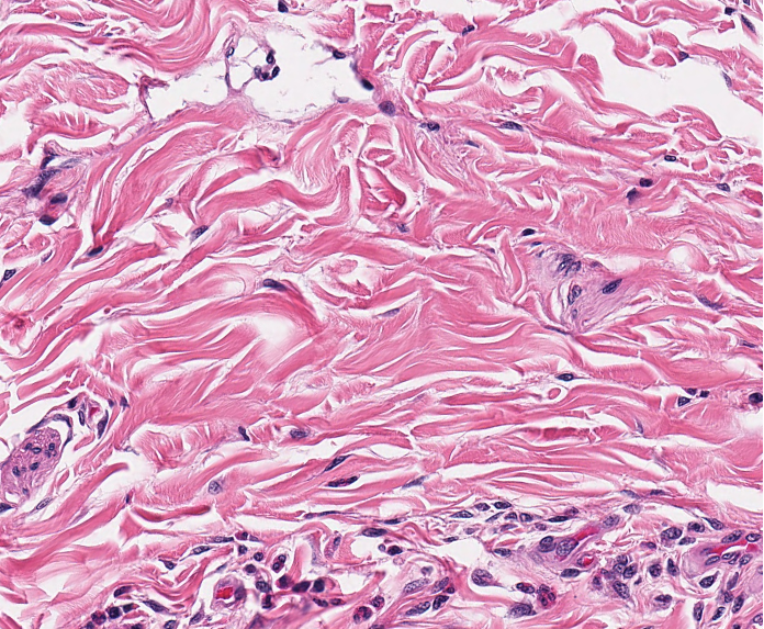
Which type of CT is this?
dense irregular CT
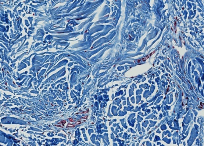
Which type of CT is this?
dense irregular
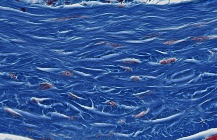
Which type of CT is this?
dense irregular
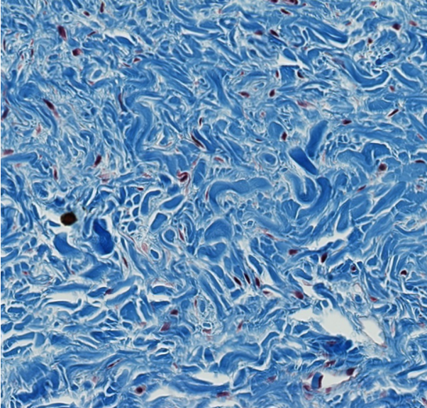
Which type of CT is this?
dense irregular
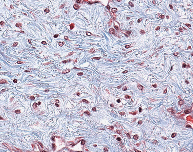
Which type of CT is this?
Loose irregular
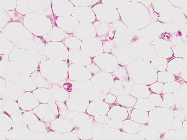
Which type of CT is this?
adipose tissue (white fat)
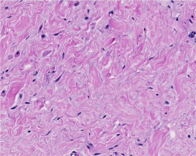
Which type of CT is this?
dense irregular
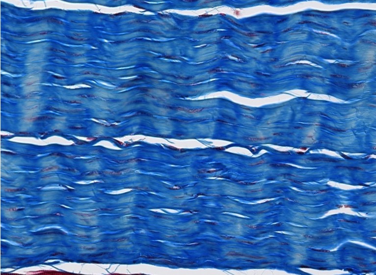
Which type of CT is this?
Dense regular
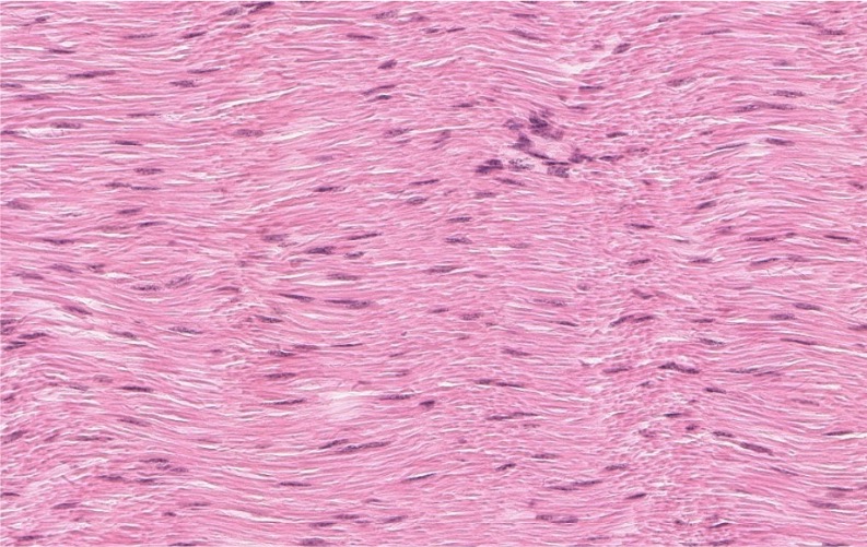
Are elastic ligaments categorized as dense regular or dense irregular CT?
Dense regular
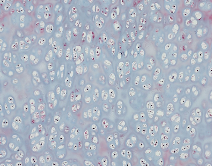
1. Which basic type of CT is this?
2. Can you be more specific?
1.Cartilage
2.Hyaline cartilage
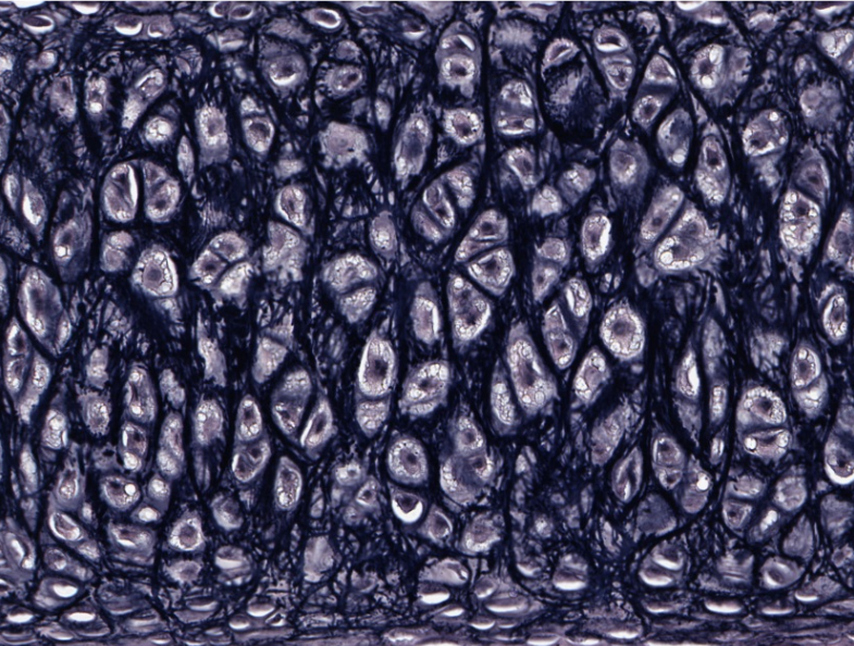
This cartilage has been stained to highlight fibers that make this tissue more flexible.
What fibers are they and what type of cartilage is this?
Elastic fibers within elastic cartilage
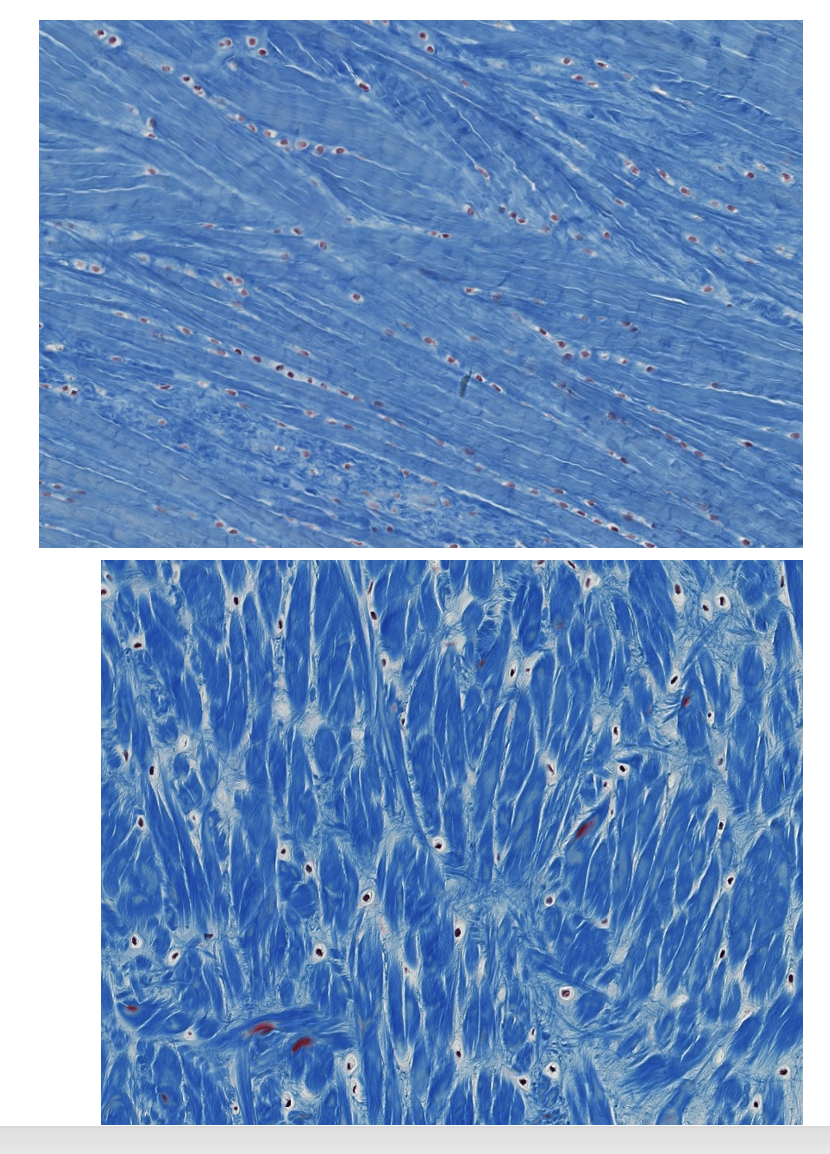
Which type of CT is this?
Fibrocartilage (the round spaces are lacunae, occupied by chondrocytes).
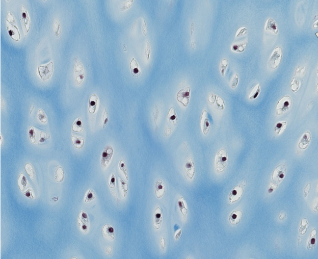
Which type of CT is this?
Be specific.
Hyaline cartilage
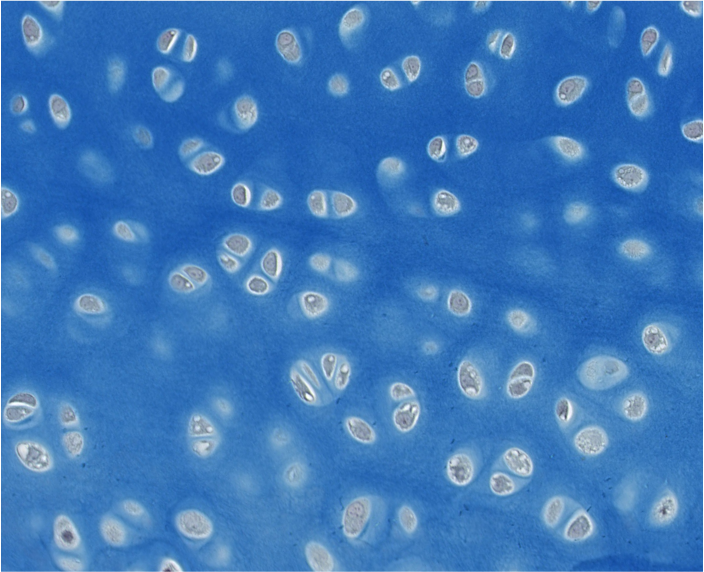
Which type of CT is this?
Be specific.
Hyaline cartilage
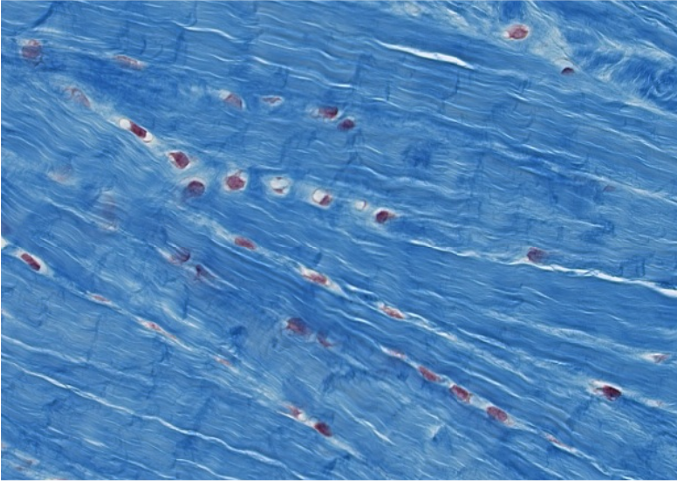
Which type of CT is this?
Fibrocartilage (has rows of lacunae).
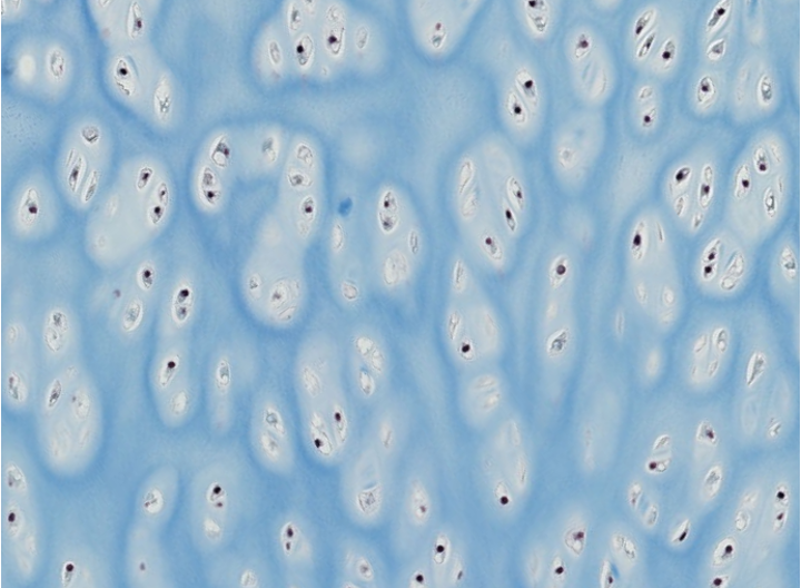
Which type of CT is this?
Hyaline cartilage
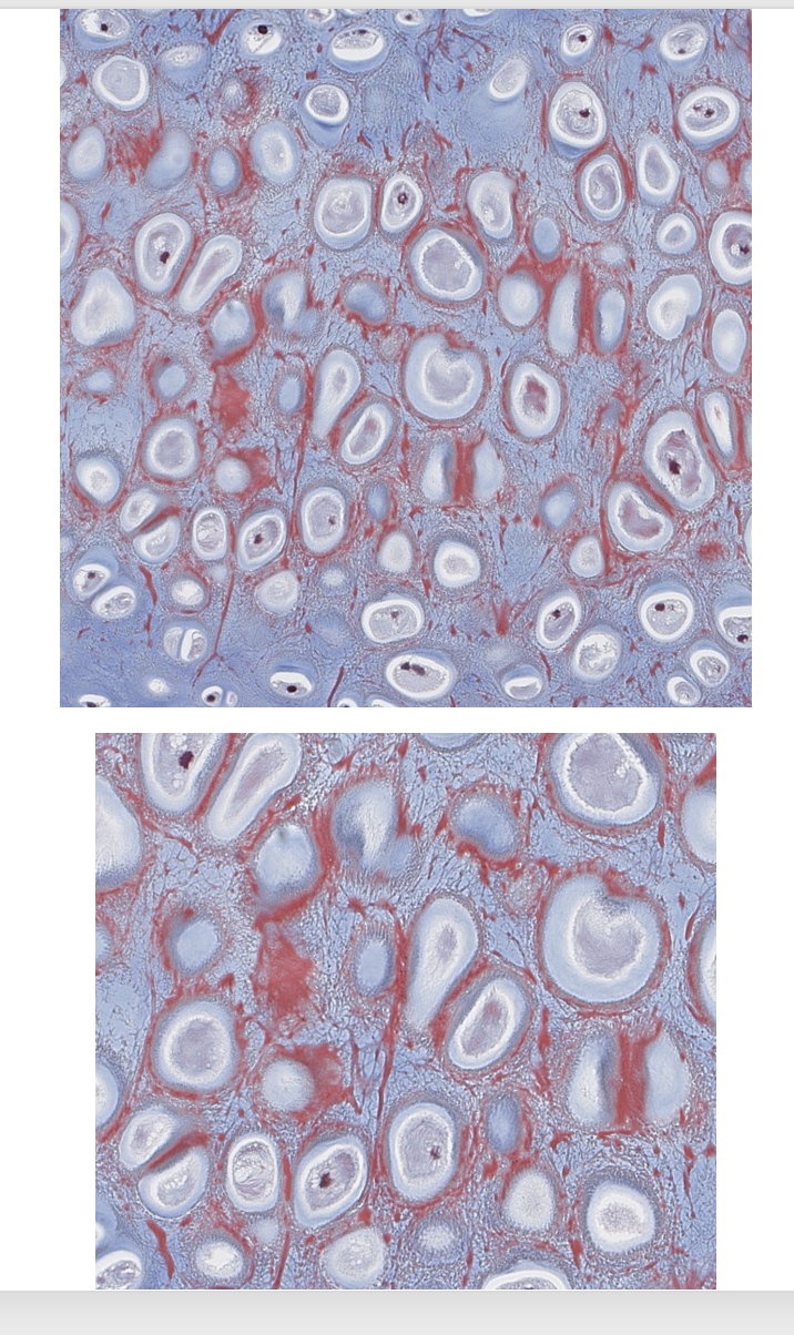
Which type of CT is this?
Elastic cartilage
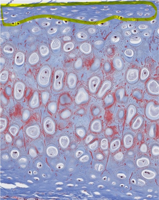
What do we call the CT capsule that surrounds hyaline & elastic cartilage?
Perichondrium
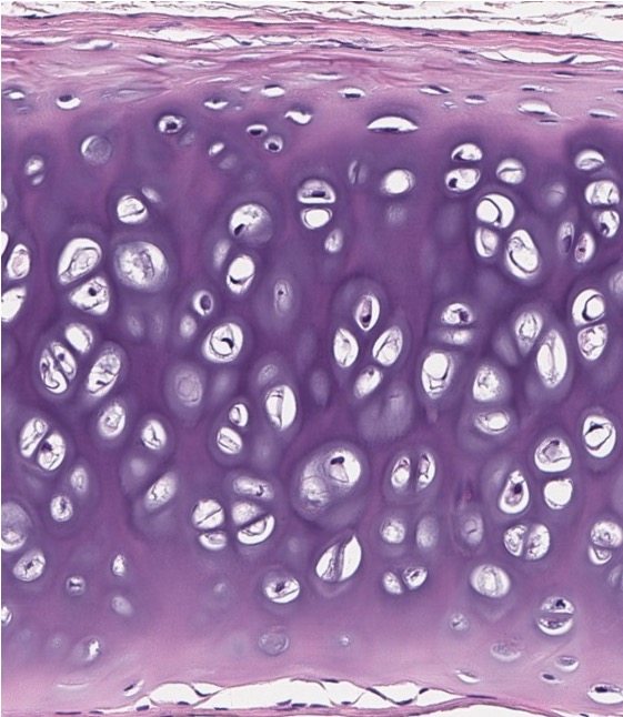
1. Which type of CT makes up the bulk of this image?
2. Does it have a perichondrium?
1.Hyaline cartilage
2.Yes
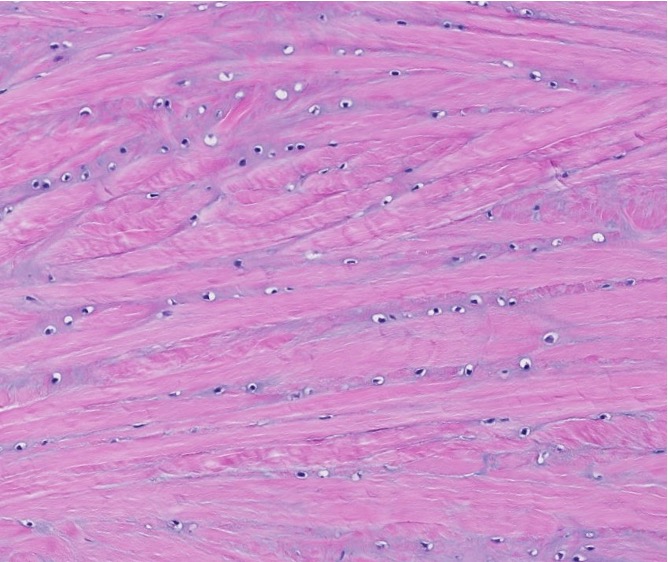
Which type of CT is this?
Fibrocartilage (the round spaces are lacunae, occupied by chondrocytes).
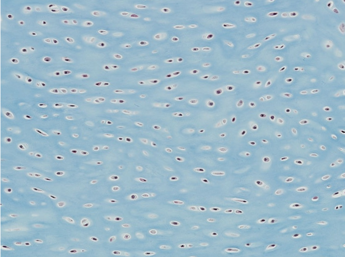
Which type of CT is this?
Hyaline cartilage
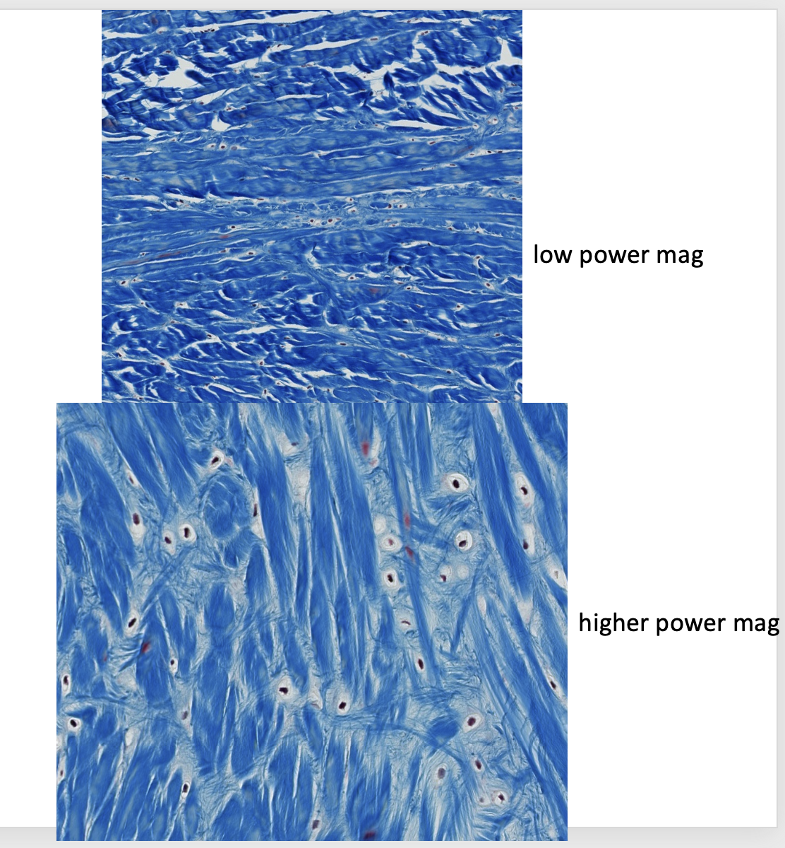
Which type of CT is this?
Fibrocartilage (the round spaces are lacunae, occupied by chondrocytes).
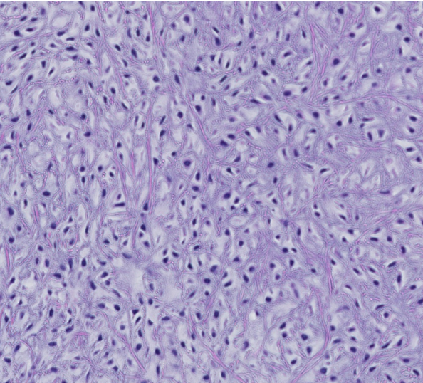
The iris diaphragm is closed to enhance naturally glowing fibers, indicating thisis which type of cartilage in the larynx?
Elastic cartilage
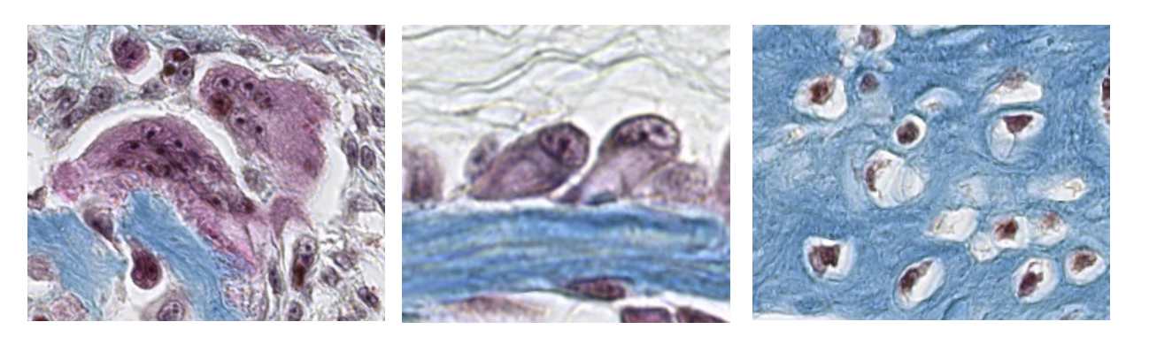
This is a high-power magnification of the three basic types of bone cells: osteoblasts, osteocytes, osteoclasts.
ID the bone cells, from left to right.
Left – osteoclasts
Middle – osteoblasts
Right - osteocytes
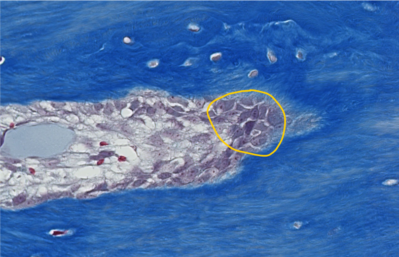
1. ID the encircled bone cells.
2. What CT layer are these cells located within?
1.Osteoblasts
2.Endosteum
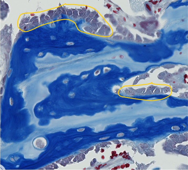
ID the encircled bone cells.
Osteoblasts (they are on the bone surface).
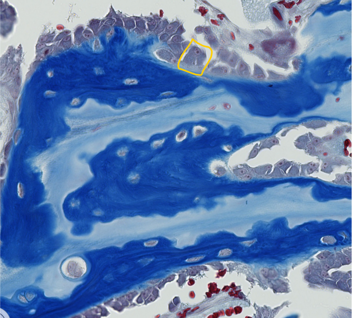
What is the name of the unmineralized matrix of collagen type I & proteoglycans secreted by the encircled bone cell?
Osteoid
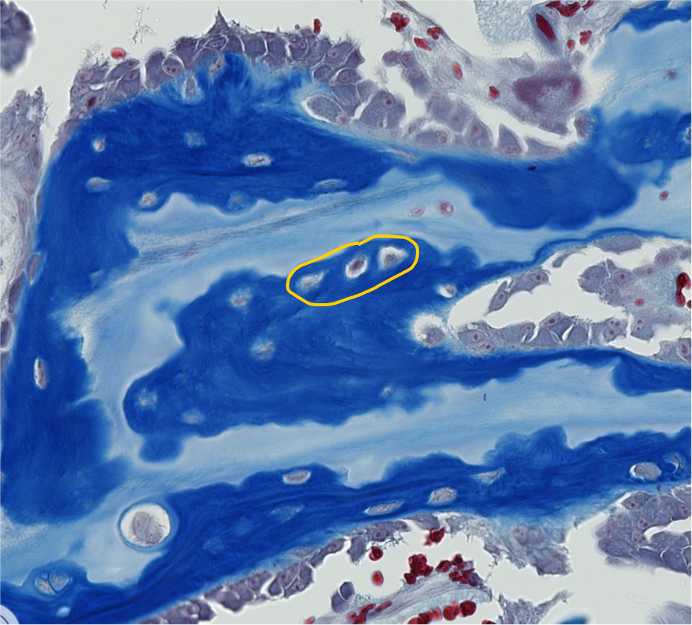
ID the encircled bone cells.
Osteocytes (they are embedded within the bone).
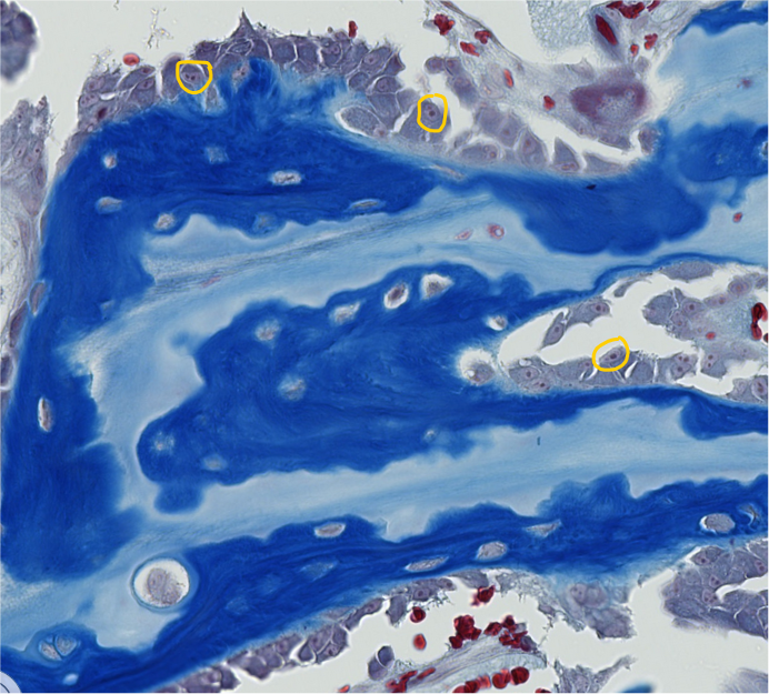
1. Which cell organelles are encircled with yellow?
2. What are the small dark circles visible inside the encircled cell organelles?
Nuclei
Nucleoli
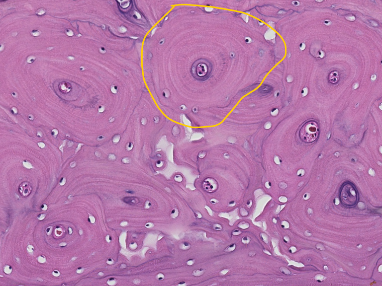
This is the diaphysis (shaft) of a long bone in transverse (cross) section.
ID the encircled structural unit of mature bone.
Osteon (Haversian system).
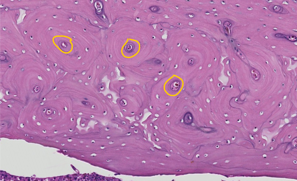
This is the diaphysis (shaft) of a long bone in transverse (cross) section.
ID the encircled region of the osteon.
Central canal
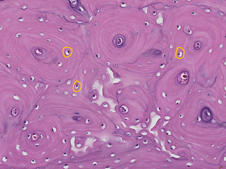
This is the diaphysis (shaft) of a long bone in transverse (cross) section.
ID the encircled structures within the osteon.
Lacunae with osteocytes
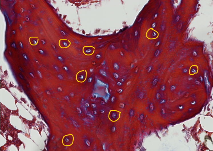
ID the encircled structures in this bone spicule.
Lacunae with osteocytes
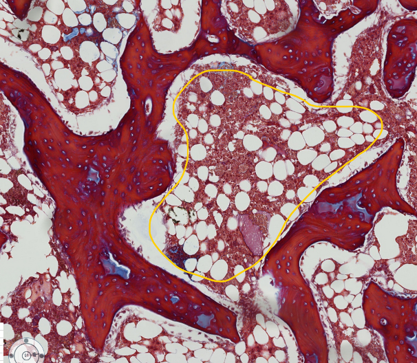
The encircled tissue is bone marrow, surrounded by bone spicules.
What do you think the clear spaces are? Hint: it is a type of adult CT proper
Adipocytes (white fat)
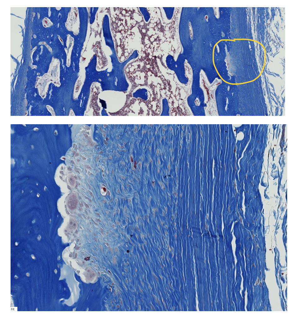
The upper image is full thickness through the shaft of a long bone, with the external surfaces on the left and right sides of the image.
The encircled area in the upper image is enlarged in the lower image.
1. What do we call the encircled external fibrous membrane (outer CT covering) of bone?
2. What does “fibrous” refer to in this context?
1.Periosteum
2.Collagen fibers
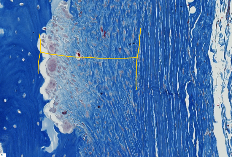
In actively growing bone, the periosteum has two visible layers.
ID the layer within the yellow bracketed portion .
(This is a high-power magnification of the periosteum of a long bone.)
Inner cellular layer
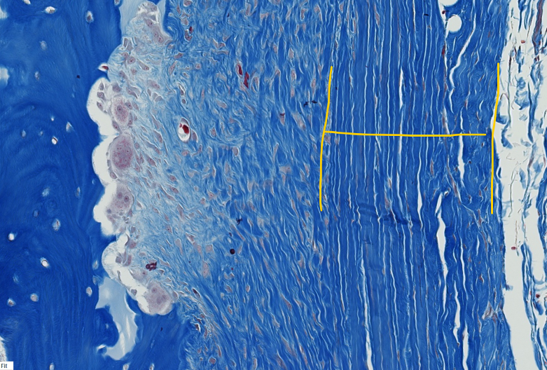
In actively growing bone, the periosteum has two visible layers.
ID the yellow bracketed layer.
Outer fibrous layer
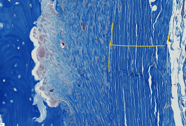
What type of collagen fibers make up the bulk of the indicated layer?
Type I collagen
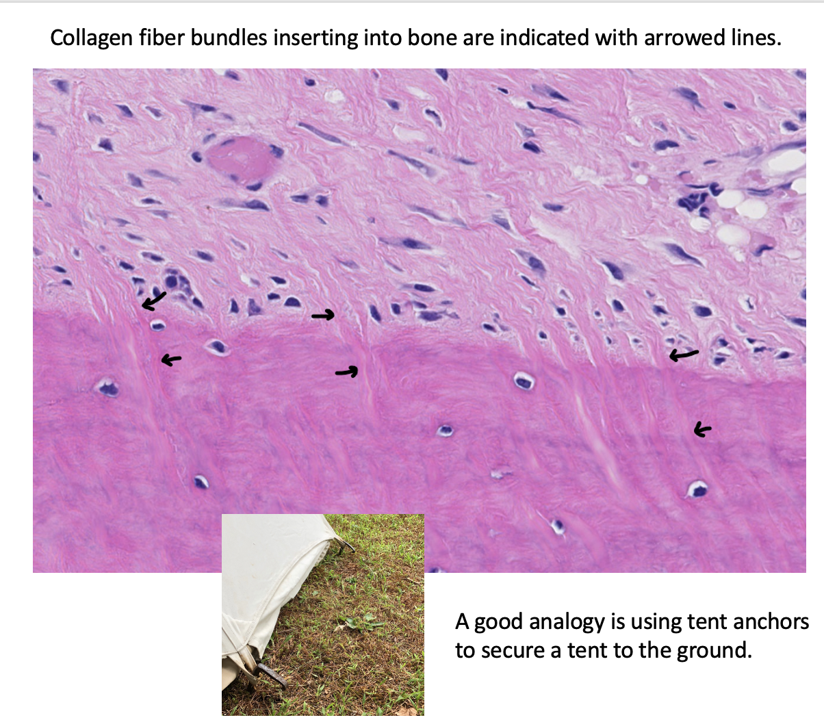
Multiple small bundles of collagen fibers from tendons, ligaments, and the periosteum are embedded within the underlying bone as a means of anchoring these structures.
What do we call these fibrous insertions?
Sharpey’s (perforating) fibers
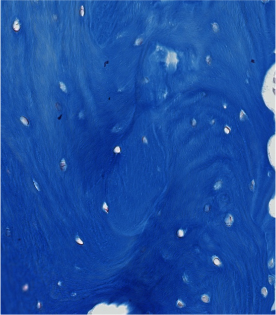
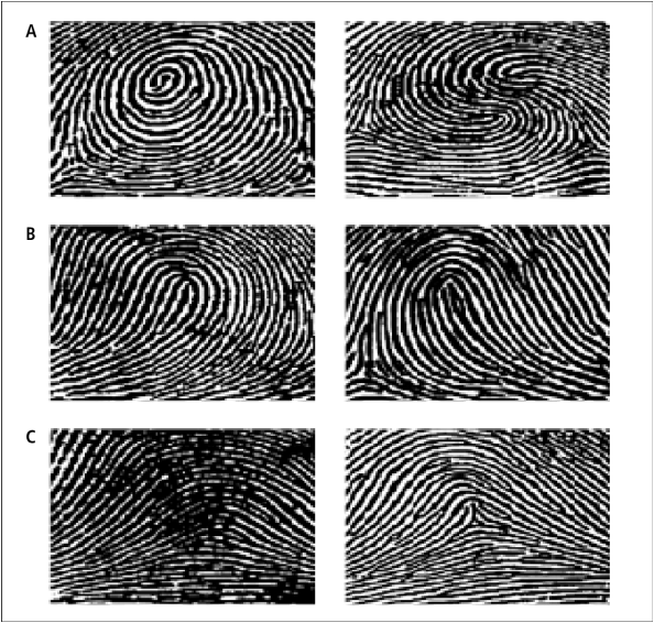
What is the microscopic architecture of woven bone (how are the collagen fibers arranged)?
The collagen fibers of immature or woven bone are randomly arranged in whorl-like layers, similar to your fingerprints.
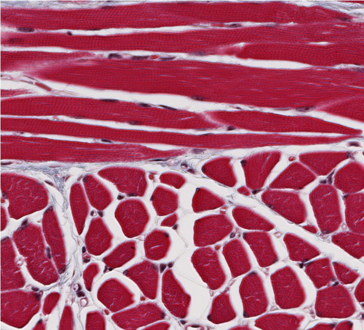
ID the type of muscle.
skeletal
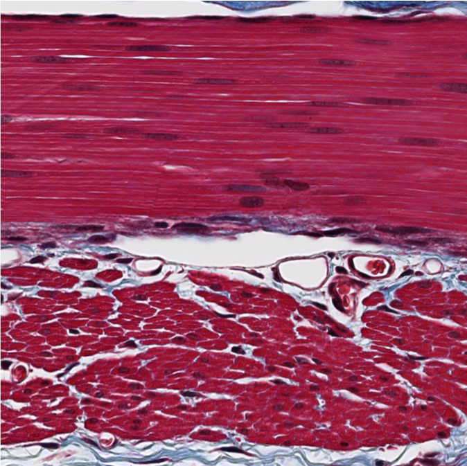
ID the type of muscle.
smooth
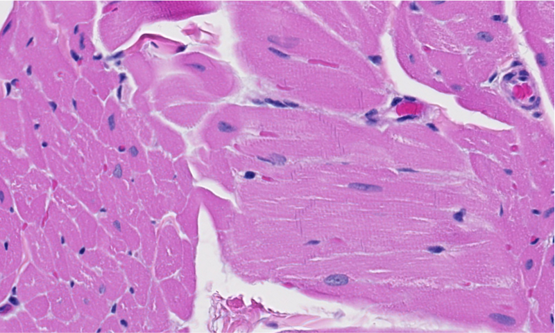
ID the type of muscle.
cardiac
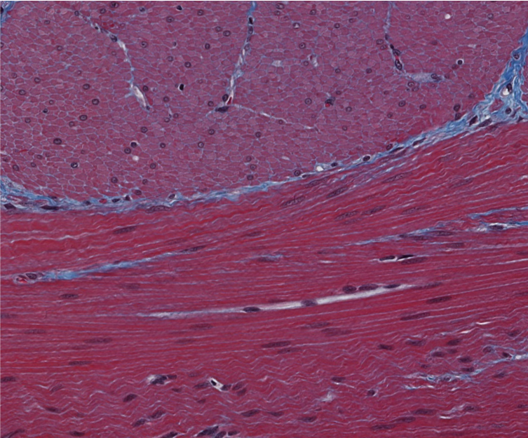
ID the type of muscle.
smooth
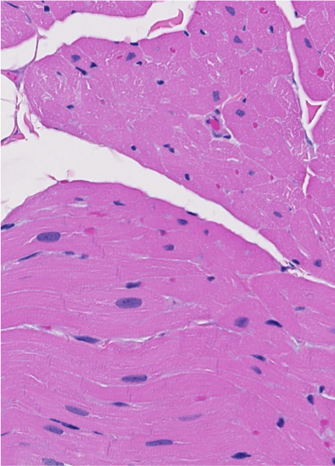
ID the type of muscle.
cardiac
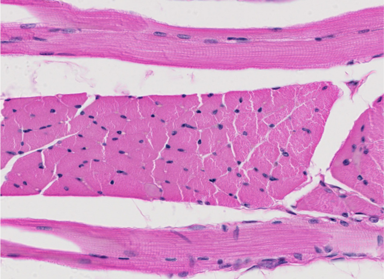
ID the type of muscle.
skeletal
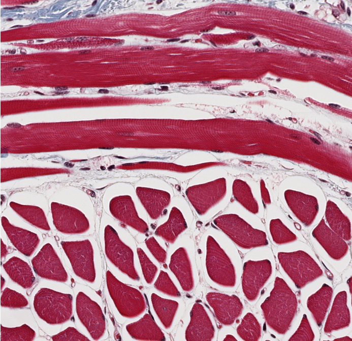
ID the type of muscle.
skeletal
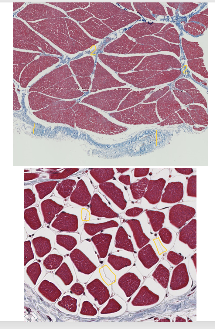
Skeletal muscle has three CT layers: epimysium, perimysium, and endomysium.
ID the numbered CT layers.
1. Epimysium
2. Perimysium
3. Endomysium
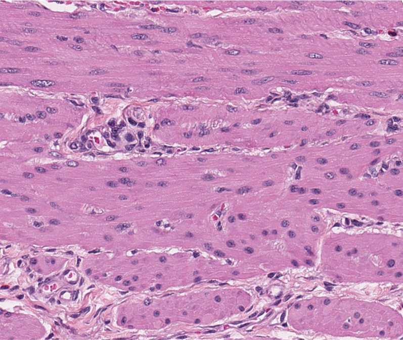
ID the type of muscle.
smooth
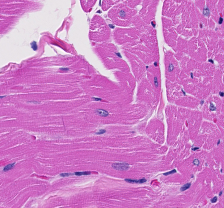
ID the type of muscle.
cardiac
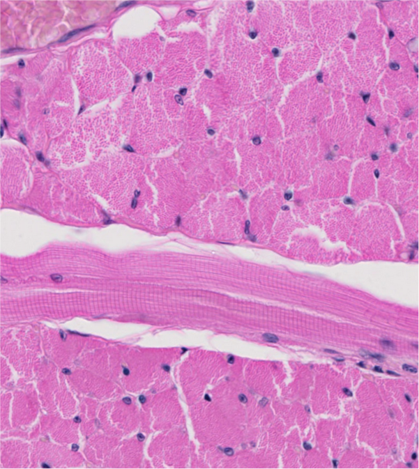
ID the type of muscle.
skeletal
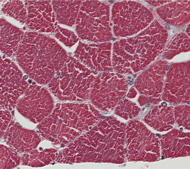
Which internal structures create the “stippling” visible inside these transversely sectioned muscle cells?
Hint: Filament << Fibril << Fiber
myofibrils
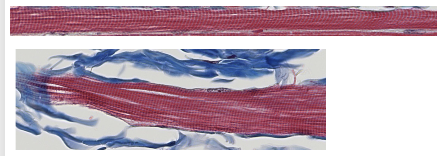
1. Which structures create the light and dark banding pattern within the myofibrils?
2. Which of these structures create the light bands? Which create the dark bands?
1.Myofilaments
2.Light I bands = actin myofilaments & dark A bands = overlapping actin & myosin myofilaments
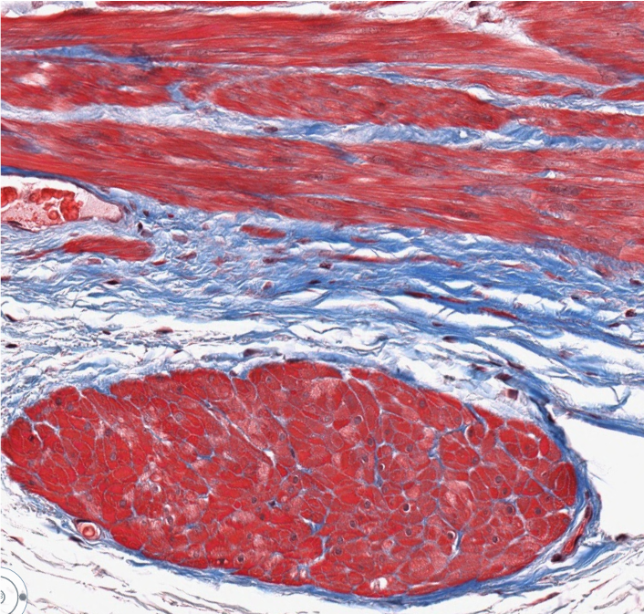
ID the type of muscle.
smooth
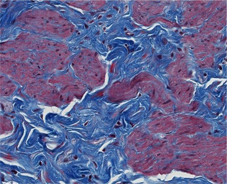
1. ID the type of muscle.
2. ID the type of CT proper that surrounds this muscle.
1.Smooth
2.Dense irregular
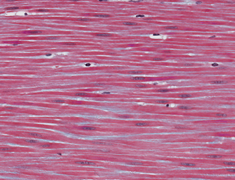
What is the shape of these smooth muscle myofibers?
Fusiform (spindle-shaped with tapered ends)
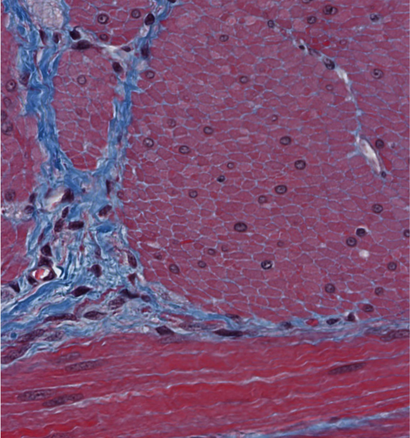
Are these myocyte nuclei centrally or peripherally located?
centrally (smooth muscle)
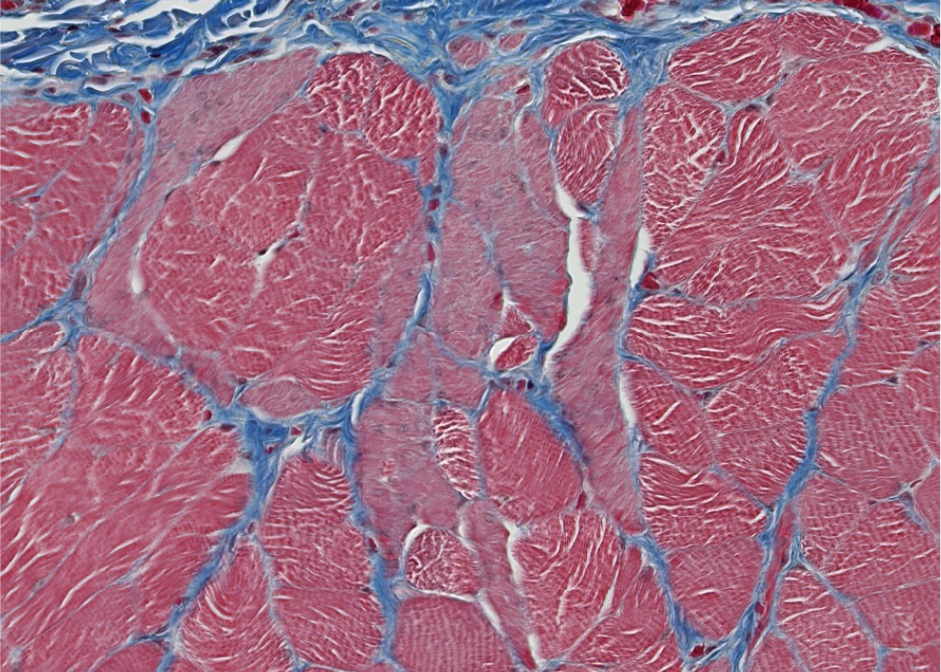
ID the type of muscle in this esophagus section.
skeletal and smooth
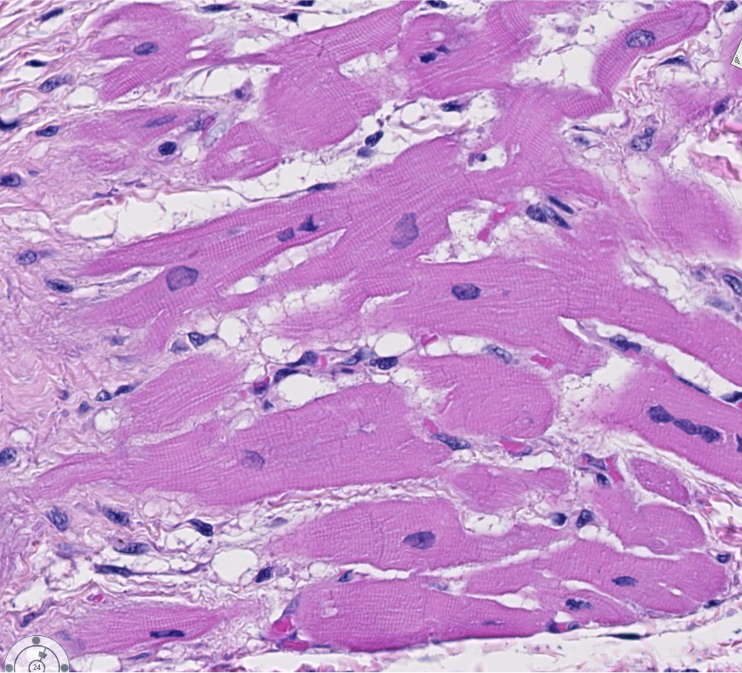
This image illustrates some of the key features of cardiac muscle.
Which key features can you identify in these longitudinally arranged cardiac myofibers?
Branching myofibers, centrally positioned nuclei, and intercalated discs.
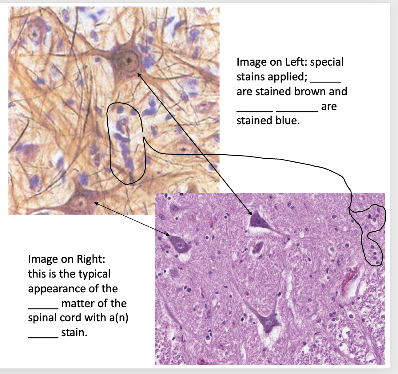
Fill in the blanks:
neurons
glial cells/glial cell bodies (aka support cells)
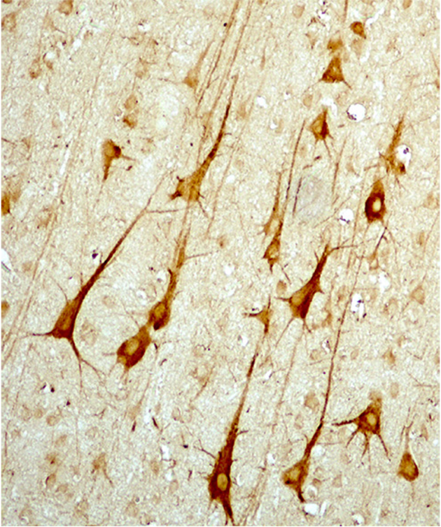
Image: Pyramidal neurons in cerebral cortex. IHC was used to stain the ___________ (cytoskeleton).
neurofilaments
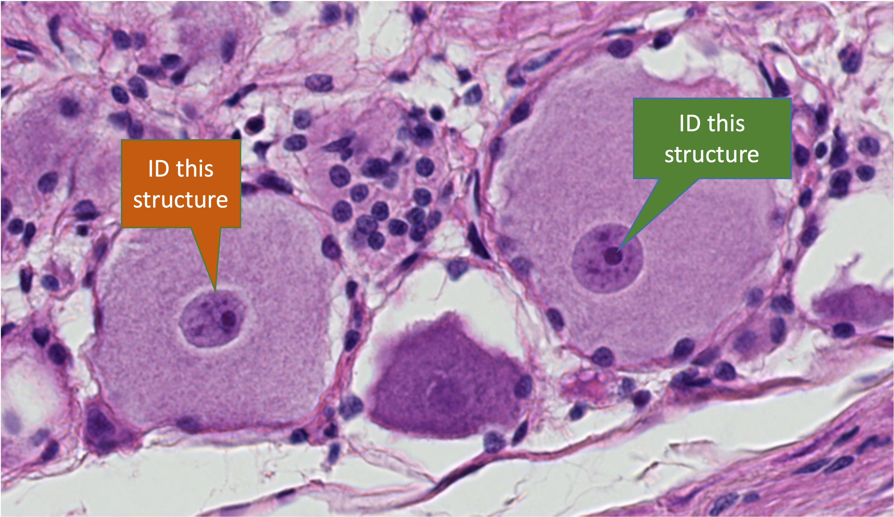
1. ID the structure indicated with the green callout box.
2. ID the structure indicated with the dark orange callout box.
Dark Green - nucleolus
Dark orange- nucleus
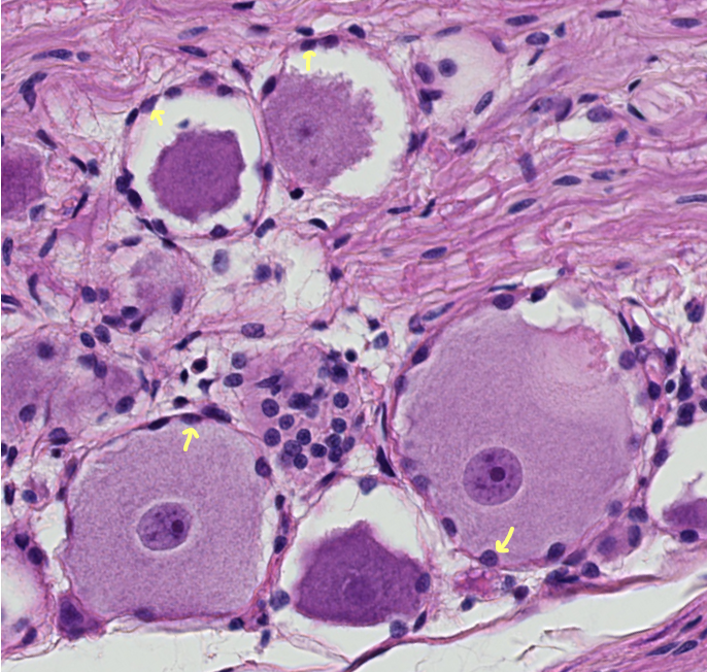
The indicated support cells are forming a protective capsule around the neuron cell bodies in this sensory ganglion.
What do we call these PNS glial cells?
Satellite cells (ganglionic gliocytes).
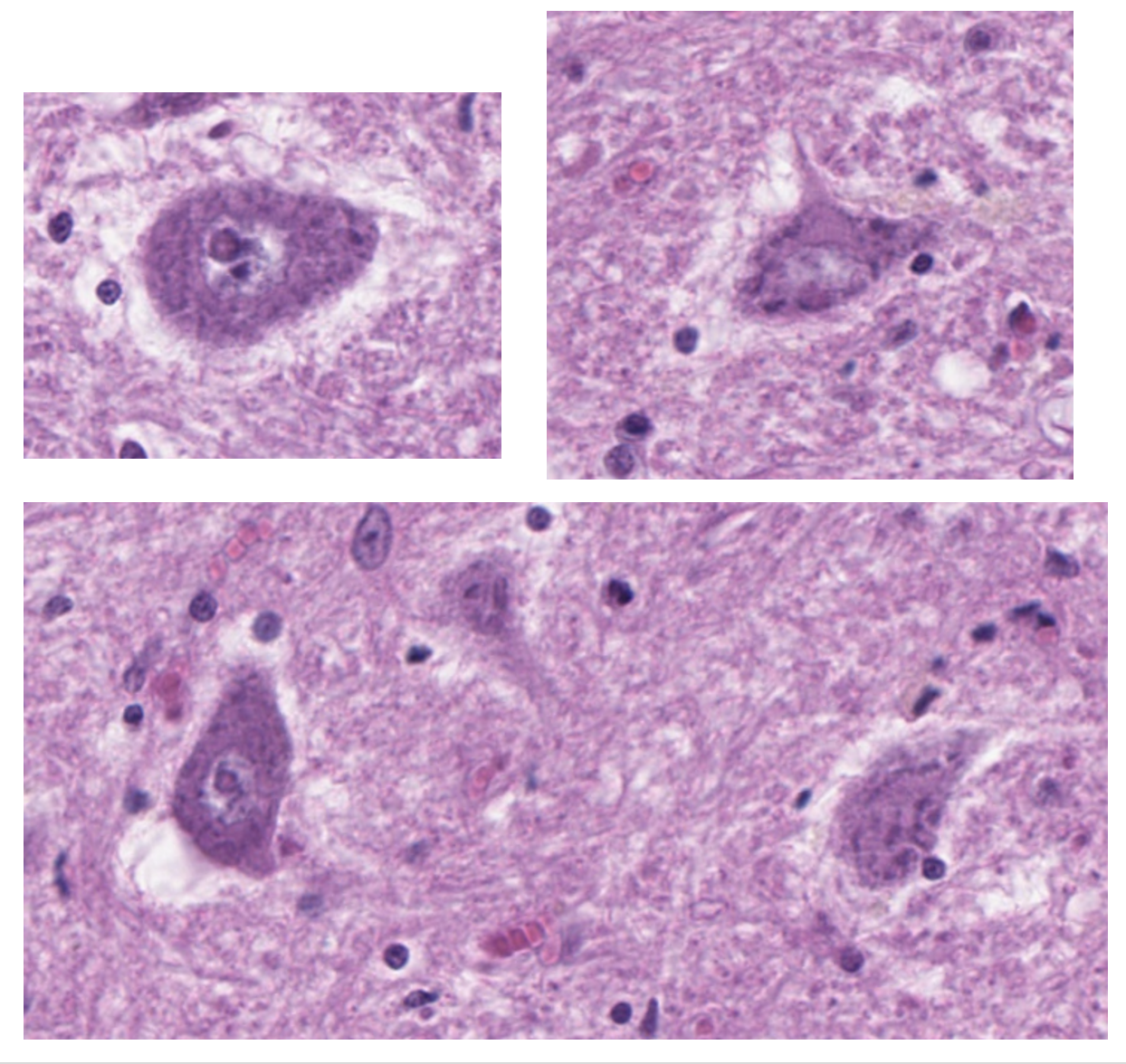
The cytoplasm of these large motor neurons has dark staining (basophilic) clumps that create a granular appearance.
What do we call these basophilic clumps?
What makes them basophilic?
Nissl (chromatophilic) substance has abundant RNA, which binds the basic tissue dye.
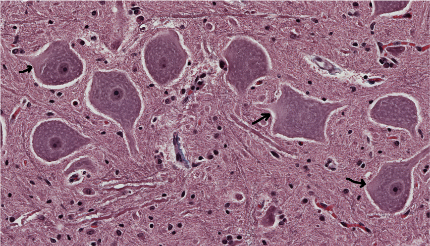
Three clear-staining areas in the cytoplasm of some of these large motor neurons are indicated with arrowed lines.
1. What are they called?
2. Why are they clear-staining?
1. They are axon hillocks, meaning the “little hills” in the cytoplasm where the action potential begins and travels out through the axon.
2. They do not contain Nissl substance.
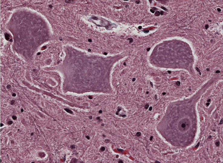
There are four large motor neurons in this image. Why does only one of them have a visible nucleus? Is there something wrong with them?
No, they are normal neurons.
The cell bodies of these motor neurons are so large, the knife can easily miss the nucleus when slicing through the neuron in a tissue section.
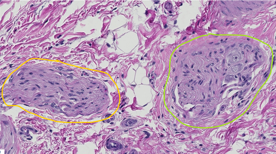
Something is different between the two structures encircled in yellow and green.
ID these structures.
Encircled with Yellow – nerve fascicle
Encircled with Green – motor ganglion (has 2-3 visible neurons)
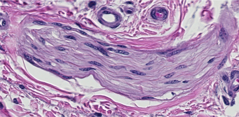
ID the structure that makes up the bulk of this image.
nerve fascicle
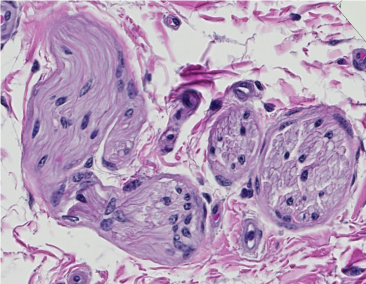
Is this nerve fascicle myelinated or unmyelinated?
Unmyelinated (no visible neurokeratin overlying the axons; can see linear portions of the endoneurium between the axons)
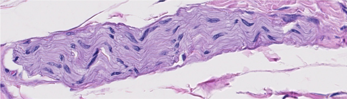
Is this nerve fascicle myelinated or unmyelinated?
unmyelinated
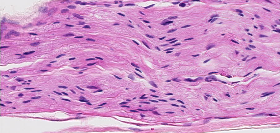
Is this nerve fascicle myelinated or unmyelinated?
Myelinated (there is visible neurokeratin)
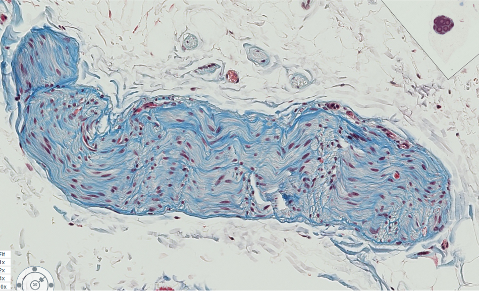
Is this nerve fascicle myelinated or unmyelinated?
Unmyelinated (no visible neurokeratin, only slightly pink axons).
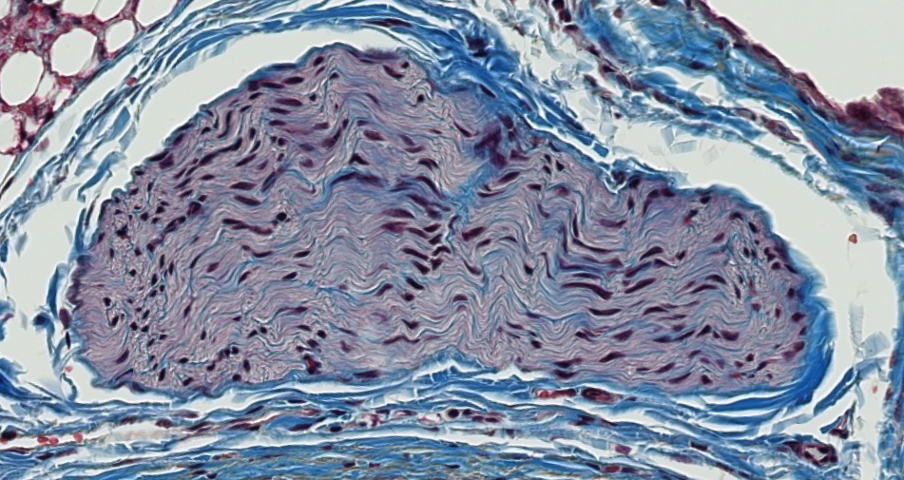
Is this nerve fascicle myelinated or unmyelinated?
Unmyelinated (the axons are pinkish grey in this example and were just gray in the previous example, but there is not visible neurokeratin overlaying the axons)
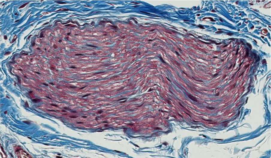
Is this nerve fascicle myelinated or unmyelinated?
Myelinated (lots of neurokeratin!)
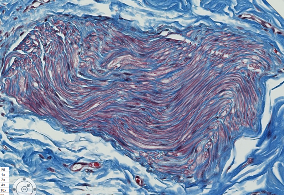
Is this nerve fascicle myelinated or unmyelinated?
Probably Mixed Myelinated & Unmyelinated (not as much neurokeratin as the previous example, and it is patchy)
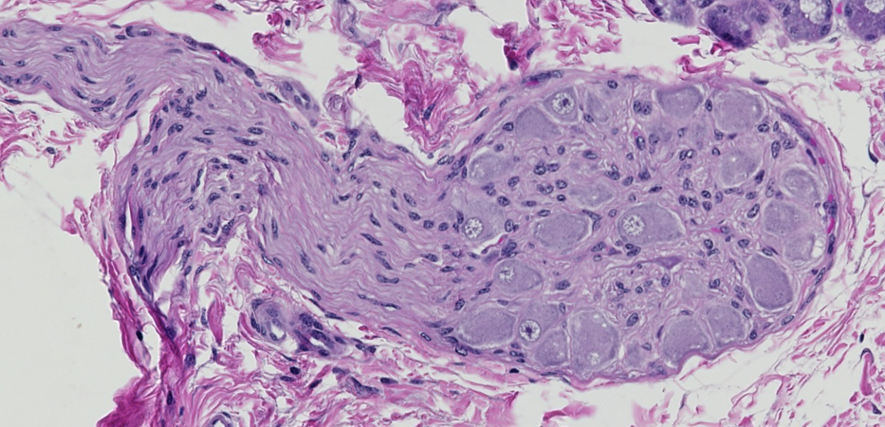
ID this structure that makes up the bulk of this image.
Motor (autonomic) ganglion (think of it as a nerve fascicle that also has neurons).
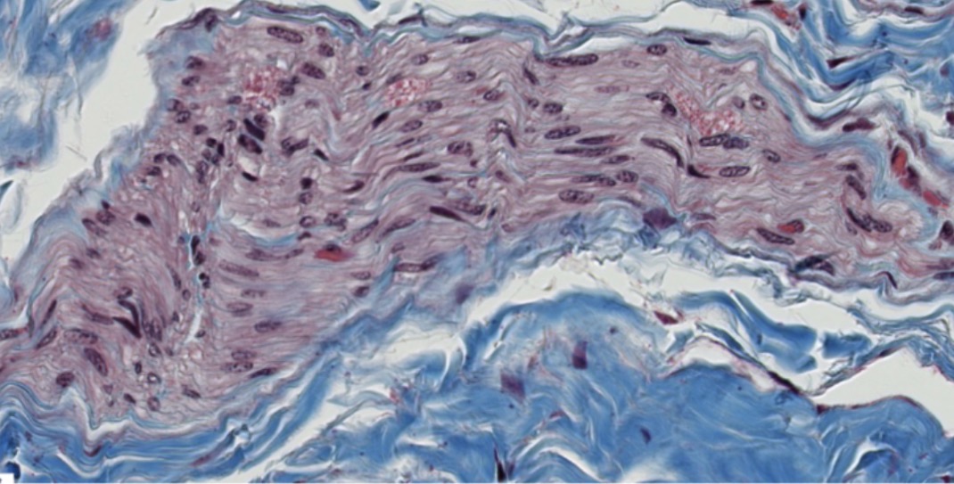
Is this nerve fascicle myelinated or unmyelinated?
Technically, this nerve is mixed (both); however, the vast majority of the axons are unmyelinated.