neuro overview
1/66
There's no tags or description
Looks like no tags are added yet.
Name | Mastery | Learn | Test | Matching | Spaced | Call with Kai |
|---|
No analytics yet
Send a link to your students to track their progress
67 Terms
what structure does the pudendal nerve provide
somatic motor innervation
somatic sensory innervation
the anal sphincter
the perineal skin
where does the pudendal nerve convey parasympathetic fibres from and to
sacral spinal cord to rectal smooth muscle
what is another thing that the pudendal parasympathetic division controls
urinary bladder contraction
what nerve is responsible for hamstring, distal limb muscle and cutaneous innervation below the stifle
the sciatic nerve
what is the largest foramen and what major structures pass through
foramen magnum
the spinal cord into the cranial cavity where it becomes continuous with the lower end of the medulla oblongata
vertebral arteries
anterior and posterior spinal arteries
spinal roots of the accessory nerve
3 foramina and nerves that exit through
foramen ovale: mandibular nerve
jugular foramen: glossopharyngeal nerve, vagus nerve, accessory nerve
foramen rotundum: maxillary nerve
a) Name two important structures that pass through the intervertebral spaces and foramina?
The articulation of the skull and the Atlas (the atlanto-occipital joint) creates a large space on the dorsal aspect. Identify this space. b) What might be the clinical significance of this space?
craniocervical junction
protets the structures transitioning from the skull to the spinal column
foramen magnum is a critical passage point
conduit for major blood vessels supplying the brain eg the vertebral arteires
white and grey matter of the brain
grey matter consists of neuronal cell bodies and dendrites and synpases and glial cells
white matter is myelinated axons
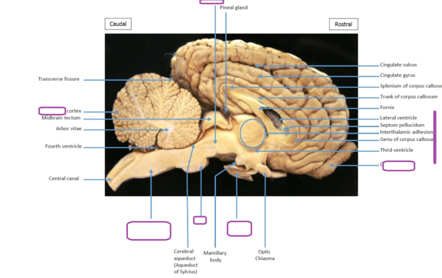
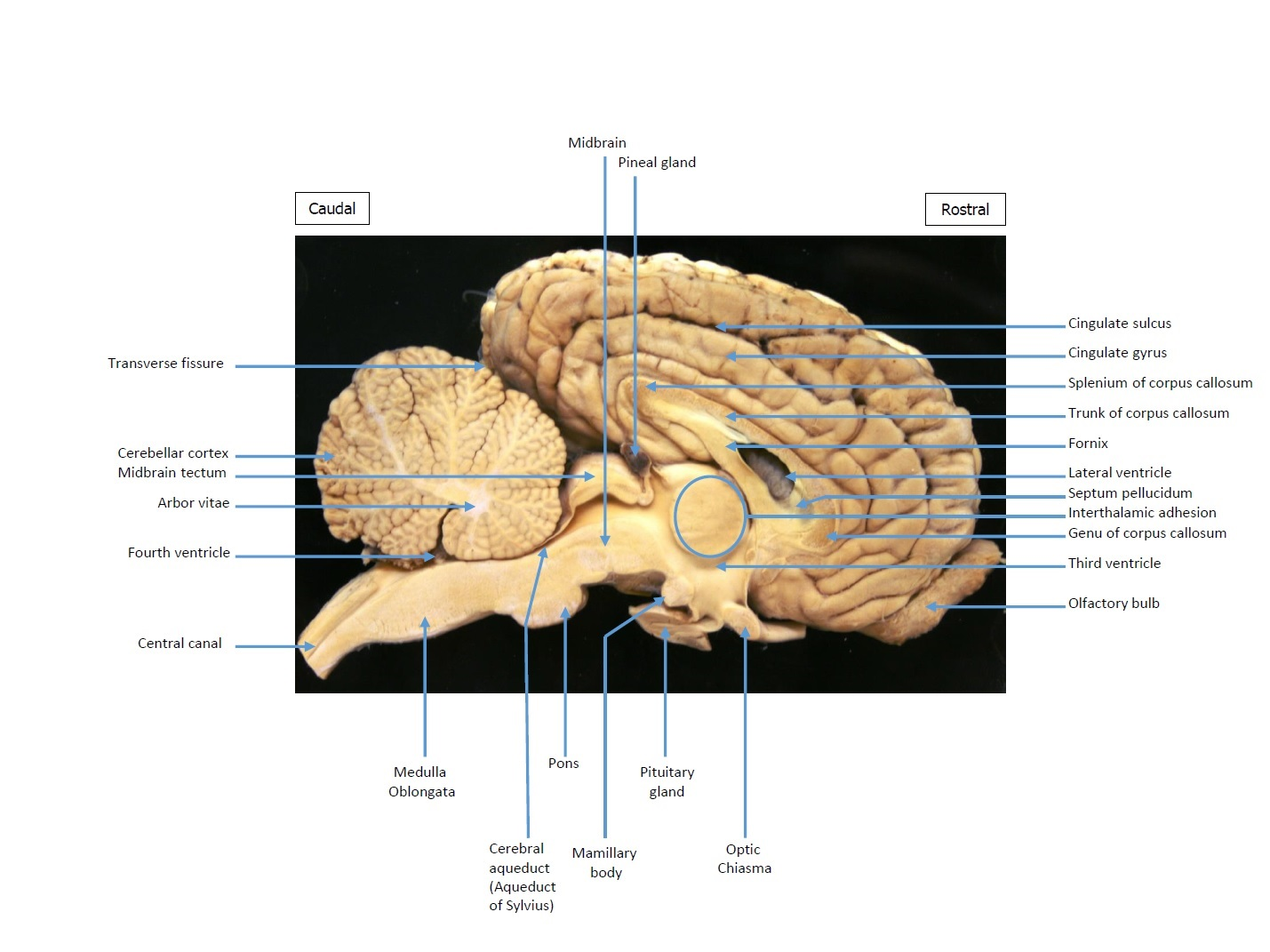
Identify and name the tough membrane that covers the outer surface of the brain.
dura mater
Name the other two layers of membranes that cover the brain.
arachnoid mater
pia mater
Briefly explain why the two membranes you have named in (b) above are not discernible over the brain surface.
what are the elevations and depressions on the brain called
gyri and sulci
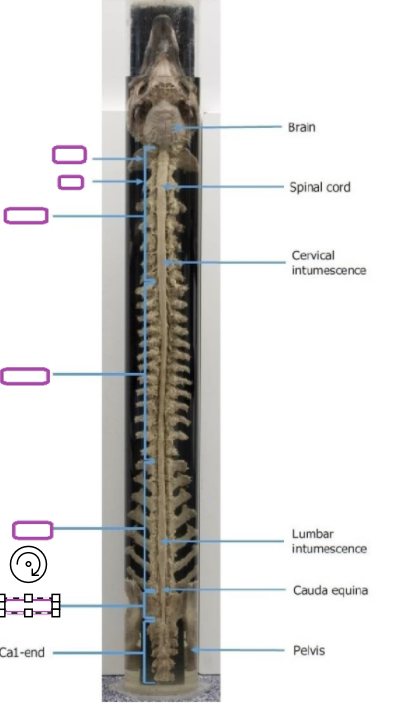
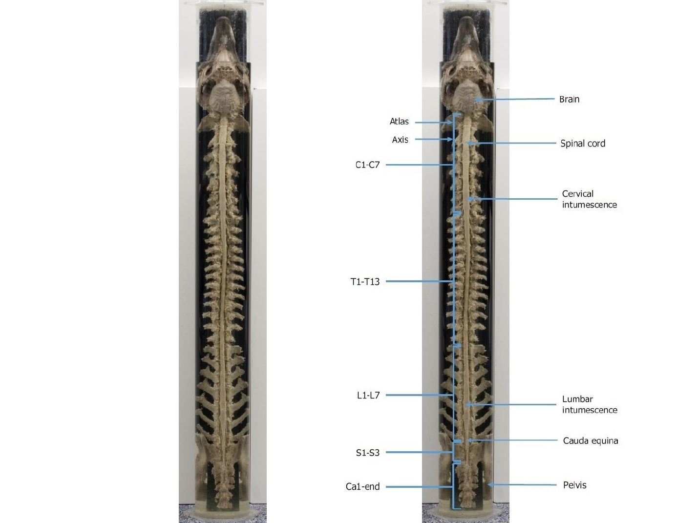
How far caudally does the spinal cord extend in the vertebral column? Use the vertebrae number (e.g. C1, T1 etc.) as a marker.
L2
here are two areas of the spinal cord where it briefly thickens, known as the intumescences. They correspond to the regions of the forelimb and hindlimb, respectively. b) What is the likely reason for these swellings?
Describe how the spinal cord terminates at its most caudal end.
where does it travel inferiorly within
what is it surrounded by
where does it taper off and what does it form
what do the spinal nerves form
it travels inferiorly within the vertebral canal surrounded by spinal meninges containing csf
at the l2 vertebrae level the spinal cord tapers off forming the conus medullaris
the spinal nerves that arise from the end of the spinal cord are bundled together forming the cauda equina
What does the DRG consist of?
a collection of neuronal cell bodies of sensory neurons
How does the location of the dura mater in the vertebral column compare to that of the cranium?
cranial dura mater
consist of an outer periosteal layer and an inner meningeal dura
periosteal is closely attached to the internal surface of skull bones
meningeal is continuous with the dura of the spinal cord
periosteal and meningeal are tightlly fused together except for where they separate to for the dural ventral sinuses
spinal
only one layer
not closely integrated with the overlying bones
there is an epidural space
Some cranial nerves travel away from the cranium to innervate structures far away from the head. In pot 361, observe the nerve passing down the neck all the way to the thorax. The same nerve can be viewed in the additional pots and laminates, running very close to the base of the heart. i) Identify this cranial nerve
What is the effect of stimulation of this nerve on the heart?
What is the effect of stimulation of this nerve on the intestines?
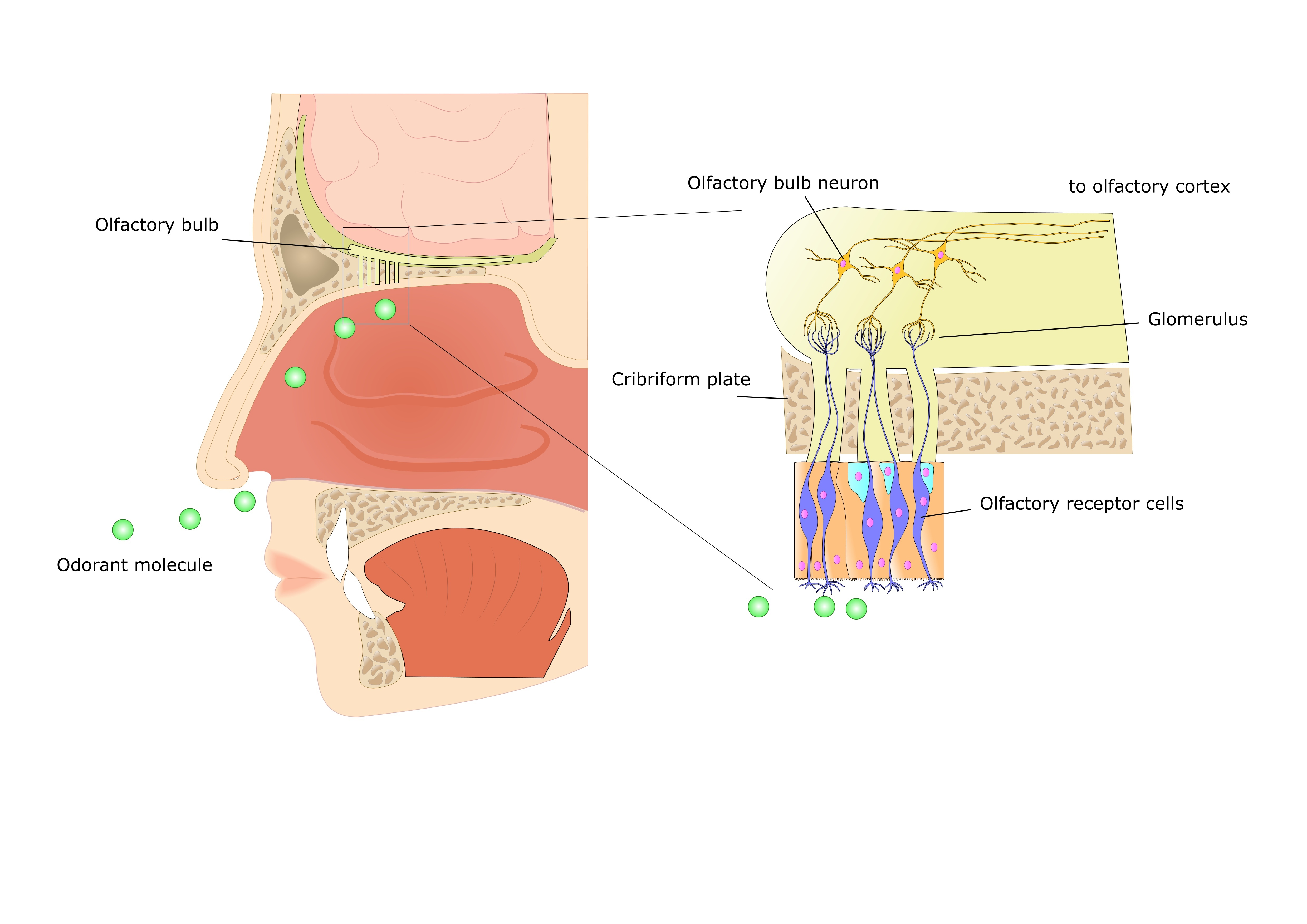
Which part of the brain connects directly to the neurons projecting the nasal cavity?
olfactory bulb
what are the 4 main nerves supplying the canine forelimb?
radial nerve
musculocutaneous nerve
median nerve
ulnar nerve
which nrve passes down the neck to the thorax and runs close to the base of the heart?
what type of nerve is it
is it afferent or efferent
effect on heart rate
effect on the intestines
vagus nerve
mainly parasympathetic autonomic
both afferent and efferent
slow heart rate
increases intestinal activity
what are the major cell types found in the retina
rods and cones
retinal ganglion cells: transmit info from the rods and cones to the brain, a bundle forms the optic nerve
list all special sense organs
nose- olfaction
vomeronasal organ- phermomone detection
tongue- taste
eye- vision
ear- hearing and balance
olfaction
which cells detect
what contain the sensory receptors
which type of neurone and where do they pass through and through which structure
what is the epithelium held by and what do they secrete
detected by special cells in muscosa of nasal cavity
mucose= olfactory epithelium, contain sensory receptors
neurons (bipolar) pass through epithelial surface to olfactory bulb in cranium after passing through bony cribiform plate (separate cranial cavity from nasal cavity)
cribiform plate has small pores
olfactory epithelium held by supporting cells that secrete lipid rich mucus, odorants dissolve in this fliod and reach the sensory receptors
signal tranduction takes place through neurons
vomeronasal organ
where is it found
what do ducts link
what type of structure are they
found on the floor of the nasal cavity
ducts link nasal and oral cavities caudal to incisor teeth
are blind ending caudal sacs
flehmen reaction- pump air in and out (sexual and social behaviour)
aroma is the combined effect of neural inputs from the sense o smell and taste
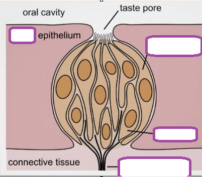
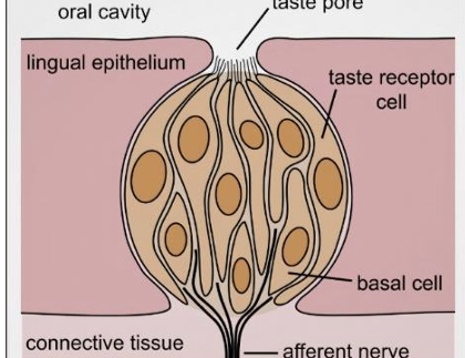
gustation
where are gustatory receptors found
features of the receptor cells
where do gustatory inputs link directly to
gustatory receptors found on the tongue mucosa
sensory neurons carry info to the brain
receptor cells have one single receptor type so each receptor can only detect one form of taste
gustatory inputs link directly to centres involving ingestion, food avoidance, insulin release, diuresis when water in pharynx
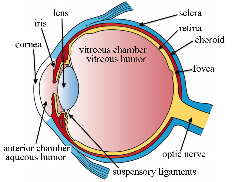
vision
what conducts light and what does this stimulate
what substance fills the eye
what is the sclera and what structure is it continuous with
eye has transparent media that conducts light to stimulate photoreceptor cells
vitreous humor is gel like substance filling the eye
sclera is a tough connective tissue to maintain integrity and strength- continuous with membrane covering the brain.
ear
outer middle and inner
the eye
what are the 3 layers
how is the light signal transmitted
what protein is present in rods and cones and what does it trigger through what and where is it transmitted
Transparent media- Cornea, aqueous humour, lens and vitreous humour direct and converge light on the photoreceptor cells on the retina
Non-Transparent media- Choroid, Uvea, Sclera- Support transparent media
The photosensitive layer - retina made up of rod and cones receptor cells. Cones for daylight vision and rods for night vision.
Light splits chemical compound -Rhodopsin in cones and rods and triggers signal transduction thru optic nerve. The signals are transmitted to the optic cortex of the brain.
outer ear
sound collected from external auditory canal to the tympanic canal and tympanic membrane
middle ear
including what structure it is connected to
maleus, incus and stapes conducts sound to oval window. also connected to eustachian tube to the nasopharynx
inner ear
oval window transmits waves to the cochlea which contain sensory receptor cells known as hair cells from where signals are transmitted to the brain via CN VIII
Ear- balance and motion
what structure detects angular movement
what detects linear acceleration
inner ear has semicircular canals that detect angular movement while the saccule and utricle (maculae) detect linear acceleration
cochlea
what structures are located in the cochlea
what causes a standing wave to travel in cochlea canals
what does this cause
basilar membrane
organ of corti
hair cells
CN VIII
fluid movement in the cochlea caused by sound vibration on the oval window cause a standing wave to travel in cochlea canals.
so hair cells on basilar membrane bends against tectorial membrane
then signals are generated and sent to the brain
hair cells bend due to fluid movement and discharge electrical signals to the brain
semi circular canal
function
innervation
cupula
hair cell
CN III
hair cells of the cupula bend due to fluid movement in the semicircular canals and discharge electrical signals to the brain
saccule and utricle
what does it detect
what angles are they at
detection of linear acceleration
hair cells
CN VIII
hair cells (otoliths) bend due to fluid movement caused by inertia in the saccule and utricle causing them to discharge electrical signals to the brain
two are in right angles to each other so can only detect linear movement in one plane
components of the somatic nervous system
nerves
what is incoming information coordinated by
(voluntary control)
afferent nerves- reception of internal and external stimuli
incoming info coordinated by spinal cord, somatomotor cortex and cerebellum
efferent transmit impulses from cns to skeletal muscles
components involved in the somatic
joints, skin, skeletal muscle, somatosensory fibers, somatomotor fibers
signal transduction route- sensory division
sensory inputs to SC and brain
SC initiate voluntary motor control
SC also sends info to brain to initiate voluntary motor control
SC and brain sends response to contract muscle
signal transduction route- motor division
spinal cord initiates response via lower motor neurons or upper motor neurons of the somatomotor cortex and brain stem pathways make contact with spinal lower neurons
somatomotor cortex initiate voluntary movement and modulate muscle/reflex activity
patella reflex
tap/bend the patella tendon
stretch receptors in muscle simulated
sensory neuron in muscle send signal spinal cord
spinal cord detects signal and sends response
motor neuron carry response to muscle from spinal cord
quads muscle contracts
meisnyer corpuscles
skin light touch
merkers disk
skin touch and texture
pacinian corpuscle
pain and deep pressure
ruffini corpuscle
stretch / kinesthesia
free nerveending
pain
components
parasympathetic, sympathetic, enteric
target tissues
cardiac, smooth, glandular tissue
what is the ans involved in
percieved sensation
involuntary inhibition or excitation of visceral and glandular tissue
homeostasis
complements the endocrine system
components of the ans
general visceral efferent motor neurons- visceromotor fibres- target tissues
general visceral afferent sensory neurons- enteroreceptors (chemoreceptors, baroreceptors)- viscero sensory fibres. Sensory fibres coveyed from the periphery to the cns via cranial nerves and somatic spinal nerves
central integration centres (brain)- hypothalamus, brain stem (pons, medulla)
anatomy of the ans
2 types of neurons
2 neuron system in the periphery
pre-ganglionic neuron- cell body in brain or spinal cord
post-ganglionic neuron-cell body lies outside cns in autonomic ganglia
which is parasympathetic and sympathetic from craniosacral outflow and thoracolumbar outflow
craniosacral- parasympathetic
thoracolumbar- sympathetic
parasympathetic
cranial parasympathetic outflow via which nerves
sacral outflow via which nerves
cranial parasympathetic outflow via:
cranial nerves III, VII, IX AND X
to visceral and glandular tissues of the head
CN X - vagus nerve
heart
most of the GIT
sacral outflow (S1-S3)
caudal GIT
urinary tract
reproductive organs
sympathetic
which spinal nerves to thoracolumnar outflow
to head and neck via which spinal nerves
thoracolumbar outflow via
spinal nerves T1 to L4
via sympathetic chain
to head and neck T1-T3
to organs of thorax abdomen and pelvis
disributed via several peripheral ganglia
autonomic vs somatic motor neurons
autonomic
pre and post ganglionic
myelinated presynaptic and unmyelinatd post synaptic
can be ach or nad
somatic
one neuron only
always excitatory
vagosympathetic tract what does it contain
parasympathetic traveling caudal direction and
sympathetic traveling cranial in neck
clinical significance of the vagosympathetic trunk
runs within the carotid sheath deep to to the jugular groove
vulnerable to needle stick injuries
control centres of autonomic
brainstem and hypothalamus