Human anatomy and physiology module 5
1/52
There's no tags or description
Looks like no tags are added yet.
Name | Mastery | Learn | Test | Matching | Spaced |
|---|
No study sessions yet.
53 Terms
What are the 6 primary functions of the skeletal system?
-Support: Skeleton creates a framework for the body that the other tissues attach to
-Protection: Skeletons provide protection to inner body parts
-Assistance in movement: Bones create joints with other bones which allow for movement
-Mineral homeostasis: Has the ability to store or release which helps them in staying within their homeostatic range
-Blood cell production: Bones have red bone marrow, which is where new blood cells are created
-Triglyceride storage: Bones have yellow bone marrow, this is a fatty tissue and stores lipids called triglyceride
What are the two primary aspects of connective tissues?
-Extracellular matrix
-Cells
What are the two primary aspects of the extracellular matrix?
-Extracellular protein
-Ground substance
What are the three cells that have the function to synthesise and secrete extracellular protein fibres?
-Osteoblasts: Cells that synthesise and secrete much of the extracellular matrix bone (osteo = bone)
-Chondroblasts: Cells that synthesise and secrete much of the extracellular matrix cartilage (chondro = cartilage)
Fibroblasts: Cells that synthesise and secrete much of the extracellular matrix of dense connective tissues (softer than bone and cartilage)
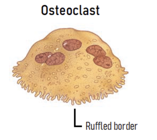
What is an osteoclast??
-Breaks down bone of the tissue
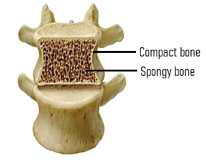
What are the 2 types of osseous tissues?
-Compact bone tissue: Very dense, hard bone
-Spongy bone tissue: Porous, honeycomb like appearance
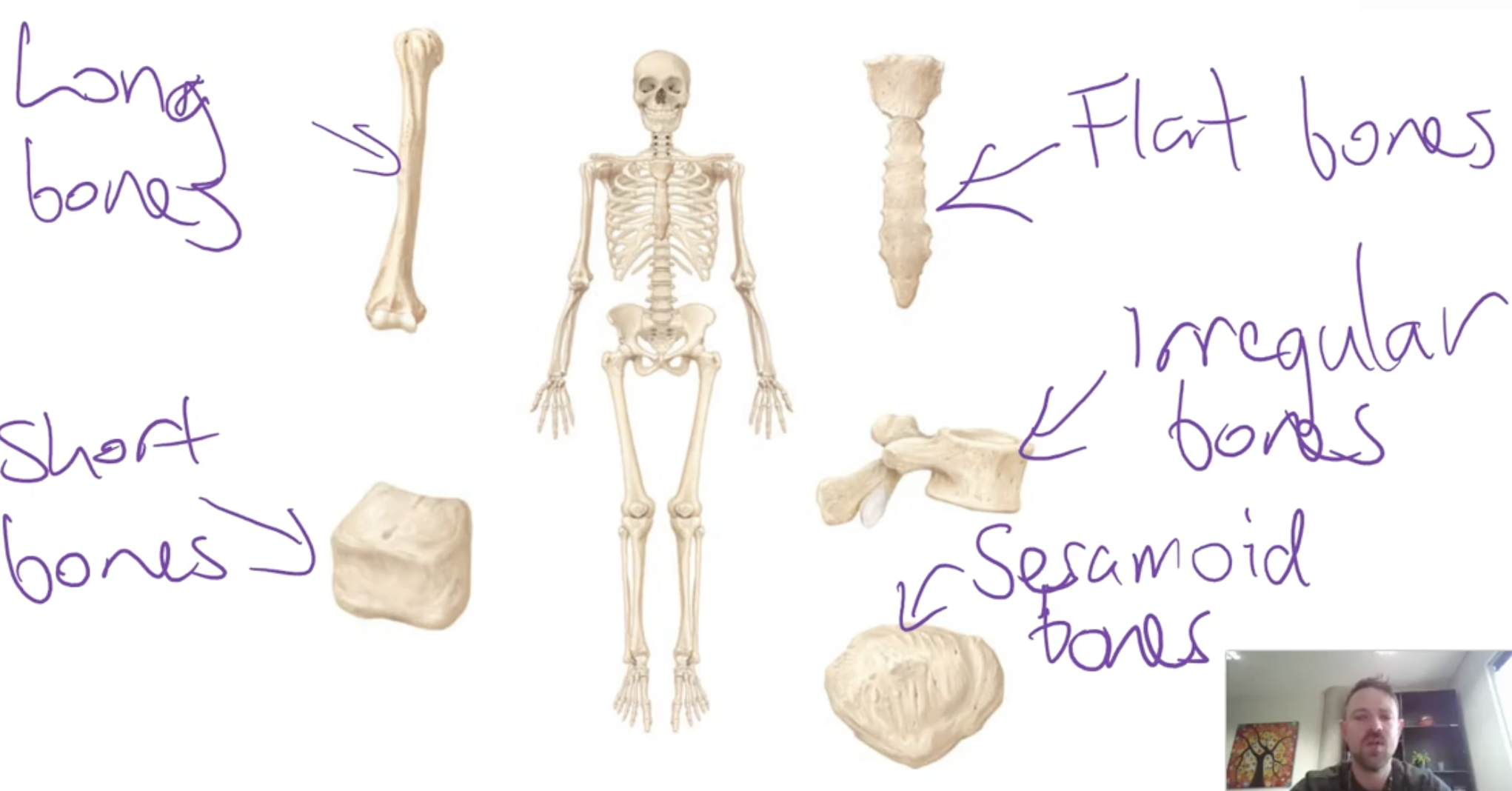
What are the classifications of the bones?
-Long bones: Longer than they are wide
-Short bones: Roughly as wide as they are long and are somewhat cuboidal shape
-Flat bones: Largely flat bones
-Irregular bones: Do not fit into any other shape category
-Sesamoid: Bones that grow inside tendons
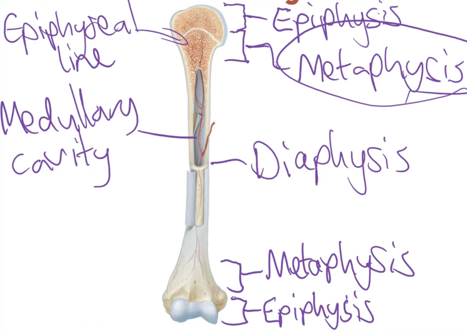
What are the features of a long bone?
-Compact bones on the outside and some spongy bone on the inside
What is a compact bone?
-More densely packed than spongy bone
-Has fewer spaces and is the stronger tissue
-Found on the outer aspect of the bone, beneath the periosteum or articular cartilage
-Consists of repeating units (osteons) which are concentric lamelle (ring of tissue) that surround a central canal which contains vessels and nerves
-Inbetween the the lamellae is the osteocytes
What is spongy bone?
-Has many spaces between it, and this osseous tissue is made by a network of spicules (trabeculae) that are joined together, due to the space between the thin trabeculae it makes the spongy bone weaker
What are the key features of a spongy bone?
1.The spaces between the trabecule makes the bone lighter than compact bone, meaning the bones move faster/easily when muscle pulls on it
2.The spaces between the trabeculae has red or yellow bone marrow
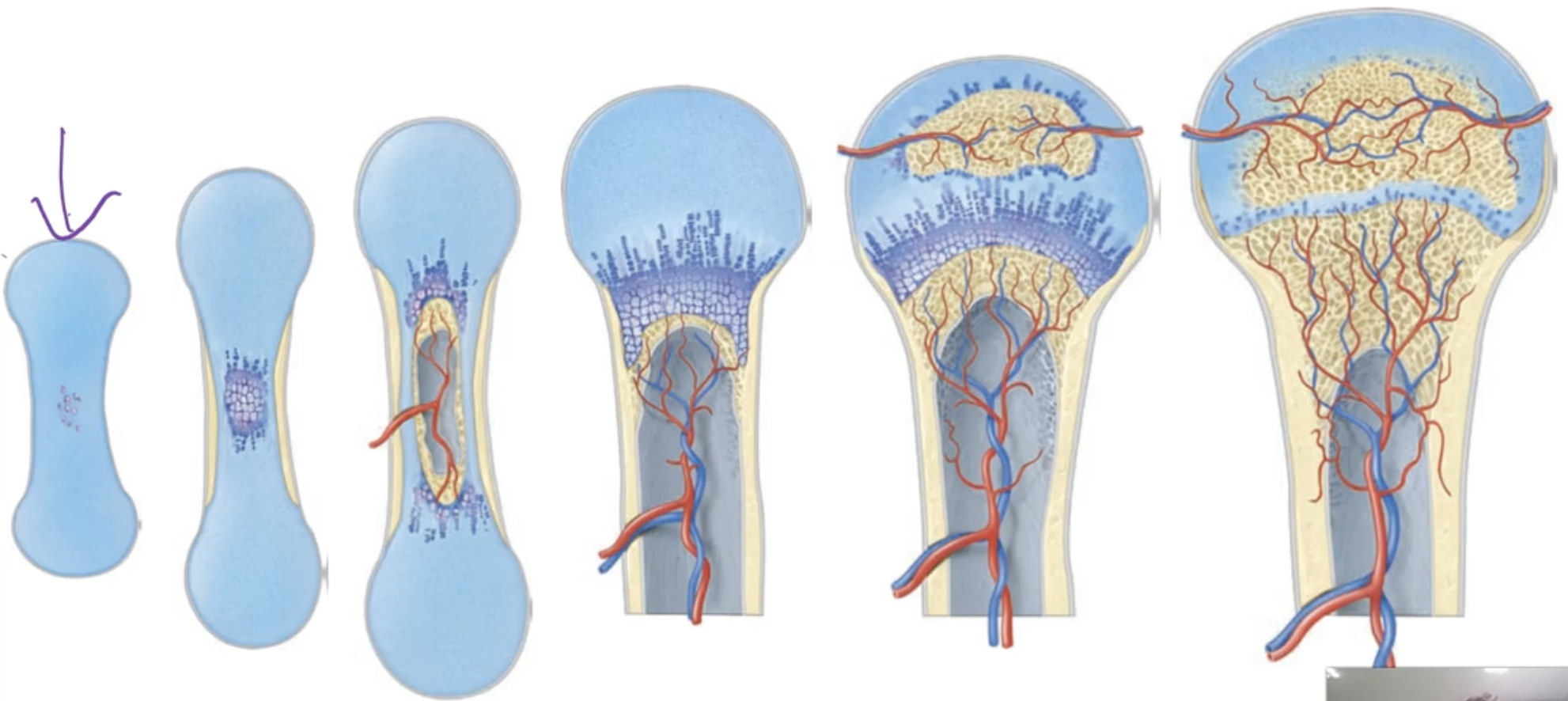
What is ossification and the 2 different processes?
-The initial formation of bones from early life
-Endochondral ossification
-Intramembranous ossification
What is endochondral ossification?
-Hyaline cartilage: Makes up the initial model/ starting process of endochondral ossification, where it slowly starts turning into a proper bone
-Middle of cartilage changes into bone first
-In the end only a bit of cartilage is left
What is bone remodelling?
-Constant process (always occuring) by which older bone is broken down and replaced with new bone
-This process involves bone resorption, which is when minerals and collaged fibres are removed by osteoclasts which have a ruffle border and attach to the bone and release acids and enzymes
-Also involves bone deposition which is the formation of new bone tissue (new collaged fibres and minerals) by osteoblasts
-This allows bones to change structural make up to match functioanl demands of the individual
Why is bone remodelling important?
-Normal growth
-Repairing injured bones
-Changes bone structure to match functional needs
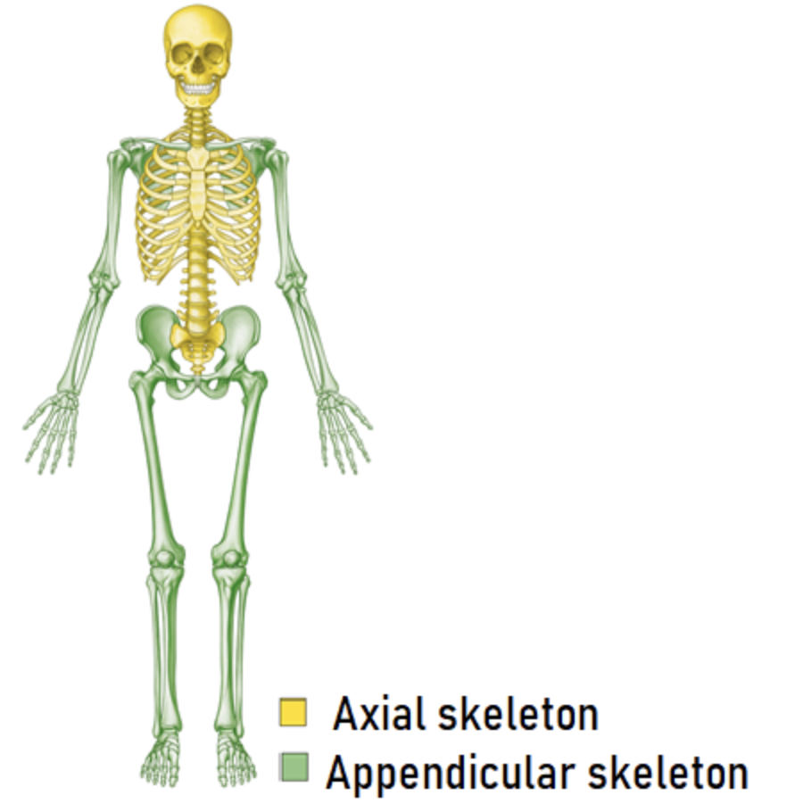
What are the 2 sections of the skeletal system?
-Axial skeleton: Consists of skull, spine, ribs and sternum
-Appendicular skeleton: Consists of the bones of the upper and lower limbs, as well as the bones from the pectoral and pelvic girdles (something that encircles something else, upper part of rib cage)
What are bony landmarks?
-Surface markings or particular aspects that allow specific functions
-They can be present at birth or they can develop in response to tendons, ligaments or other elements pushing or pulling the bone
-Projections: Any sort of bumps on the bone
-Depressions: Divot in the bone
-Openings: Any holes in the bone
What are the major bones of the axial skeleton?
Thoracic cage:
Ribs and associated cartilage, thoracic spine and the Sternum
Skull:
-Cranial bones: any bones that makes up part of the wall of the cavity where the brain sits
-Facial bones: Any other bones that apart of the skull that dosent contribute to making the walls of the house where the brain sits
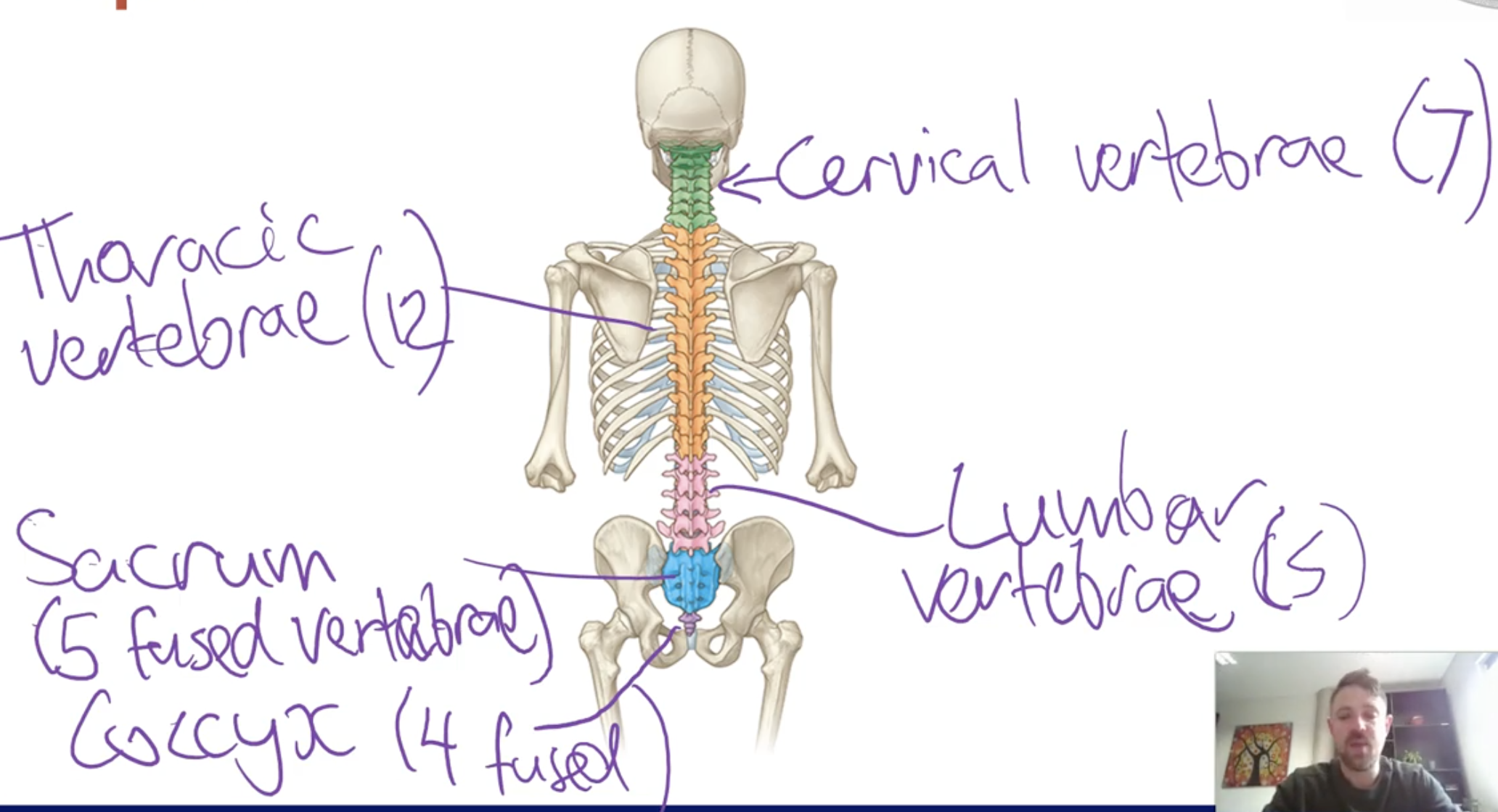
Spine:
-Made up of vertebrae's
-Cervical vertebrae (7 vertebrae)
-Thoracic vertebrae (12 vertebrae, corresponds to 12 ribs)
-Lumbar vertebrae (5 vertebrae)
-Sacrum (5 fused vertebrae)
-coccyx (4 fused vertebrae)
What are the 2 parts to the vertebrae?
-Arch
-Body
Which both fuse together to protect the spinal cord
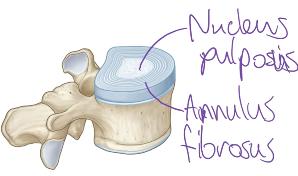
What is the intervertebral disc?
-Discs that sit in between the vertebrae and connect the vertebrae in most of the spine
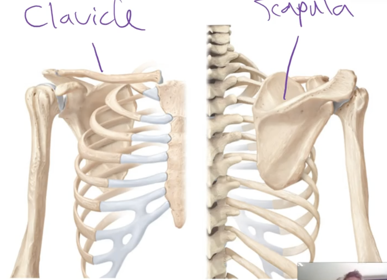
What is the pectoral girdle?
-Consists of the clavicle (collar bone) and the scapula (shoulder blade)
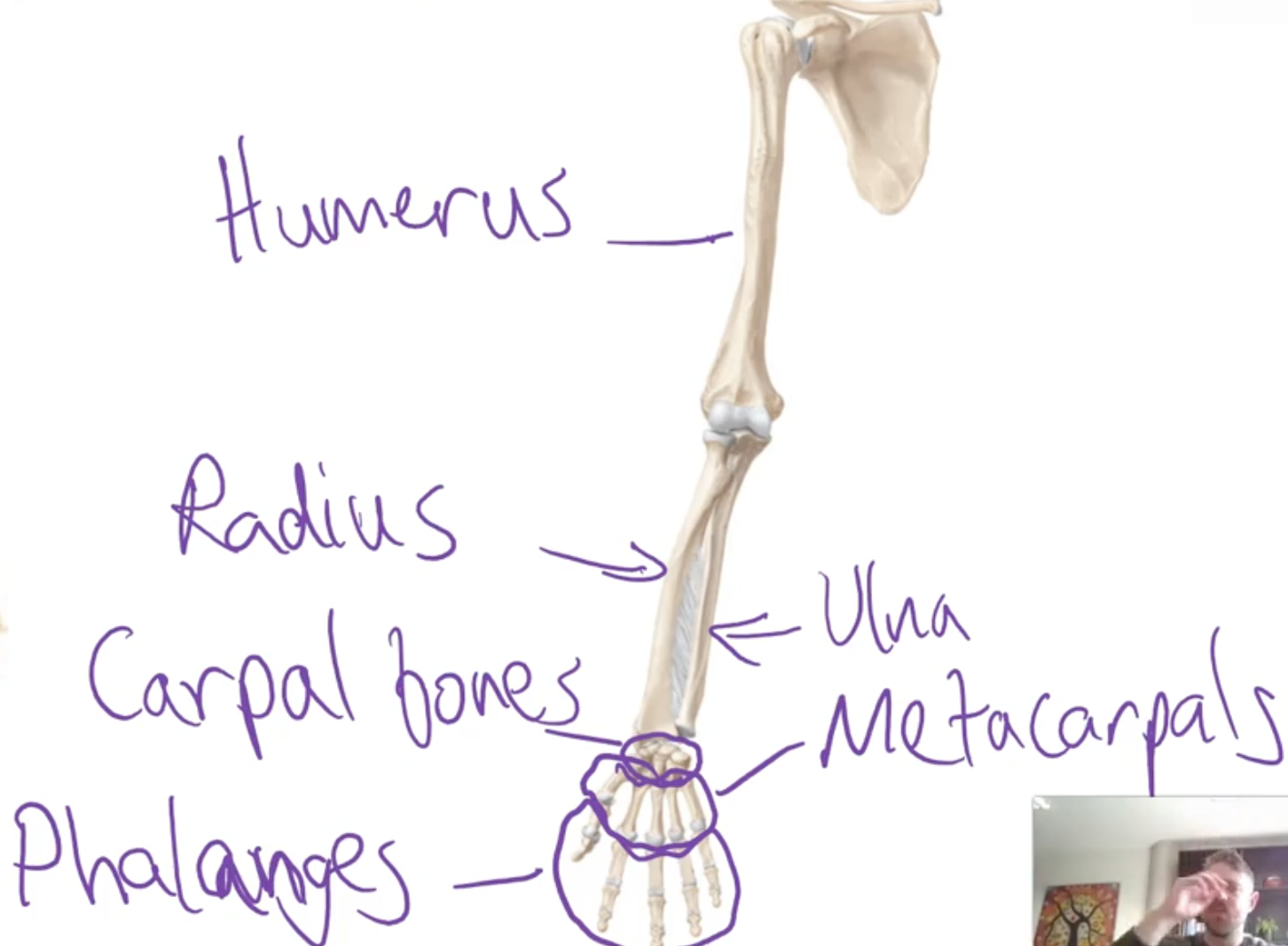
What is the upper limb bones?
-Humerus, Radius and ulna (forearm. bones), carpal bones, metacarples (hand bones), Phalanges (finger bones)
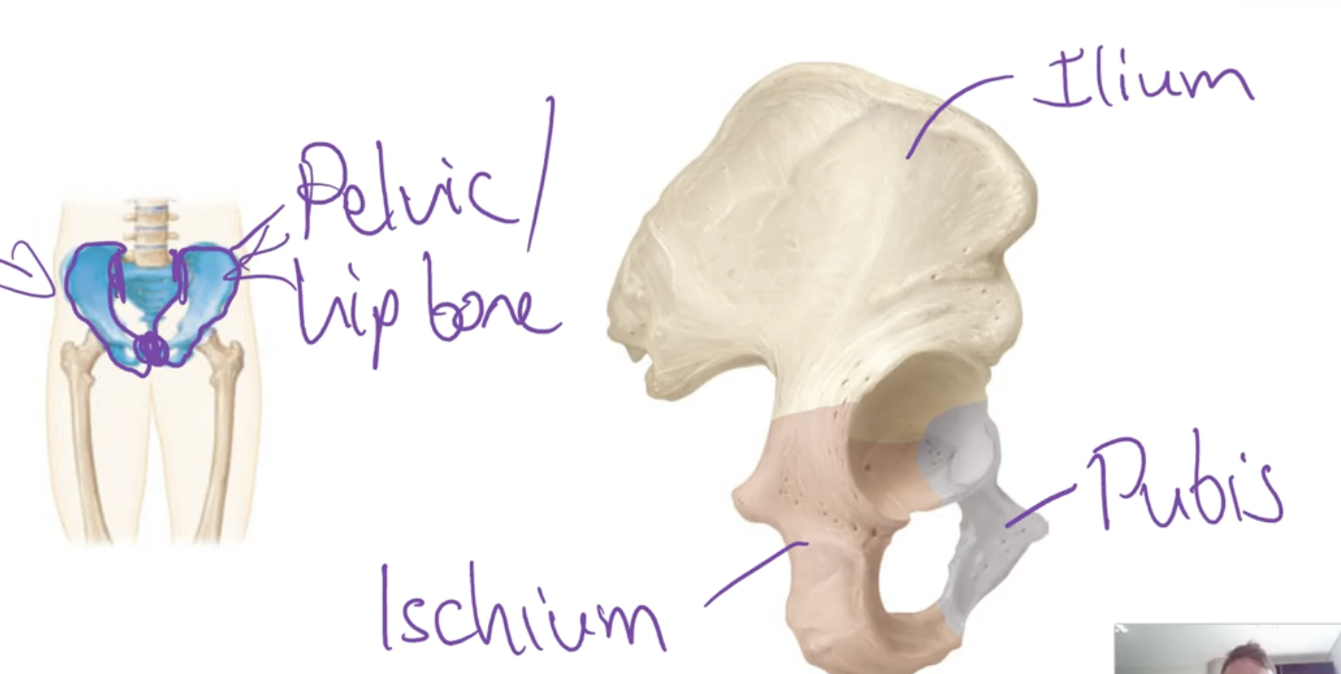
What are the pelvic girdle bones?
-Pelvic/hip bones, one on each side, attached posteriorly to the sacrum and attached anterior to each other
-Each hip bone is made from 3 fused bones:
-Ilium (superior bone), Ischium (posterior, inferior bone), Pubis (inferior anterior)
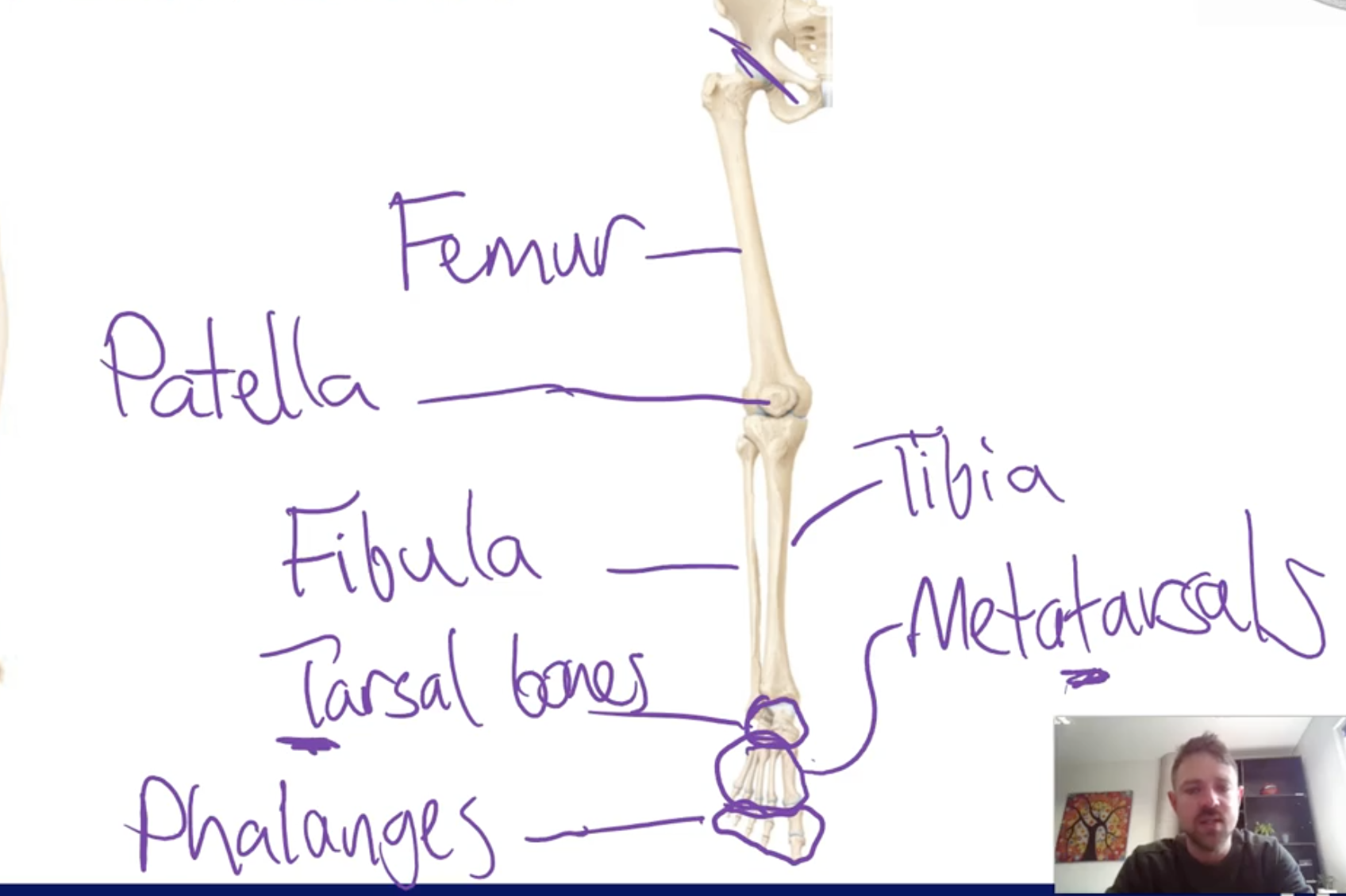
What are the lower limb bones?
-Thigh bone: Femur
-Lower leg: Tibia (larger) , Fibula (smaller)
-Feet bones: Tarsel bones (back foot), metatarsals (mid foot), phalanges (toe bones)
What are the types of cartilages?
Hyaline cartilage: Shiny, smooth white and is the most abundant cartilage in the body. It is flexible but weak
Fibrocartilage: Very strong and rigid cartilage. Multiple thick collages fibres give it strong property
Elastic cartilage: Somewhat rigid, yet flexible and elastic. The elastic fibres present give it this property
What are joint classifications and the 3 types?
-Functional classifications relate to how much movement is available for the joint
-Structural classification relate to whether or not a joint cavity space is present the type of tissue that binds to the joints if theres no cavity
-Fibrous joints
-Cartilaginous joints
-Synovial joints
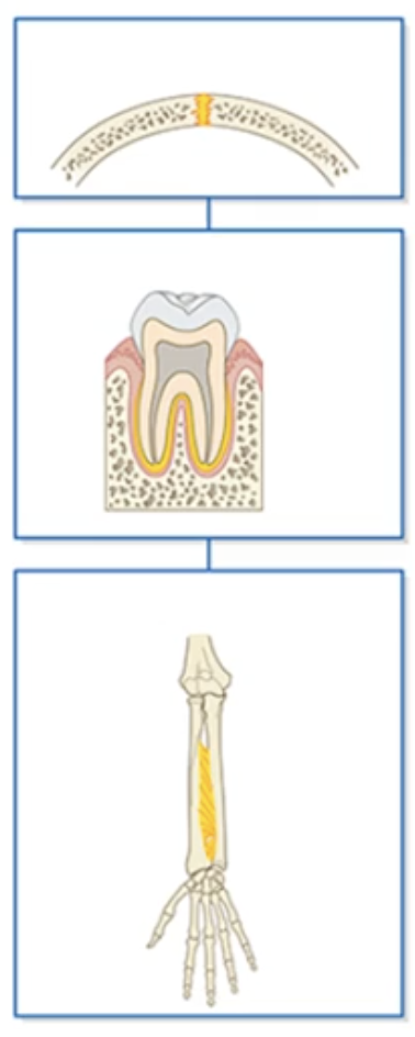
What are fibrous joints and the 3 types?
-2 bits of bone united by a sheet of dense connective tissue
Suture joint: Located in the skull, and hold skull bone together
Gomphosis joint:The joint that holds the tooth into the tooth socket
Syndesmosis joint: The joint tat holds together 2 bits of bone that are more further away such as the forearm. also called interosseous membrane/ligament
What are cartilaginous joints and the 2 types?
Primary cartilaginous /Synchondrosis joint:
-A joint within a bone that holds different parts of the bone together and its only present in growing bones
Secondary cartilaginous /Symphysis joint:
-A joint that unites 2 different bones together and they do using a plate of cartilage
What are synovial joints and the types?
-All synovial joints fit together very nicely, where bones shape matches the other bones shape perfectly
Plane/Gliding joint: 2 articulating bones of 2 flat areas that are on one another and can move/glide in any direction
Hinge joint: Movement of bone forwards or backwards
Pivot Joint: Projection of bone that sits inside a ring
Ball and socket joint: Spherical shaped projection that fits inside a round spherical shaped hole, causing spinning occurring
Condyloid joint:
Saddle joint: Movement that occurs to the sides
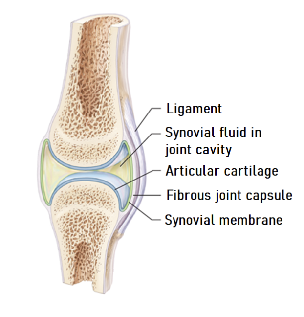
What are the features of a synovial joint?
-Synovial joints connect 2 or more bones
-Articular cartilage covers the bone ends
-A joint capsule which has inner synovial membrane which synthesises and secretes synovial fluid and an outer fibrous capsule which is flexible encloses the joint cavity filled with synovial fluid(reduces friction between articulating surfaces and nourished articular cartilage) and ligaments provide support
-Many also have extra structures to enhance stability and function
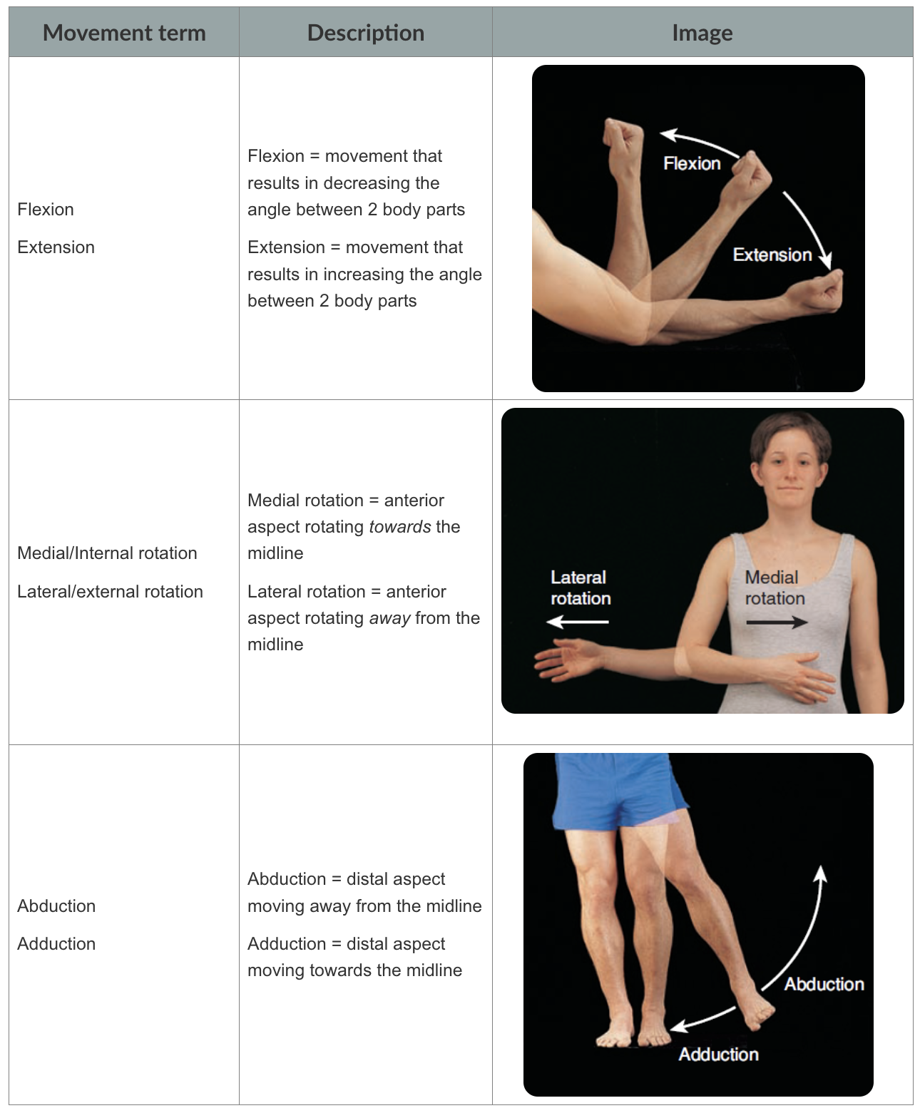
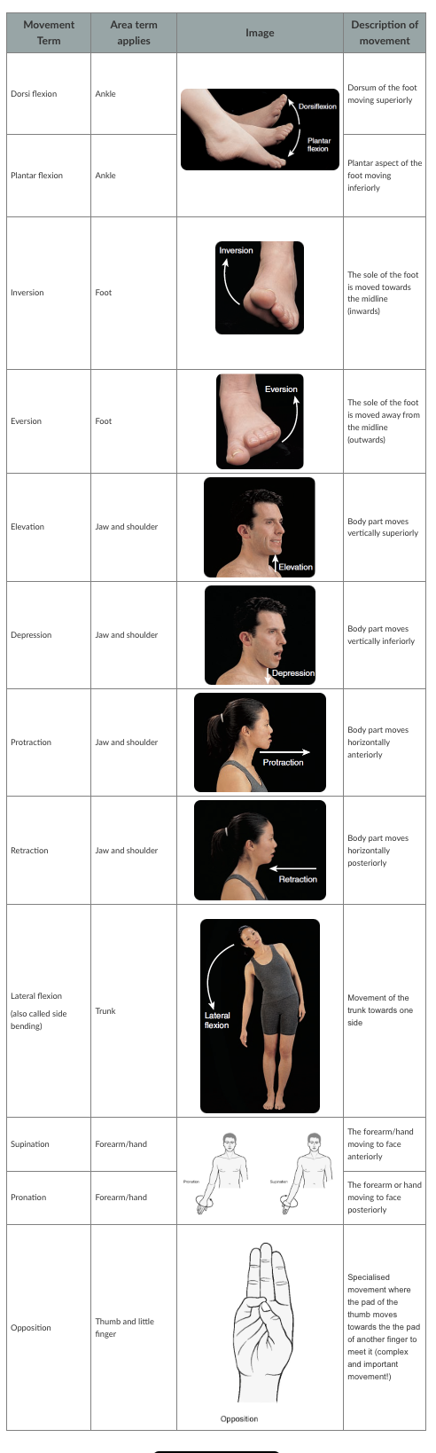
What aer the synovial joint movement terms?
Flexion: decreasing angle b
What are the functions of the muscular system?
Producing body movements: When skeletal muscles activate they generate force that pulls on bones, creating the capacity to move joints
Stabilising body positions: Skeletal muscle contraction can also stabilise joints. Eg: When standing up gravity is trying to collapse us, however our muscles are resisting the force of gravity to stabilise our posture and keep us upright
Storing substance in the body: Muscles work to close off the opening of an organ to ensure substances are properly stores in the organ until its time for htem to be released. Eg the bladder, the Urethral sphincter is a ring of muscle that surrounds a bladder opening, when the muscle is contracted the opening is closed off, when the muscle relaxes the opening opens
Moving substances around the body: The cardiac muscle of the heart pumping blood around the body eg; heart muscle contracts which forces blood through body vessels
Generating heat: Chemical reactions involve in overall muscle contraction process released by heat as a by product. causing heat when finshing exercise
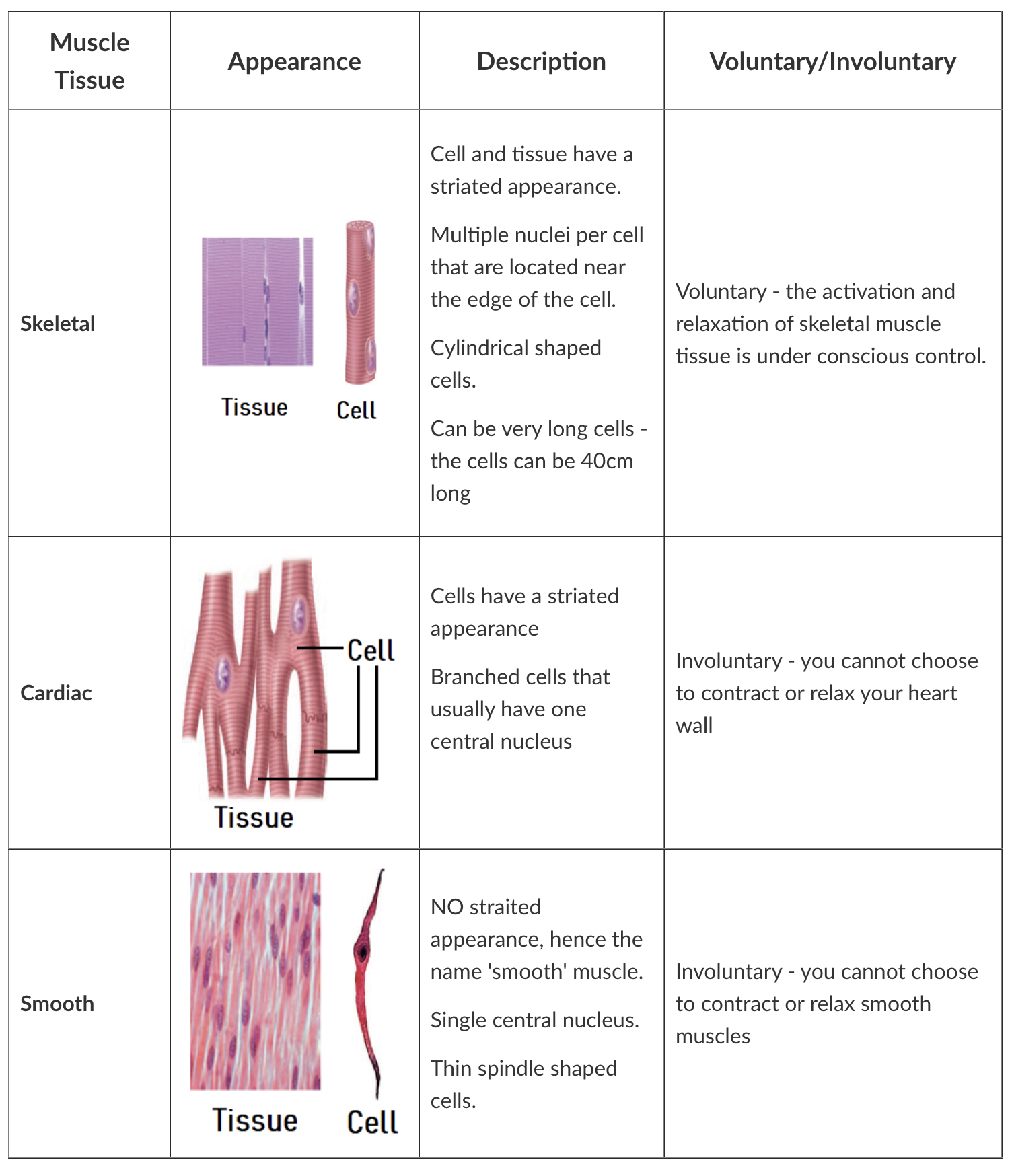
What are common muscle tissue properties?
1.Electrically excitable: Plasma membrane has the ability to establish an action potential that can move across the entire plasma membrane
2.Contractility: Once excited, muscle cells have the ability to generate force by significantly altering their shape and size
3.Extensibility: Muscle tissues have ability to be stretched to a point without being damaged
4.Elasticity: Muscle tissues spring back to original length once the stretch force is removed
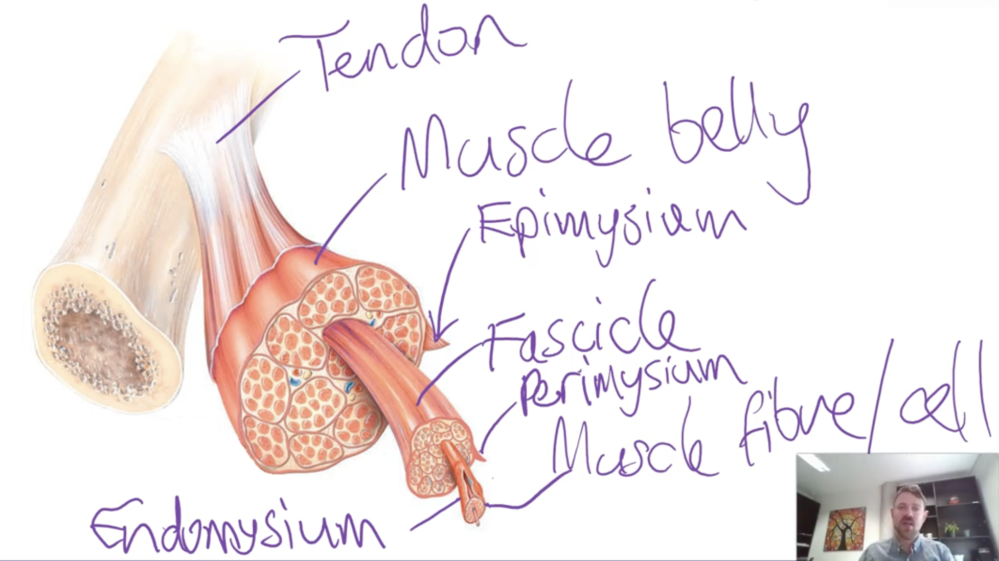
What is gross anatomy of skeletal muscles?
-Muscle belly and tendons.
-Muscle belly has muscle fibres which are cells capable of activating
-Tendons are connective tissue structures that attach the muscle belly to a bone
-Muscle belly and its fibres are arranged into structures called fascicles
Epimysium - surrounds the whole muscle
Perimysium - surrounds a fascicle
Endomysium - surrounds each individual muscle fibre
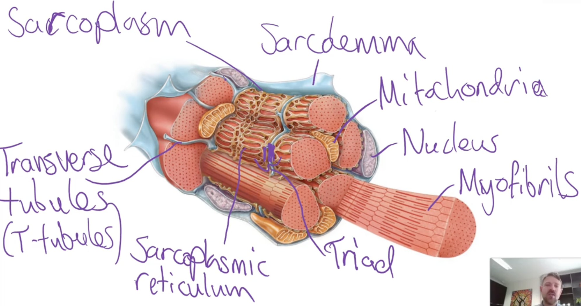
What are skeletal muscle fibre cells and their important elements?
-Functional unit of a skeletal muscle
-Are long cells that have ability to generate pulling force when excited by motor neuron
-Multinucleated with lots of mitochondira
Sarcolemma - the plasma membrane
Transverse tubules - tube-like extensions of the plasma membrane that penetrate through the fibre
Sarcoplasm - the cytoplasm of the muscle fibre
Sacroplasmic reticulum - fluid filled membranous sacs that store calcium ions (Ca2+)
Terminal cisterns - enlarged parts of the sacroplasmic reticulum that are nestled against the transverse tubules
Triad - a transverse tubule with a terminal cistern on either side
Myofibrils - microfilament components that move to generate the pulling force by changing the shape of a muscle fibre
What are sarcomeres?
Skeletal muscle has a striped (striated) appearance due to repeating units called sarcomeres in each cylindrical myofibril. Each sarcomere contains thin filaments (attached to the Z-discs at each end) and thick filaments (attached to the M-line in the center). The overlapping of these filaments creates the striated pattern.
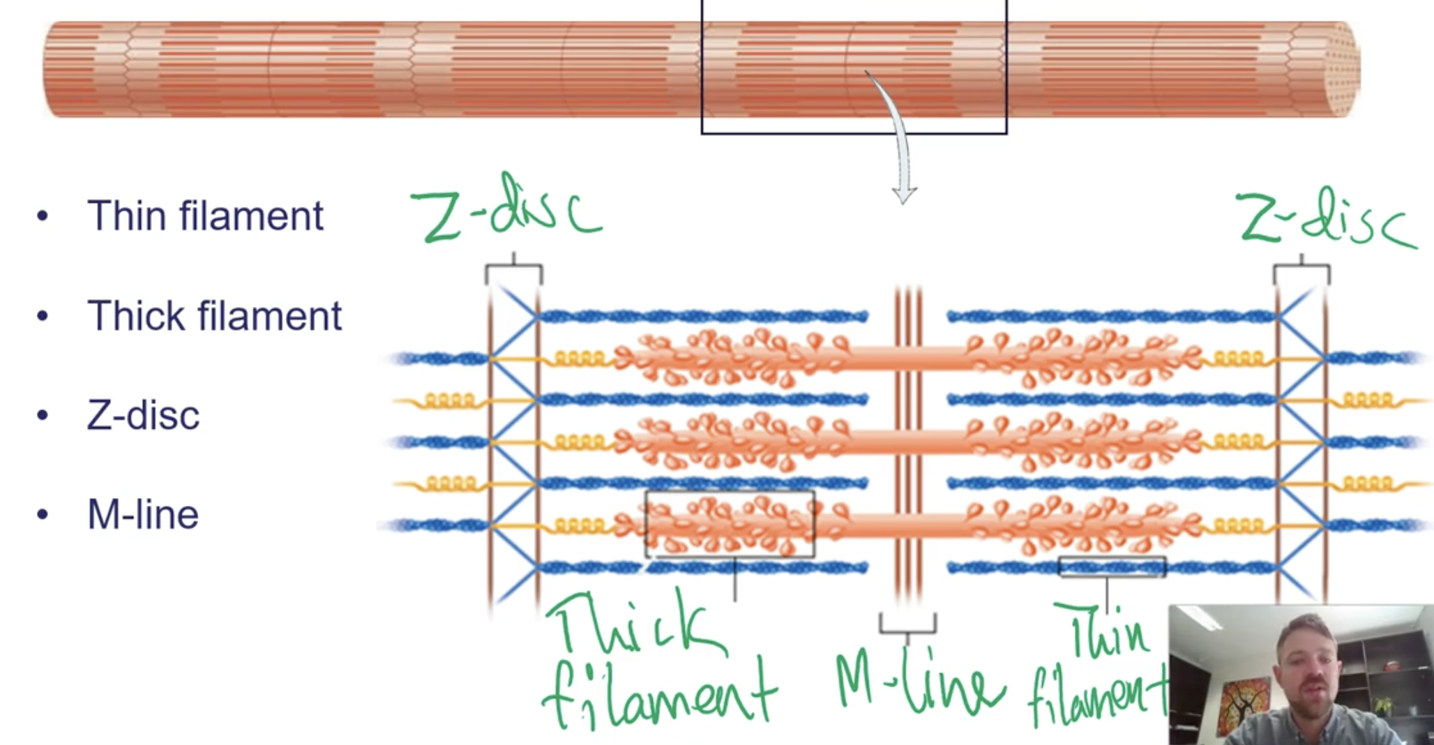
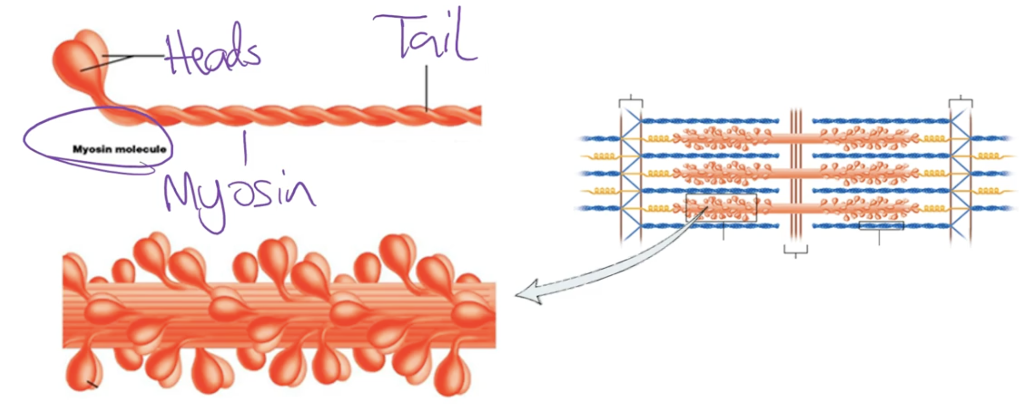
What is thick filament?
Thick filaments are mainly made of myosin, a contractile protein that converts ATP energy into mechanical force during muscle contraction. Each myosin molecule has two twisted tails and a head that extends toward nearby thin filaments
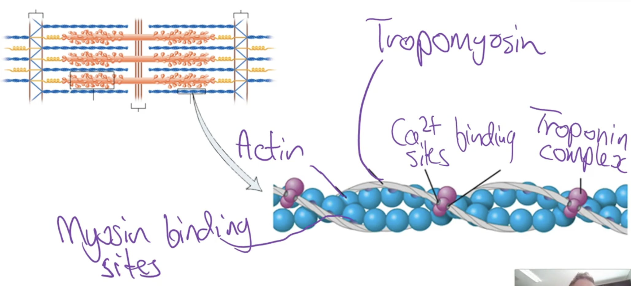
What is thin filament?
thin filament has a contractile protein component which is called actin. However the thin filament also contains other parts we need to know of including the troponin complex and tropomyosin
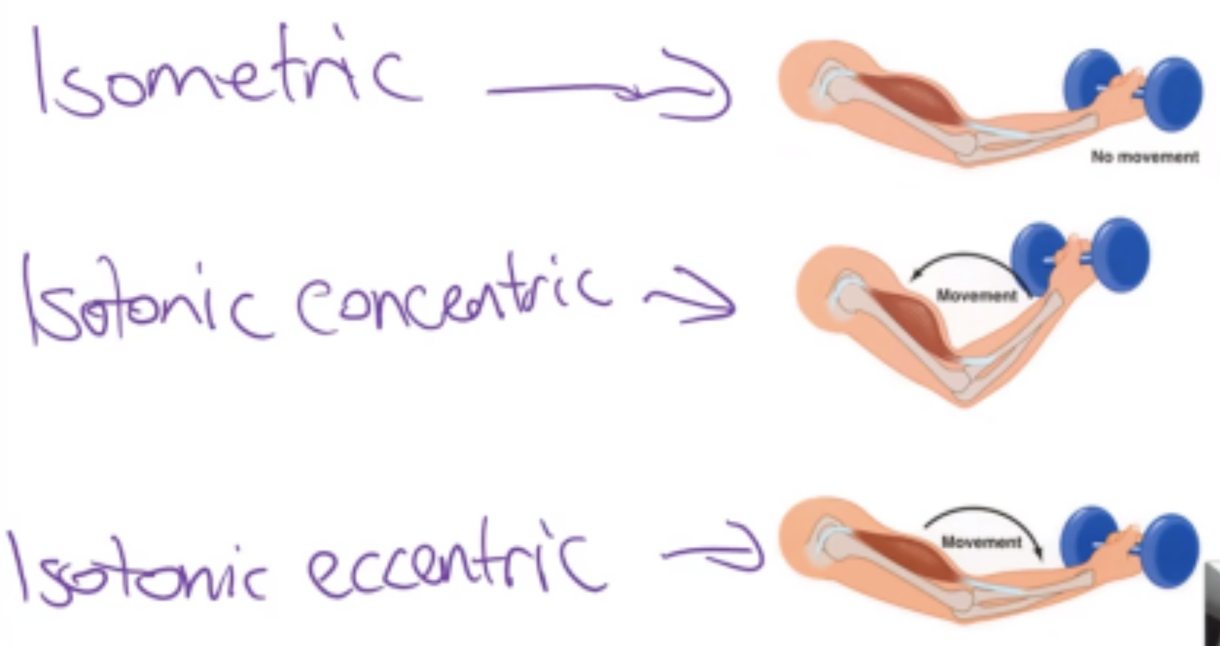
What are the types of muscle contractions?
Isometric activation - the muscle stays the same length as it activates
Isotonic concentric activation - the muscle shortens as it is activating
Isotonic eccentric activation - the muscle lengthens as it is activating
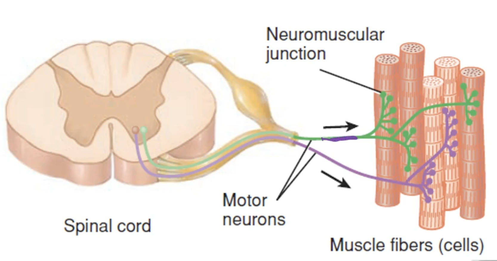
What is the neuromuscular juction?
-Where a neuron meets a muscle cell, at this junction an action potential is transferred from a motor neuron to a muscle fibre electrically exciting it
What is the process of excitation contraction couple?
It’s the process that links a nerve signal to muscle contraction. A nerve impulse triggers the release of calcium from the sarcoplasmic reticulum. Calcium binds to troponin, which moves tropomyosin and exposes binding sites on actin. Myosin heads then attach to actin and pull, causing the muscle to contract.
What is the sliding filament mechanism?
The sliding filament mechanism works when myosin heads on the thick filaments walk along the thin filaments, pulling them toward the M-line. This draws the Z-discs closer together, shortening the sarcomere and causing muscle contraction.
What is the cross bridge cycle?
The cross-bridge cycle is the process where myosin heads bind to actin and pull the thin filament, creating muscle contraction. Actin (part of the thin filament) has binding sites blocked by tropomyosin at rest. When calcium (Ca²⁺) is released, it binds to troponin, shifting tropomyosin and exposing the binding sites. Myosin uses energy from ATP hydrolysis to attach, pull, and release—repeating this cycle for contraction.
What is a motor unit?
-Somatic motor neuron with all the muscle fibres it supplies
-If 1 motor neuron is stimulated than all of the muscles fibres will contract
-All or none principle either all muscle fibres are stimulated or none of them
How can the body change the strength of contraction?
Stimulating more or less motor units
Changing the frequency of motor unit stimulation
Different muscles also have different sized motor units which allows the contraction strengths to be tailored to certain muscles.
What are the major head muscles?
-Masseter
-Temporalis
-Sternocleidomastoid
What are the major trunk muscles?
Trapezius
Rhomboids
Latissimus dorsi
Erector spinae
Pectoralis major
Pectoralis minor
Serratus anterior
Rectus abdominis
What are the major upper limb muscles?
Deltoid
Biceps brachii
Triceps brachii
Forearm flexor muscles
Forearm extensor muscles
Rotator cuff muscles:
Teres minor
Infraspinatus
Supraspinatus
Subscapularis
What are the major lower limb muscles?
Pelvic floor muscles
-Gluteal Muscles:
Gluteus maximus
Gluteus medius
Gluteus minimus
Piriformis
-Quadriceps femoris muscle heads:
Rectus femoris
Vastus lateralis
Vastus medialis
Vastus intermedius
-Hamstring muscles:
Biceps femoris
Semitendinosus
Semimembranosus
-Calf muscles:
Gastrocnemius
Soleus