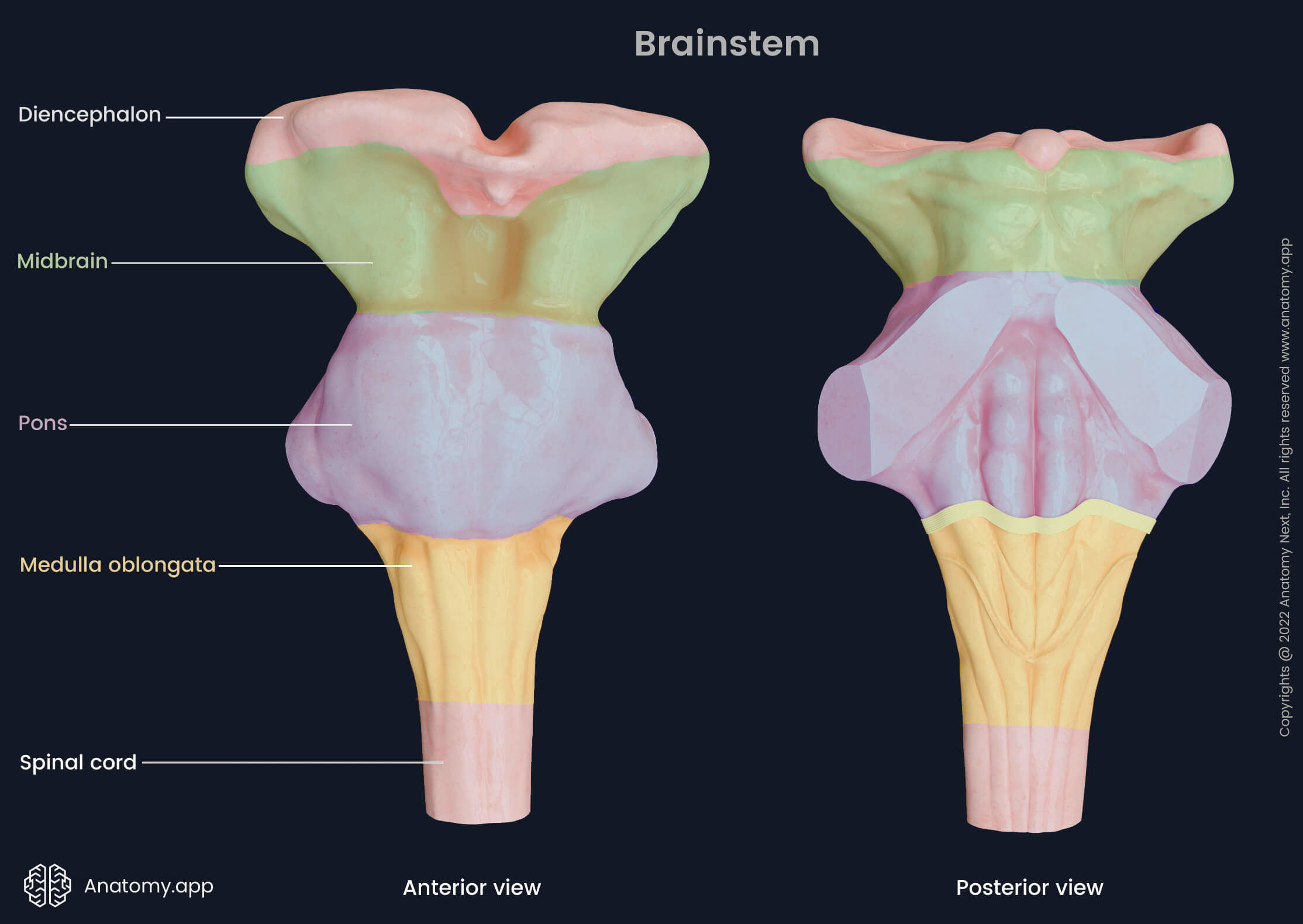
Head and neck things to remember
1/87
There's no tags or description
Looks like no tags are added yet.
Name | Mastery | Learn | Test | Matching | Spaced |
|---|
No study sessions yet.
88 Terms
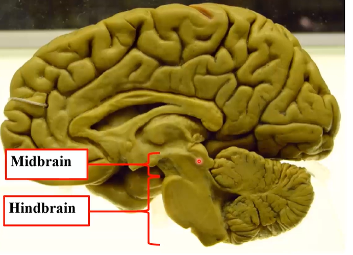
what constitutes the brainstem?
the midbrain and the hindbrain (pons and medulla oblongata)
what constitutes the hindbrain?
the pons, medulla oblongata and the cerebellum
is the thalamus included in the brainstem?
typically no
what does the pons extend back into?
the 4th ventricle
what structure marks the limits of the midbrain?
the cerebral aqueduct - the channel that links the third ventricle above with teh fourth ventricle below and the two bumps at the back - colliculi
where is the open medulla?
above the closed medulla
what are the functions of the brainstem? 7
Conduit for ascending and descending pathways
Conduit for cerebellar connections - communicates with the cerebellum
Houses most cranial nerve nuclei
Chemoreception, salivation, mastication, swallowing and gag reflex
Reticular formation – arousal; cardiovascular and respiratory centres – vital life-supporting role
Raphe, locus coeruleus nuclei - mood, sleep
Substantia nigra – movement control
what is the raphe, locus coeruleus and substantia nigra?
grey matter: monoaminergic centres
the raphe is located on the midline of the brainstem and is the source of all the serotonergic centres in the body - mood and sleep
the locus coeruleus has its nuclei in the pons and is the location for all the noradrenergic pathways - mood and sleep
the substantia nigra is most of the dopaminergic pathways - important in movement - parkinson’s - the rest come from the tegmentum

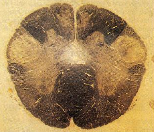
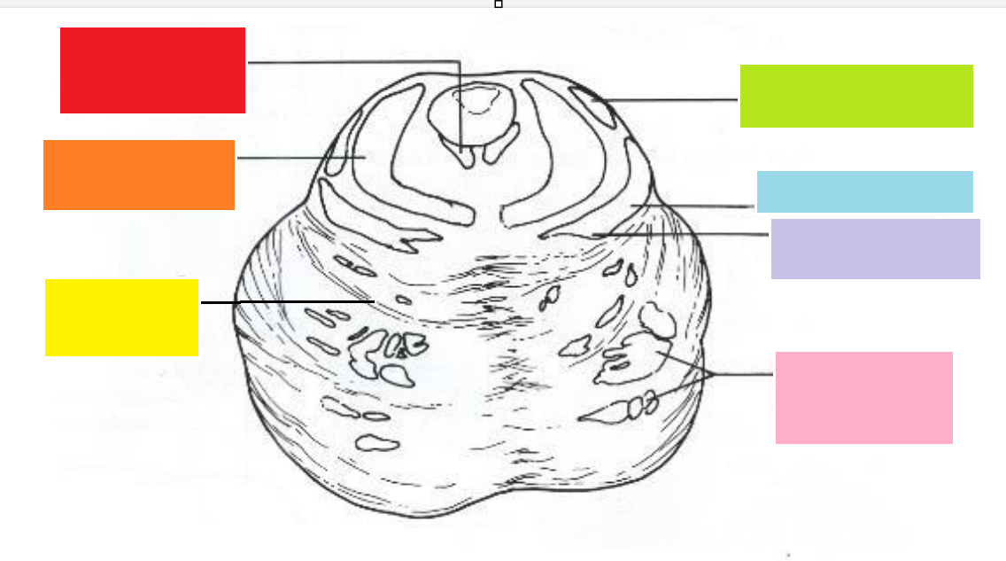
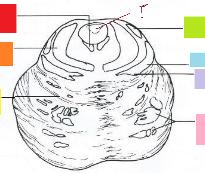
what is this? and what is the dorsal side?
section through the closed medulla, dorsal is TOP


how would the image differ if it was the open medulla?
the dorsal side would be concave because of the floor of the FOURTH ventricle

red
closed medulla - fasciculus gracilis - ascending pathway , carrying fine touch, conscious proprioception and vibrational information - remember nerve fascicles

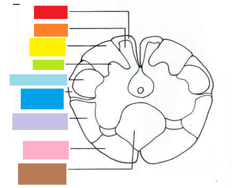
orange
closed medulla - nucleus gracilis - at this point the ascending fibres are synapsing with their target fibres


yellow
closed medulla - fasciculus cuneatus - also an ascending pathway carrying fine touch, conscious proprioception and vibrational information
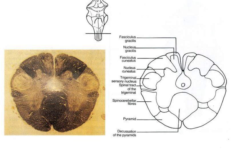

green
closed medulla - nucleus cuneatus


sky blue
trigeminal sensory nucleus - closed medulla - the long one from the medulla to the midbrain


lilac
closed medulla - spinocerebellar fibres


pink
closed medulla - pyramid - contain axons of the cortiospinal tract - axons are named from their origin to where they are going - these axons are going from the cortex to the spinal cord - DECENDING PATHWAY


brown
closed medulla - decussation of the pyramids - this is where they cross over to the opposite side - they cross in swaves


what is the structure in the centre?
central canal of the medulla - continuous with the 4th ventricle above and the central canal of the spinal cord below

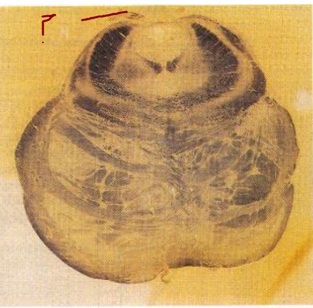
what is significant about this stain?
myelin is stained black
this is nuclei for the spinal accessory nerve

which side is ventral?
the bottom is ventral

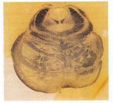
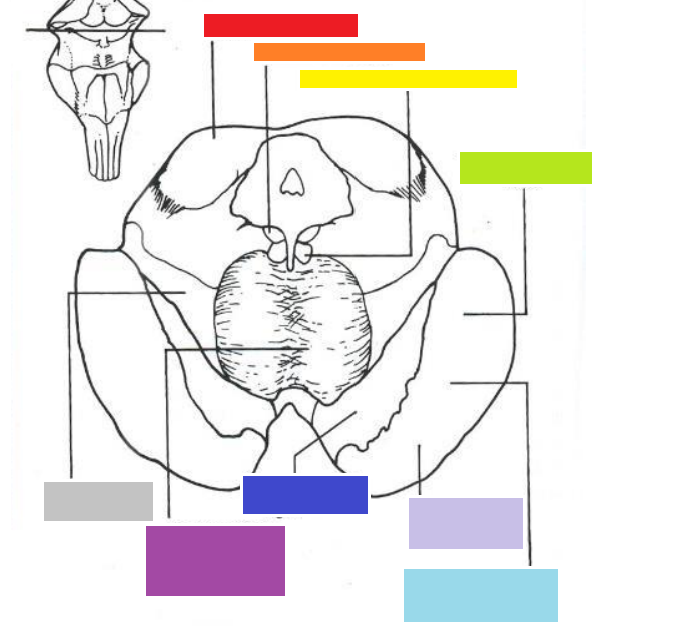
what is this?
pons - notice the ventral bulge at the bottom

red
pons - medial longitudinal fasiculus

orange
pons - superior cerebellar peduncles

yellow
pons - pontocerebellar fibres
green
pons - lateral meniscus

blue
pons - spinothalamic tract

lilac
medial lemniscus

pink
pons - upper motor neurones corticospinal (from cortex to spine) and corticobulbar fibres (they terminate at cranial nerve motor nuclei)- these with gather into the medullary pyramids soon
start of the cerebral aqueduct - remember this is the pons - around this is grey matter, peri-aqueduct grey matter
dorsally - trochlear is the only cranial nerve that exits
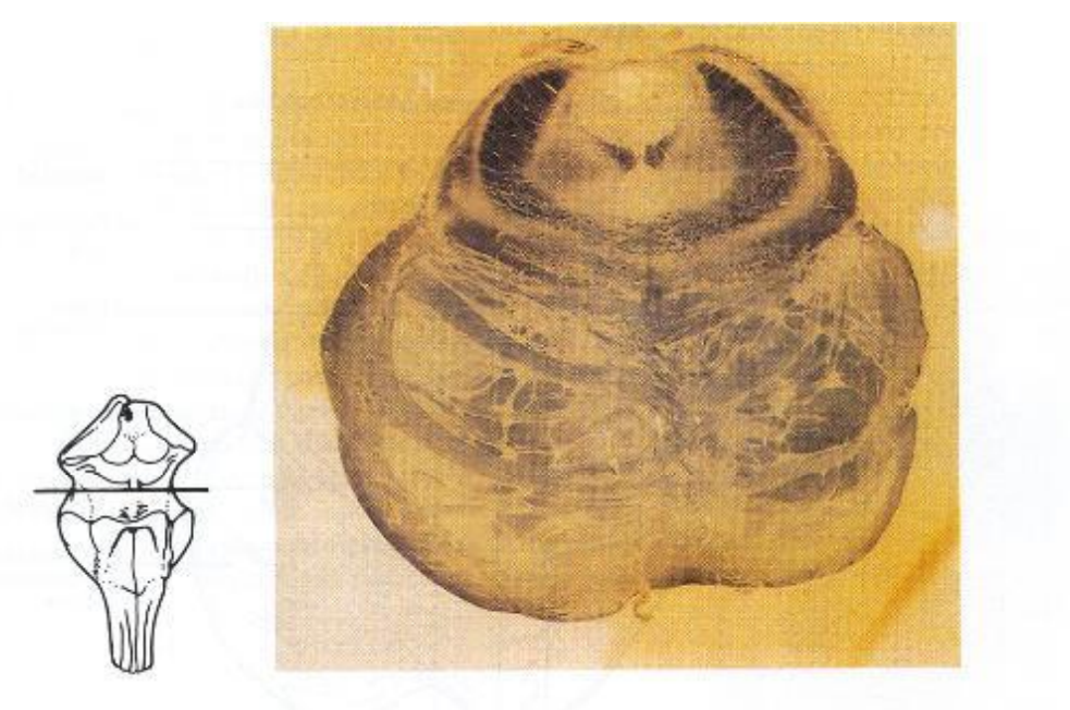
what is this and what is the dorsal surface?
this is the midbrain, with the dorsal surface at the top


red
midbrain, inferior colliculi, along with the superior colliculi, they are responsible for reflex extraocular muscles, the superior are visual stimuli reflex and the inferior are the auditory reflex

orange
midbrain - trochlear nucleus

yellow

green
midbrain, temporopontine fibres
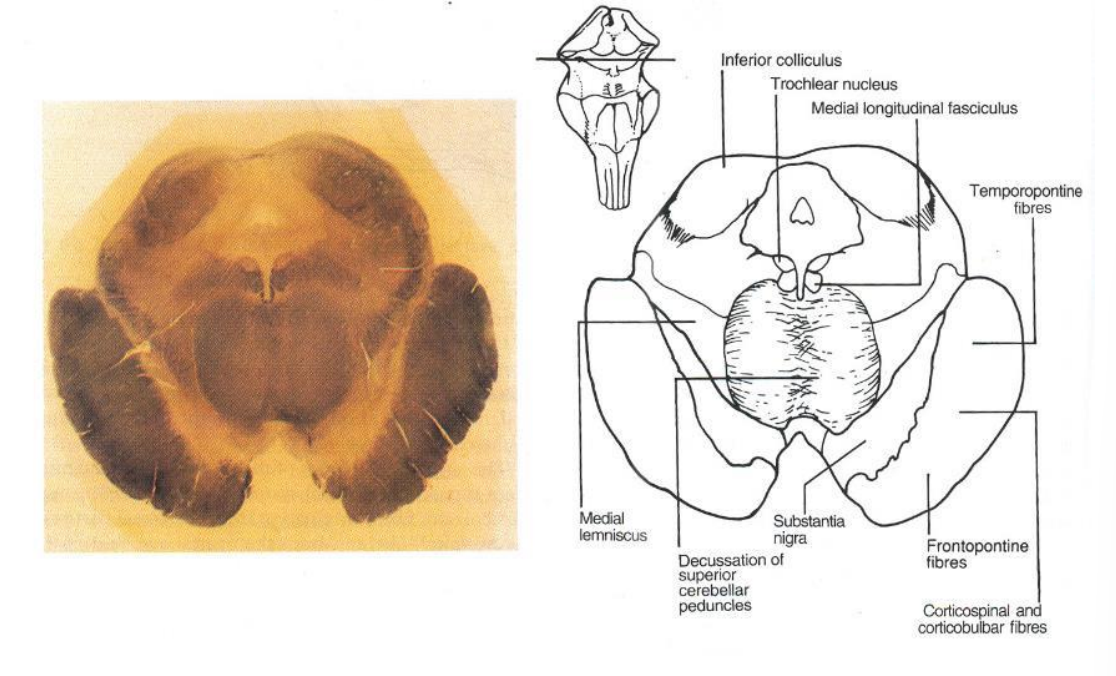

blue
cerebral peduncles - midbrain big white matter tract - large number of descending tracts - corticospinal and corticobulbar tracts

lilac
front pontine fibres - midbrain

navy
midbrain - substantia nigra - neuromelanin
purple
decussation of superior cerebellar peduncles - midbrain

grey
medial lemniscus- midbrain

what is the hole in the dorsal medial ?
start of the cerebral aqueduct- midbrain

what are the two bumps on the dorsal surface?
hills - inferior colliculus - midbrain
what is special about the trochlear nerve?
all cranial nerves exit the brain on the ventral surface except the trochlear, which exits dorsally and wraps back round
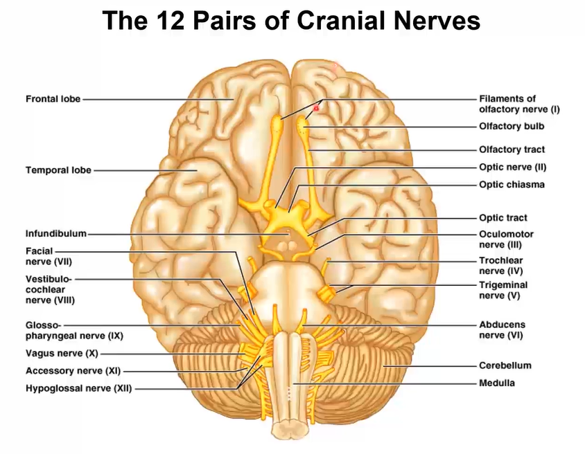
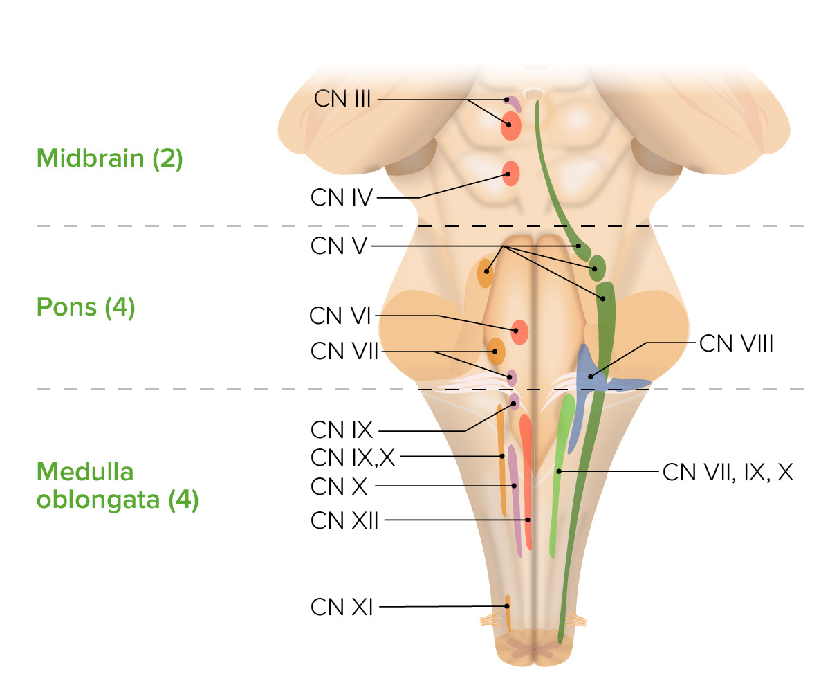
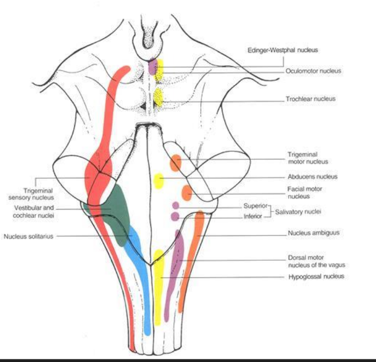
location of the cranial nerve exit points
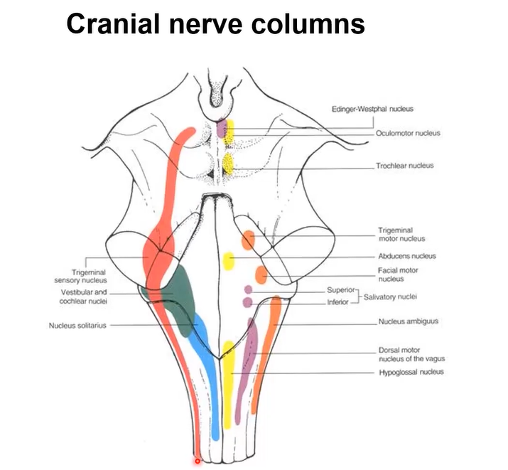
cranial nerve columns
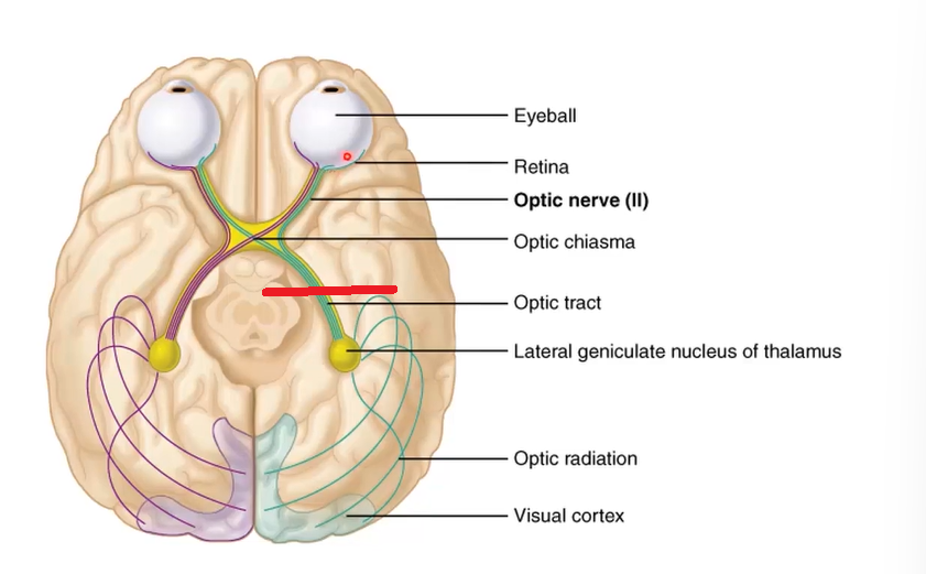
optic nerve vs tract
since the retina is part of the cns, anything coming from it is a tract but we call the axon between the retina and the optic chiasm the optic nerve, note that it doesn’t synapse at the optic chiasm

where do special sensory optic nerves synapse?
in the lateral geniculate nucleus of the thalamus - to visual cortex
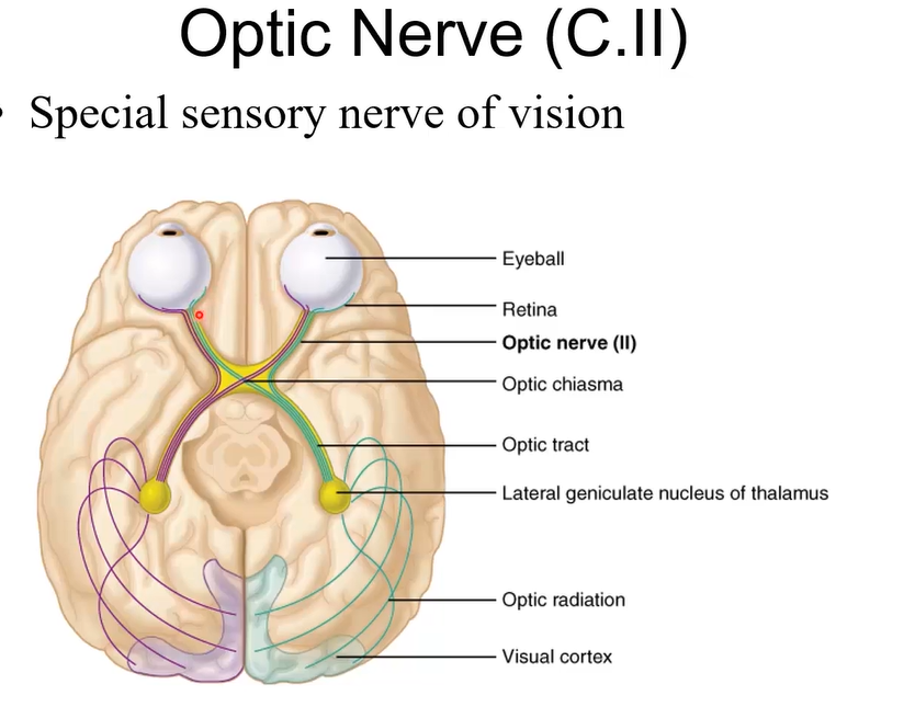
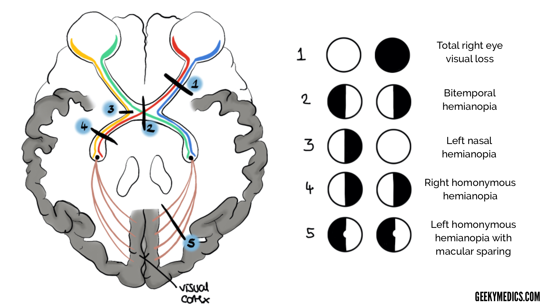
what part of the pathway cross over?
nasal retinae, midline
as opposed to temporal - stay ipsilateral

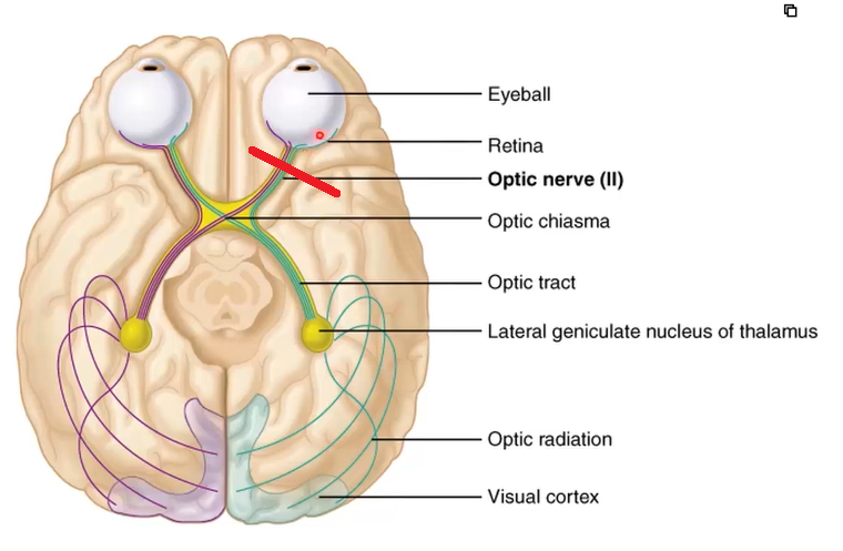
complete blindness in left eye

half blind in both eyes
what happens at the edinger whesphal nuclei?
It receives input from the pretectal nucleus and sends parasympathetic fibers via cranial nerve III to the ciliary ganglion, controlling pupillary constriction. These fibers innervate the iris sphincter muscle, causing the pupil to constrict in response to light.
list the trigeminal nuclei 4
Motor nucleus (muscles of mastication)
Spinal nucleus (sensory) – thermal/nociception from head and neck
Principal nucleus (sensory) –touch, proprioception
Mesencephalic nucleus –jaw reflexes

A
Trigeminal ganglion

B
trigeminal principle nucleus - sensory - proprioception and touch

C
thalamus - VPM

D
cerebral cortex
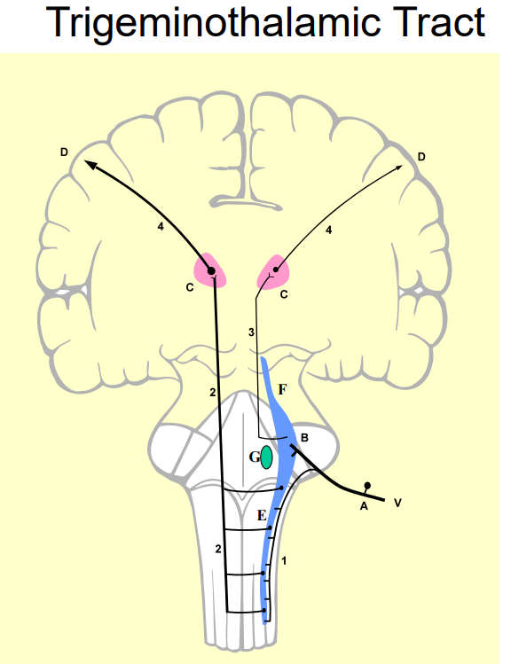
E
trigeminal spinal nucleus - thermal and nociception from the head and neck

F
trigeminal mesencephalic nucleus - jaw reflexes

G
trigeminal motor nucleus - muscles of mastication

1
spinal tract of trigeminal

2
ventral trigeminothalamic tract

3
dorsal trigeminothalamic tract

4
coronal radiata
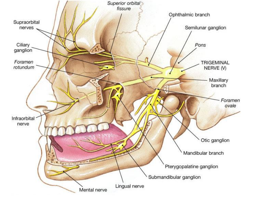
what is the trigeminal ganglion?
where the cell bodies are of the sensory component - like DRG
how is the facial nerve general somatic sensory?
skin of the external auditory meatus and the tympanic membrane/eardrum
what are mimetic muscles?
muscles of facial expressions
where does the facial nerve innervate taste?
anterior 2/3rds of the tongue and the palate
what cranial nerve innervates the lacrimal gland?
facial - parasympathetic
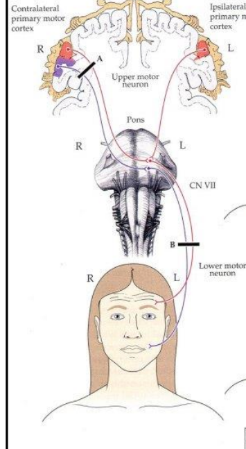
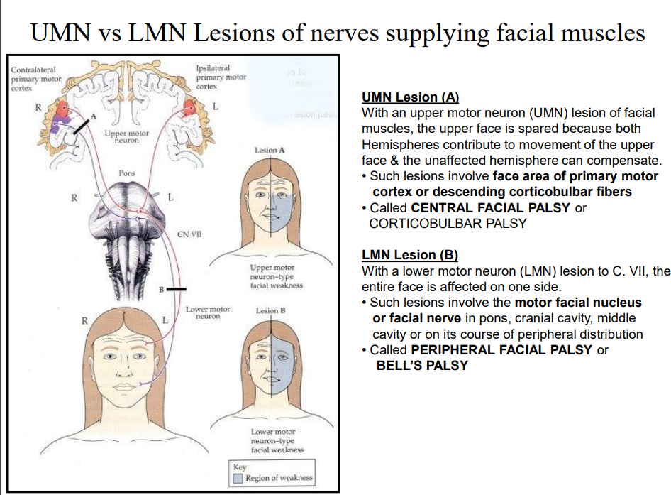
lesion A
as both hemispheres contribute to the facial nuclei for upper face innervation, an upper motor lesion will not affect the upper face, but as the lower face is innervated only by the nerve on the contralateral, the lower opposite half of the face is paralysed
• Called CENTRAL FACIAL PALSY or CORTICOBULBAR PALSY
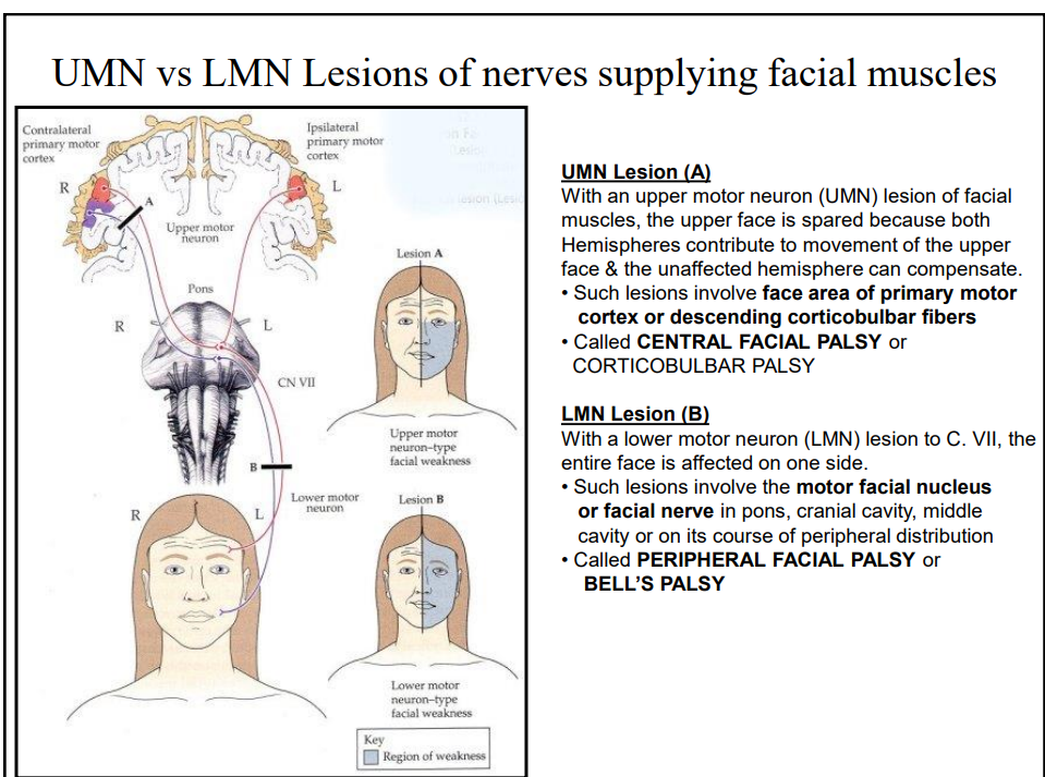
lesion B
there are no neurones from the contralateral that contribute to a side’s innervation after the nuclei in the pons
contralateral face is paralysed,
• Called PERIPHERAL FACIAL PALSY or BELL’S PALSY
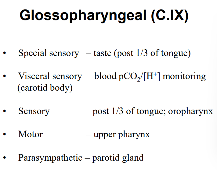
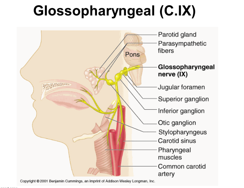
glossopharyngeal motor component
upper pharynx
glossopharyngeal visceral sensory component
– blood pCO2 /[H+ ] monitoring (carotid body)
glossopharyngeal sensory component
post 1/3 of tongue; oropharynx
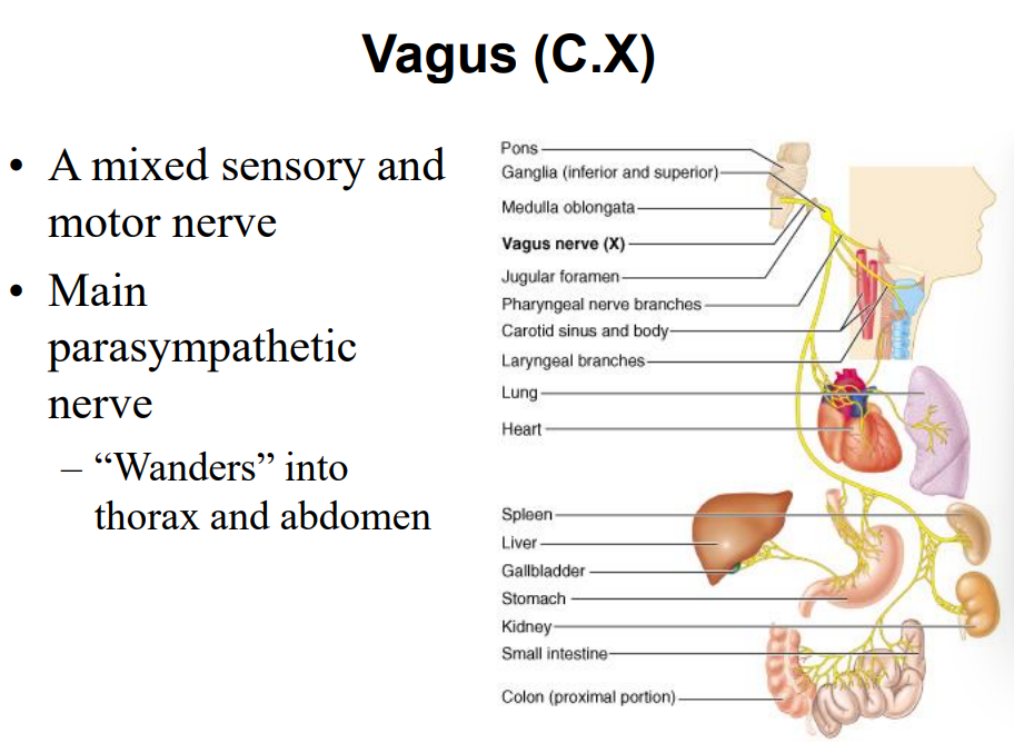
vagus somatic sensory
- mucous membranes of laryngopharynx, larynx and upper trachea
glossopharyngeal takes pharynx
vagus visceral sensory
- trachea, lungs, carotid sinus, abdominal veins, gut (to splenic flexure)
vagus somatic motor
- lower pharynx, upper oesophagus (swallowing and vomiting)
vagus parasympathetic motor
- cardiac muscle (control of heart beat), smooth muscle in GIT (GI motility), trachea and bronchi (airway diameter)
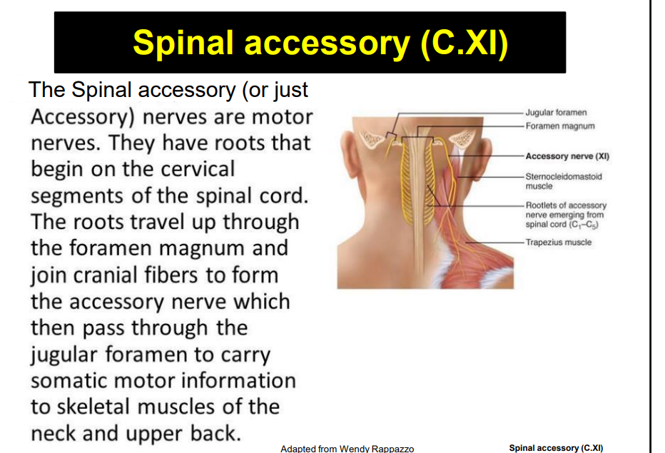
Note that there is probably only a spinal root of C.XI.
Earlier texts additionally refer to a cranial root but these are now thought to be aberrant fibres of C.X. but also has origins in cervical 1-4
what cranial nerves are in the nucleus ambiguous?
glossopharyngeal and vagus
what is the function of the nucleus ambiguous?
Motor: larynx, pharynx, upper oesophagus
-Vomiting, swallowing, modulation of phonation
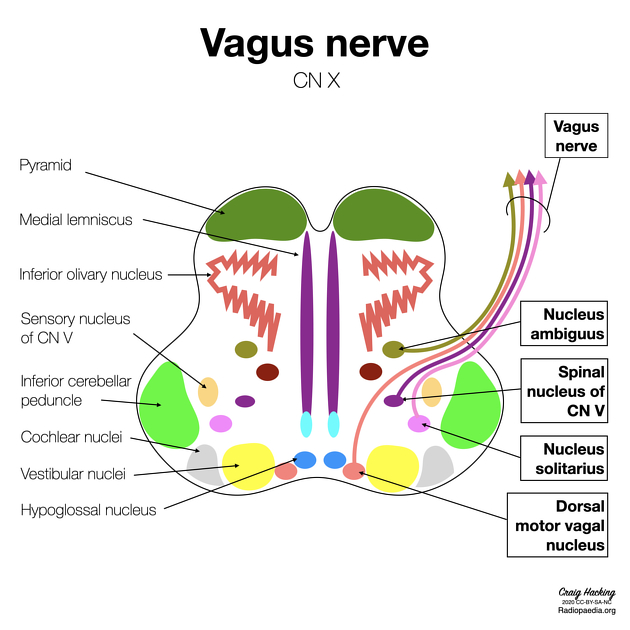
what cranial nerves make up the nucleus tractus solitaris?
facial, glossopharyngeal, vagus
(Solitary Nucleus) Visceral Sensory: tongue, carotid body, carotid sinus, GIT, trachea, bronchi
- Chemoreception (inc taste, pCO2/[H+] monitoring, gut distension, blood pressure monitoring
![<p><strong>facial</strong>, glossopharyngeal, vagus </p><p>(Solitary Nucleus)<strong><u> Visceral Sensory:</u></strong> <strong><em><u>tongue</u></em></strong>, carotid body, carotid sinus, GIT, trachea, bronchi </p><p>- <strong><em>Chemoreception </em></strong>(inc taste, pCO2/[H+] monitoring, gut distension, blood pressure monitoring</p>](https://knowt-user-attachments.s3.amazonaws.com/32eb2cd7-81c1-4d41-8170-97fd9567eecc.png)