Muscles of upper limb
1/23
There's no tags or description
Looks like no tags are added yet.
Name | Mastery | Learn | Test | Matching | Spaced |
|---|
No study sessions yet.
24 Terms
Pectoral region
Located in the anterior chest wall
Contains the breast and four muscles
Pectoralis major, pectoralis minor, serratus anterior and subclavius
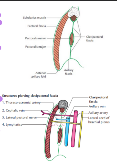
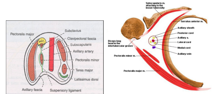
Clavipectoral fascia
Fibromuscular sheet situated deep to the clavicular portion of pectoralis major
Vertically- superiorly splits to enclose subclavius muscle and attached to clavicle
Horizontally- medially attached to 1st rib, costocoracoid ligament
Laterally- attached to coracoid process and blends with coracoclavicular ligament
Pierced by- lateral pectoral nerve, cephalic vein, thoracoacromial vessels and lymphatics
Muscles of pectoral region
ORIGIN | INSERTION | ACTION | INNERVATION | |
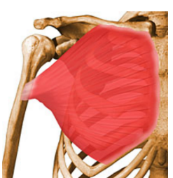
Pectoralis major | ||||
Clavicular head Sternocostal head | Anterior surface of medial ½ of clavicle Anterior surface of sternum, 2nd to 6th costal cartilages | Lateral lip of bicipital groove | Adduction and medial rotation of shoulder | Lateral and medial pectoral nerve |
Pectoralis minor | From 3rd to 5th rib near the costochondral junction | Medial border and superior surface of coracoid process | Draws scapula forward, depresses the point of the shoulder | Lateral and medial pectoral nerve |
Subclavius | 1st rib at costochondral junction | Subclavian groove of inferior surface of clavicle | Steadies clavicle during movement | Nerve to subclavius |
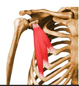
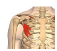
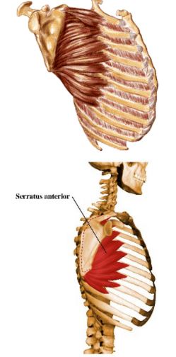
Serratus anterior | 8 digitations from the outer surface of the upper 8 ribs | 1st attached to superior angle 2-3 attached to lateral border last 4-5 attached to inferior angle | Protracts scapula around chest wall | Long thoracic nerve |
Axilla
Has apex, base and 4 walls
Apex
In front clavicle
Behind superior border of scapula
Medially outer border of 1st rib
Anterior wall
Pectoralis major and minor, clavipectoral fascia and subclavius
Posterior wall
Subscapularis, teres major and latissimus dorsi
Medial wall
Upper 4 ribs with their intercostal muscles
Lateral wall
Formed by upper part of shaft of humerus and coracobrachialis
Contents of axilla
Axillary artery
Axillary vein
Infra-clavicular part of brachial plexus
Axillary lymph nodes (anterior, posterior, lateral, central, apical)
Long thoracic nerve
Intercostobrachial nerve
Axillary pad of fat
Brachial plexus
Formed from ventral rami C5 to T1
Pre-fixed, C5 large, C4 present, T1 small and T2 absent
Post-fixed, T1 large, T2 present, C5 small and C4 absent
Branches from cord
Lateral cord
Lateral pectoral nerve
Lateral root of median nerve
Musculocutaneous nerve
Medial cord
Medial root of median nerve
Medial cutaneous nerve of arm
Medial cutaneous nerve of forearm
Medial pectoral nerve
Ulna nerve
Posterior cord
Upper subscapular
Lower subscapular
Thoracodorsal nerve
Radial nerve
Axillary nerve
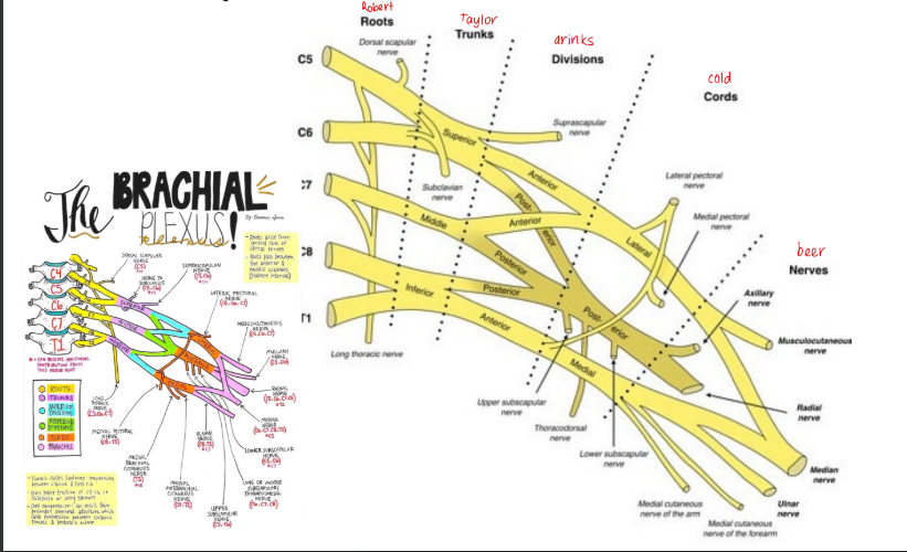
Erb’s point
Consists of C5, C6 ventral and dorsal division of upper trunk, suprascapular and nerve to subclavius
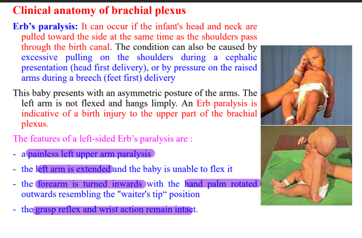
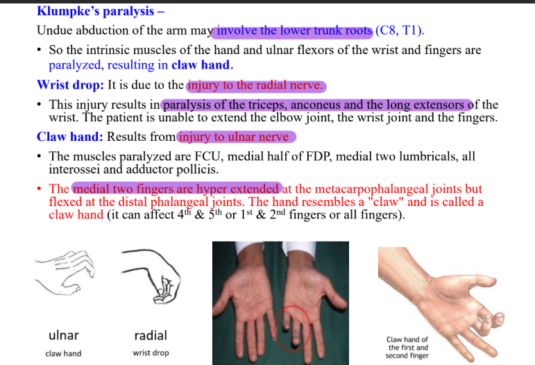
Clinical anatomy of brachial plexus
Muscles of back
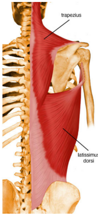
Superficial- latissimus dorsi and trapezius
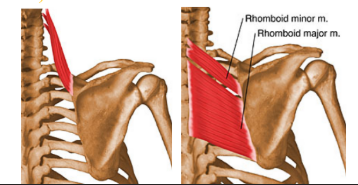
Deep- levator scapulae, rhomboid major and minor
ORIGIN | INSERTION | ACTION | INNERVATION | |
Trapezius | Ligamentum nuchae, superior nuchal line, external occipital protuberance, spinous process of C7 to T12 | Lateral 1/3 of clavicle, medial margin of acromion, upper lip of crest of spine of scapula | Superior fibres elevate Middle fibres retract Inferior fibres depress | Spinal part of accessory nerve |
Latissimus dorsi | Spinous process of lower 6 thoracic vertebrae, inferior angle of scapula, posterior 1/3 of outer lip of iliac crest, posterior layer of thoracolumbar fascia | Floor of bicipital groove | Extends, adducts and medially rotates humerus | Thoracodorsal nerve |
Levator scapulae | Posterior tubercle of transverse process of C1 to C4 | Superior surface of medial border of scapula | Elevates scapula and tilts glenoid cavity inferiorly | Dorsal scapula nerve |
Rhomboid | Minor- lower ligamentum nuchae, spinous process of C7 & T1 Major- spinous process of T2-T5 | Medial border of scapula from level of spine to inferior angle | Retracts scapula and/ rotates it to depress glenoid cavity | Dorsal scapular nerve |
Muscles of scapular region
ORIGIN | INSERTION | ACTION | INNERVATION | |
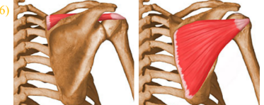
Supraspinatus | Supraspinous fossa | Superior facet of greater tubercle | Initiates and assists deltoid with abduction | Suprascapular nerve |
Infraspinatus | Infraspinous fossa | Middle facet of greater tubercle | Laterally rotates arm | Suprascapular nerve |
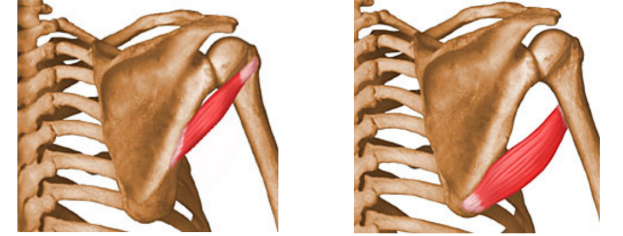
Teres minor | Superior part of lateral border of scapula | Inferior facet of greater tubercle | Laterally rotates arm | Axillary nerve |
Teres major | Dorsal surface of inferior angle of scapula | Medial lip of bicipital groove | Medially rotates and adducts | Lower scapular nerve |
Subscapularis | Subscapular fossa | Lesser tubercle | Medially rotates and adducts | Upper and lower scapular nerve |
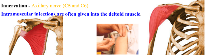
Deltoid | Acromion process, anterior border of lateral 1/3 clavicle, lower lip of crest of spine | Deltoid tuberosity | Anterior flexes and medially rotates Middle abducts Posterior extends and laterally rotates | Axillary nerve |
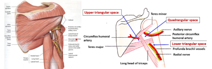
Intermuscular spaces
Formed by scapular muscles, humerus and long head of triceps
Quadrangular space
Upper triangular space
Lower triangular space
Quadrangular space
Superior- subscapularis, capsular ligament of shoulder joint, teres minor
Inferior- teres major
Medial- long head of triceps
Lateral- surgical neck of humerus
Contents- axillary nerve and posterior circumflex humeral vessels
Upper triangular space
Medial- teres minor
Lateral- long head of triceps
Inferior- teres major
Contents- circumflex scapular artery
Lower triangular space
Medial- long head of triceps
Lateral – medial border of humerus
Superior – teres major
Contents – radial nerve and profunda brachii vessels
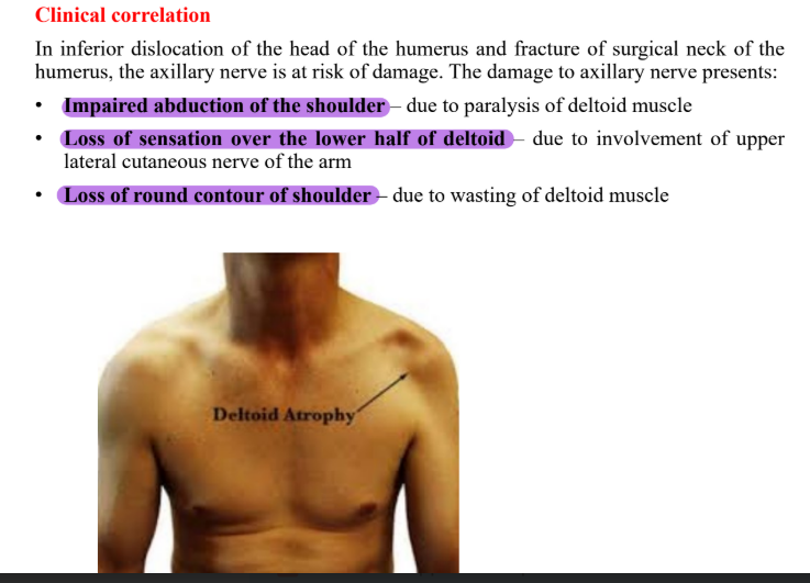
Clinical correlation of shoulder
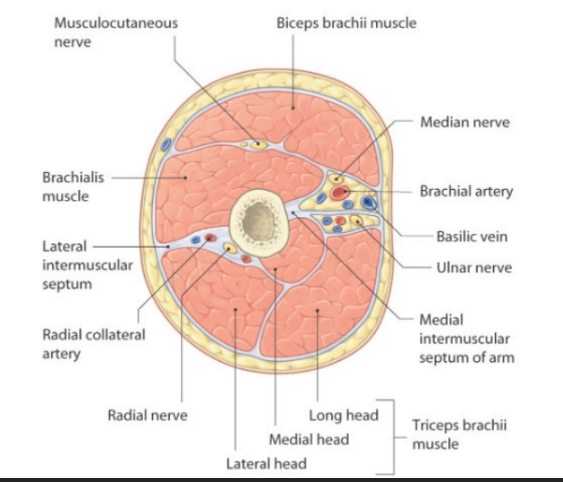
Compartments of arm
Medial and lateral intermuscular septae arises from deep fascia and divides arm into flexor and extensor compartment
Contents of flexor compartment
Muscles- biceps brachii, coracobrachialis and brachialis
Nerve- musculocutaneous
Artery- brachial
Contents of posterior compartment
Muscles- triceps
Nerve- radial
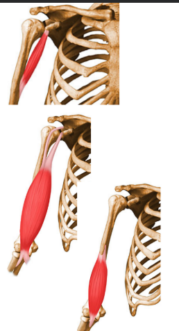
Muscles of arm
Flexor compartment
ORIGIN | INSERTION | ACTION | INNERVATION | |
Coracobrachialis | Tip of coracoid process | Middle 1/3 of medial border of humerus | Flexes and adducts arm | Musculocutaneous nerve |
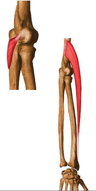
Biceps brachii | Short head- tip of coracoid process Long head- supraglenoid tubercle | Tuberosity of radius and deep fascia of forearm | Supinator of forearm Flexor of elbow | Musculocutaneous nerve |
Brachialis | DIstal half of anteromedial and anterolateral surface of humerus Medial and lateral intermuscular septae | Coronoid process and tuberosity of ulna | Major flexor of forearm | Musculocutaneous nerve |
Posterior compartment
ORIGIN | INSERTION | ACTION | INNERVATION | |
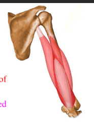
Triceps brachii | Long head- infraglenoid tubercle Lateral head- posterior surface of humerus, superior to radial groove Medial head- posterior surface of humerus, inferior to radial groove | Posterior part of superior surface of olecranon process of ulna | Chief extensor of forearm | Radial nerve |
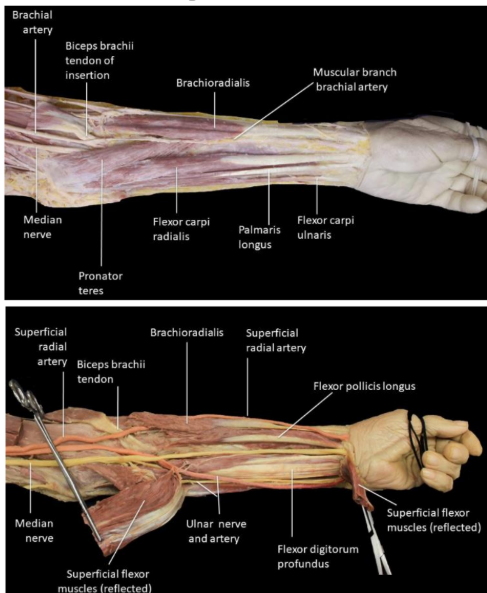
Cubital fossa
Triangular hollow region situated in front of the elbow joint
Boundaries
Laterally- medial border of brachioradialis
Medially- lateral border of pronator teres
Base- directed upwards and formed by an imaginary line joining two epicondyles
Apex- directed downwards and formed by overlapping of medial and lateral boundaries
Roof- skin, superficial fascia with its contents, deep fascia
Floor- brachialis above and supinator below
Contents
Median nerve
Tendon of biceps brachii
Termination of brachial artery
Ulnar artery
Radial artery
Radial nerve
Forearm
Contains anterior and posterior compartment
Anterior compartment
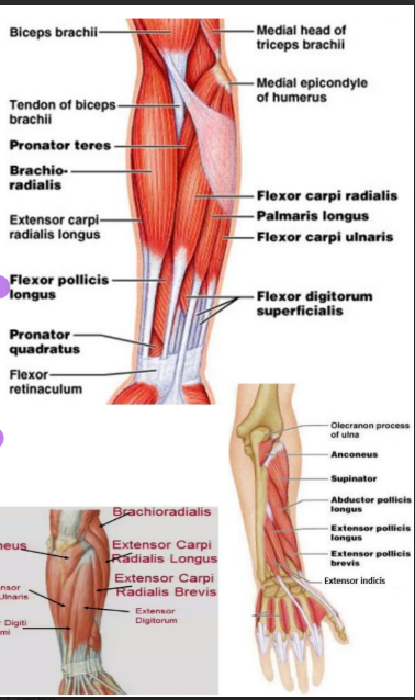
Superficial- pronator teres, flexor carpi radialis, palmaris longus, flexor digitorum superficialis, flexor carpi ulnaris
Deep- flexor policis longus, flexor digitorum profundii, pronator quadratus
Radial and ulnar arteries
Median, ulnar and radial nerves
Posterior compartment
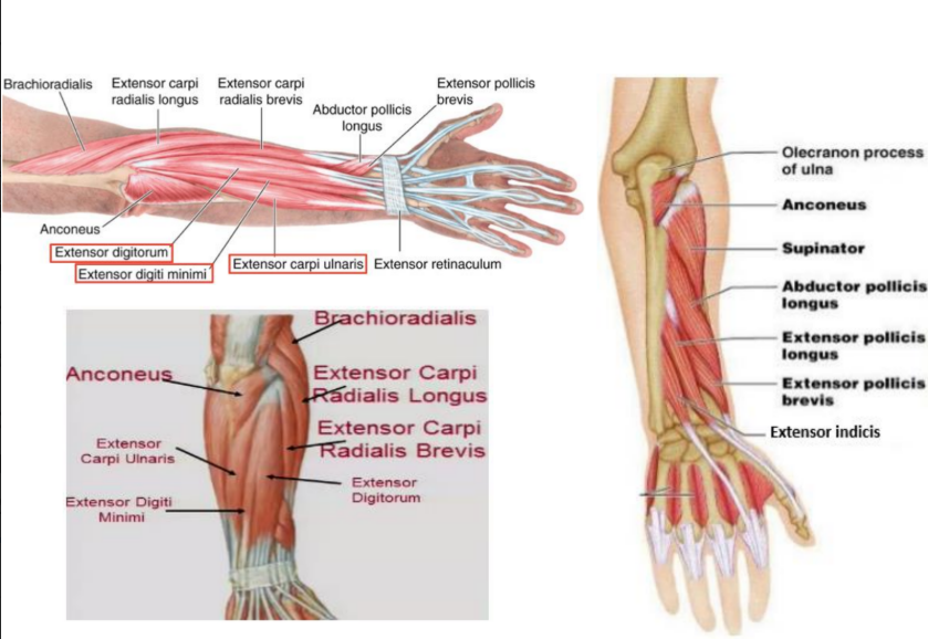
Superficial- anconeus, brachioradialis, extensor carpi radialis longus, extensor carpi radialis brevis, extensor digitorum, extensor digiti minimi, extensor carpi ulnaris
Deep- supinator, abductor pollicis longus, extensor policis brevis, extensor indicis
Flexor muscles of forearm
ORIGIN | INSERTION | ACTION | INNERVATION | |
Pronator teres | Medial epicondyle and coronoid process of ulna | Middle 1/3 of lateral surface of radius | Pronates and flexes forearm at elbow | Median nerve |
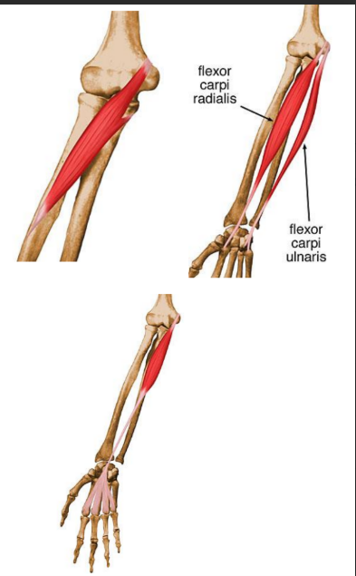
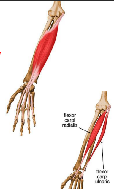
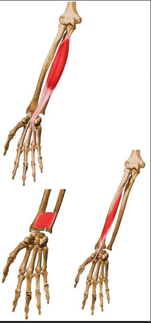
Flexor carpi radialis | Medial epicondyle | Base of 2nd and 3rd metacarpal bone | Flexes and abducts hand at wrist | Median nerve |
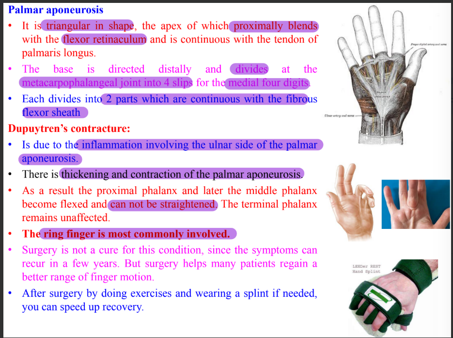
Palmaris longus | Medial epicondyle | Distal half of flexor retinaculum and apex of palmar aponeurosis | Flexes hand at wrist and tightens palmar aponeurosis | Median nerve |
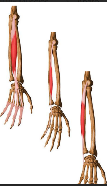
Flexor digitorum superficialis | Humeroulnar- medial epicondyle, ulnar collateral ligament and coronoid process of ulnar Radial- upper half of anterior border of radius | Bodies of phalanges 2-3 | Flexes middle phalanges at proximal interphalangeal joint | Median nerve |
Flexor carpi ulnaris | Humeral- medial epicondyle Ulnar- olecranon and posterior border of ulnar | Pisiform bone, hook of hamate and 5th metacarpal | Flexes and adducts at wrist | Ulnar nerve |
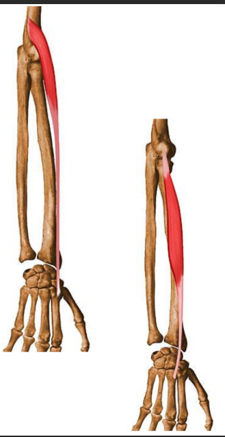
Flexor digitorum profundus | Proximal ¾ of medial and anterior surface of ulnar and interosseous membrane | Base of distal phalanx 2-5 | Flexes distal phalanges at distal interphalangeal joint | Medial- ulnar nerve Lateral- anterior interosseous branch of median nerve |
Flexor pollicis longus | Anterior surface of radius and interosseous membrane | Base of distal phalanx of thumb | Flexes phalanges of 1st digit | Anterior interosseous nerve from median nerve |
Pronator quadratus | Distal ¼ of anterior surface of ulna | Distal ¼ of anterior surface of radius | Pronates forearm | Anterior interosseous nerve from median nerve |
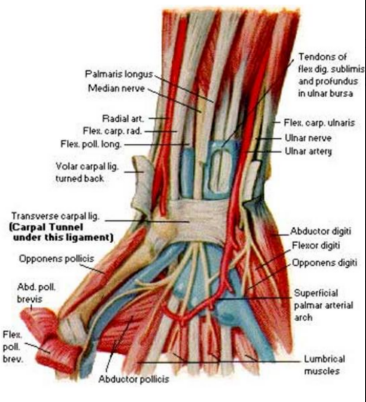
Flexor retinaculum
Fibrous band which bridges anterior concavity of carpals and creates a tunnel
Attachment
Medially- pisiform bone and hook of hamate
Laterally- tubercle of scaphoid and crest of trapezium
Structures superficial
Tendon of palmaris longus
Palmar cutaneous branch of median and ulnar nerve
Ulnar vessels
Structures deep
Median nerve
Ulnar bursa
Tendons of FDS, FPL, FDP
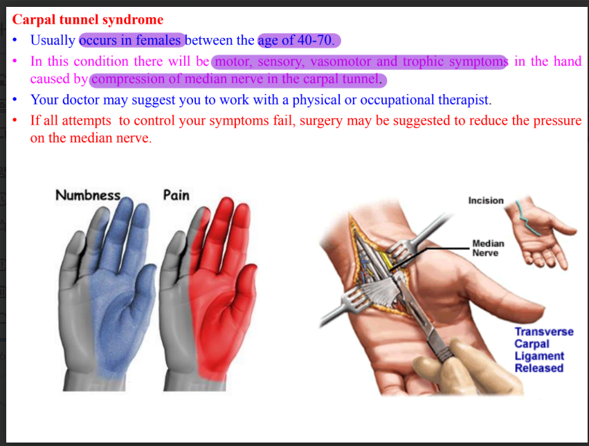
Carpal tunnel syndrome
Extensor muscles of the forearm
ORIGIN | INSERTION | ACTION | INNERVATION | |
Aconeus | Posterior aspect of lateral epicondyle | Lateral surface of olecranon and superior part of posterior surface of ulna | Weak extensor of elbow, stabilizes joint | Radial nerve |
Brachioradialis | Proximal 2/3 of lateral supracondylar ridge of humerus and lateral intermuscular septum | Lateral surface of distal end of radius just above styloid process | Flexor of forearm | Radial nerve |
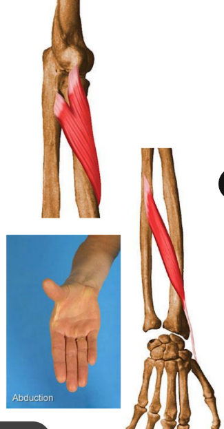
Extensor carpi radialis longus | Lower 1/3 of lateral supracondylar ridge of humerus, lateral intermuscular septum | Dorsal surface of base of 2nd metacarpal bone | Extension and abduction of hand at wrist | Radial nerve |
Extensor carpi radialis brevis | Common extensor origin and radial collateral ligament below elbow | Dorsal aspect of base of 2nd and 3rd metacarpals | Extension and abduction of hand at wrist | Posterior interosseous nerve |
Extensor digitorum | Common extensor origin | Extensor expanasion of medial 4 | Extension of interphalangeal, metacarpophalangeal and wrist joint | Posterior interosseous nerve |
Extensor digiti minimi | Common extensor origin | Extensor expansion of 5th | Extends 5th digit at metacarpophalangeal and interphalangeal joint | Posterior interosseous nerve |
Extensor carpi ulnaris | Common extensor origin and posterior border of ulna | Medial side of base of 5th | Extends and adducts at wrist | Posterior interosseous nerve |
ORIGIN | INSERTION | ACTION | INNERVATION | |
Supinator | Lateral epicondyle, radial collateral and annular ligaments, supinator fossa and crest of ulna | Lateral, posterior and anterior surfaces of proximal 1/3 of radius | Supination of forearm | Posterior interosseous nerve |
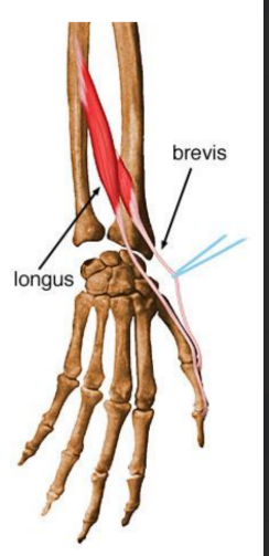
Abductor pollicis longus | Posterior surfaces of ulnar, radius and interosseous membrane | Base of 1st metacarpal and trapezium | Abducts thumb and extends it at carpometacarpal joint | Posterior interosseous nerve |
Extensor pollicis brevis | Posterior surfaces of radius and interosseous membrane | Base of proximal phalanx of thumb | Extends proximal phalanx of thumb at carpometacarpal | Posterior interosseous nerve |
Extensor pollicis longus | Posterior surface of middle 1/3 of ulna and interosseous membrane | Base of distal phalanx of thumb | Extends distal phalanx of thumb at carpometacarpal and interphalangeal joints | Posterior interosseous nerve |
Extensor indicis | Distal 1/3 of posterior surface of ulna and interosseous membrane | Middle and distal phalanx of 2nd digit | Extends index finger | Posterior interosseous nerve |
Extensor retinaculum
Thick fibrous band present on back of wrist joint
Attached laterally to the lower part of the anterior border of the radius, medially to the styloid process of the ulna, triquetral and pisiform bones.
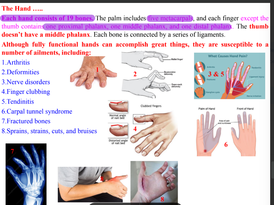
Clinical correlation of hand
Intrinsic muscles of hand
Four thenar muscles
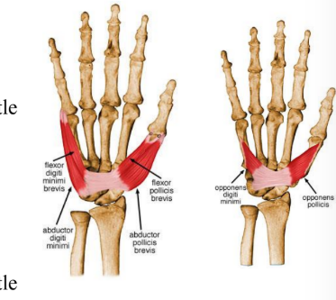
Abductor pollicis brevis
Flexor pollicis brevis
Opponens pollicis brevis
Adductor pollicis brevis
Four hypothenar muscles
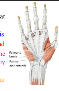
Palmaris brevis
Abductor digiti minimi
Flexor digiti minimi
Oppenens digiti minimi
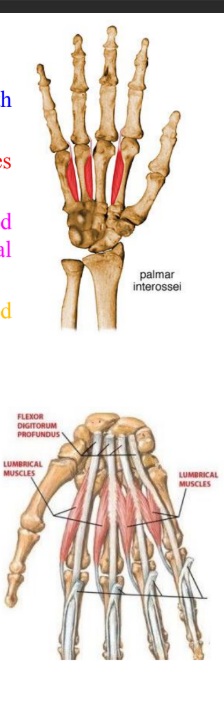
Four lumbricals
Three palmar interossei
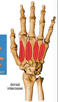
Four dorsal interossei
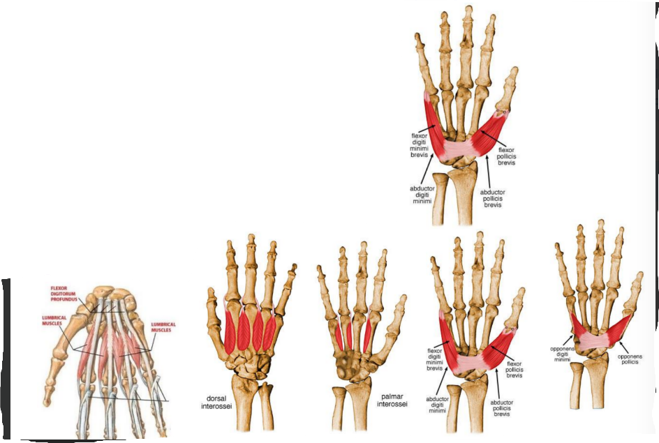
Thenar muscles
ORIGIN | INSERTION | ACTION | INNERVATION | |
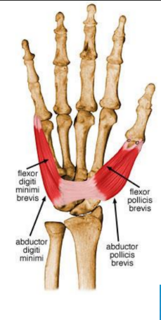
Abductor pollicis brevis | Flexor retinaculum and tubercle of scaphoid and crest of trapezium | Lateral of base of proximal phalanx of thumb | Abducts the thumb at metacarpophalangeal and carpometacarpal joint | Median nerve |
Flexor pollicis brevis | Superficial head- flexor retinaculum and crest of trapezium Deep- trapezoid and capitate bone | Lateral side of base of proximal phalanx | Flexes thumb | Median nerve |
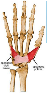
Opponens pollicis | Flexor retinaculum and crest of trapezium | Lateral side of 1st metacarpal | Draws 1st metacarpal to oppose thumb | Median nerve |
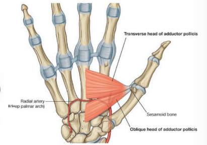
Adductor pollicis | Oblique- base of 2nd and 4rd metacarpals, capitate and adjacent carpals Transverse- palmar surface of 3rd metacarpal | Medial side of base of proximal phalanx of thumb | Muscle adducts thumb from flexed or abducted position | Deep branch of ulnar nerve |
Hypothenar muscles
ORIGIN | INSERTION | ACTION | INNERVATION | |
Abductor digiti minimi | Pisiform bone | Medial side of base of proximal phalanx of little finger | Abducts little finger | Deep branch of ulnar nerve |
Flexor digiti minimi | Hook of hamate a/nd flexor retinaculum | Medial side of base of proximal phalanx of little finger | Flexes little finger at metacarpophalangeal joint | Deep branch of ulnar nerve |
Opponens digiti minimi | Hook of hamate and flexor retinaculum | Medial border of shaft of 5th metacarpal | Draws 5th metacarpal anteriorly and rotates it laterally, opposes thumb | Deep branch of ulnar nerve |
Palmaris brevis | Flexor retinaculum and palmar aponeurosis | Skin along medial border of hand | Helps with gripping | Superficial branch of ulnar nerve |
Interossei, palmar and lumbricals
ORIGIN | INSERTION | ACTION | INNERVATION | |
Dorsal interossei (4) | Adjacent sides of two metacarpals | Extensor expansions and base of proximal phalanges of digit 2-4 | Abduct digits from axial line Act with lumbricals to flex metacarpophalangeal joint and extend interphalangeal joint | Deep branch of ulnar nerve |
Palmar interossei | Palmar surfaces of 2nd, 4th and 5th metacarpals | Extensor expansions of digit and bases of proximal phalanges | Adduct digits towards axial line Assist lumbricals in flexing metacarpophalangeal joints and extending interphalangeal joints | Deep branch of ulnar nerve |
Lumbrical muscles | From tendon of flexor digitorum profundus | Dorsal digital expansion | Flex the metacarpophalangeal joints and extend the interphalangeal joints | 1st and 2nd lumbricals by median nerve 3rd and 4th lumbricals by deep branch of ulnar nerve |