ILS 2: Embryology-Conception
 Call Kai
 Learn
Learn Practice Test
 Spaced Repetition
 Match
Match Flashcards
 Knowt Play
1/72

There's no tags or description
Looks like no tags are added yet.
Name | Mastery | Learn | Test | Matching | Spaced | Call with Kai | Chat |
|---|
No analytics yet
Send a link to your students to track their progress
73 Terms
Primary oocytes are arrested at
Prophase I
After puberty, oocyte is arrested at
Metaphase II
Secondary oocytes are released during
ovulation
When do secondary oocytes complete Meiosis II?
After fertilization
when do spermatogonium start 1st meiotic division?
after puberty
total length of spermatogenesis
74 days
site of fertilization
ampulla of uterine tubes
ovaries produce
estrogen & progesterone
hormones that cause ovarian follicles to mature to a Graafian follicle
FSH & LH
what event causes primary oocyte to complete M1 and begin M2 as a secondary oocyte?
LH surge
Ovulation is driven by these hormones
Prostaglandins and LH [expel oocyte from mature follicle]
a yellow body is also known as
corpus luteum [follicle remaining after oocyte expelled during ovulation]
function of corpus luteum
secretes progesterone & some estrogen to help endometrium prepare for implantation
Before being able to fertilize oocyte, spermatozoa must under these 3 steps:
Capacitation
Acrosome rxn
Polyspermy block induced
What is capacitation?
epithelial interactions btwn sperm and mucosal surface of uterine tube [~7hrs], stripping sperm cells
What is the acrosome rxn?
occurs after binding to zona pellucida;
induced by zona proteins
release enzymes [acrosin] needed to penetrate zona pellucida
Phases of Fertilization [3]
Penetration of corona radiata
Penetration of zona pellucida
Fusion of oocyte & sperm cell membranes
What happens in slow block of polyspermy?
mechanical block [hyaline layer] hardens
internal SER stores of calcium ions diffuse thru oocyte membrane to promote fusion of egg & release cortical granules
permanent!
16-cell structure of developing embryo is known as
Morulla / Early Blastocyst
What is the embryoblast?
inner cell mass → embryo
What is the trophoblast?
outer cell mass → placenta
When does an early blastocyst/morulla become a blastocyst?
when fluid penetrates zona pellucida into the inner cell mass
fluid-filled cavity forms
zona pellucida disappears and implantation begins
~day 5
Implantation occurs …
when?
where?
5-15 days after fertilization
posterior superior wall of uterus in endometrial layer
What is AVE?
Anterior Visceral Endoderm
dorsal-ventral polarity established in embryo
Which cells form AVE?
Hypoblast cells
-migrate to cranial end of embryo
AVE cells are classifed as which germ layer?
endoderm
secrete signals to specific cranial end of embryo
Where do primitive streak and node begin formation?
at caudal end
Gestational Age
counted from 1st day of the last LMP (last mentrual period)
more accurate
accounts for 2 weeks leading up to fertilization
Embryologic Age (Conception)
day-day events that begin @ time of fertilization
How to calculate fetus due date?
LMP
count back 3 calendar months
add 1 yr & 7 days
What occurs in 2nd week of development?
bilaminar germ disc
2 cell masses → 2 cell types each
2 cell cavities
Trophoblast differentiates into which 2 layers?
Cytotrophoblast
Synctiotrophoblast
Cytotrophoblast
inner layer of mononucleated cells (mitosis)
Synctiotrophoblast
outer multinucleated zone WITHOUT distinct cell boundaries (no mitosis)
comes from cells in cytotrophoblast
invades maternal endometrium & becomes placenta
makes hCG
Embryoblast differentiates into which 2 layers?
Hypoblast
Epiblast
Hypoblast
small cuboidal cells adjacent to blastocyst activity
Epiblast
tall columnar cells adjacent to amniotic cavity
differentiate into amnioblast cells & line amniotic cavity [secrete amniotic fluid]
What is the exocoelomic (Heuser) membrane?
hypoblast cells differentiate to form this thin membrane
lines inner surface of cytotrophoblast
AKA exocoelomic cavity AKA primitive yolk sac
Lacunae
blood network formed by syncytiotrophoblasts to form utero-placental circulation
penetration opening in surface epithelium of uterus [from implantation] is sealed by what?
fibrin coagulum/plug
hCG produced by syncytiotrophoblasts —> ?
stimulate progesterone production by corpus luteum
*maintains pregnancy until WEEK 8, when placenta takes over
hCG levels detected in maternal blood at day __
maternal urine at day __
8
10
low levels of hCG suggest
spontaneous abortions
ectopic pregnancy
high levels of hCG suggest
pregnancy with multiples
hydatidiform mole
gestational neoplasias & tumors
Primitive yolk sac cells form
extraembryonic mesoderm
extraembryonic mesoderm is located between which 2 things?
inner surface of cytotrophoblast
outer surface of exocoelomic cavity
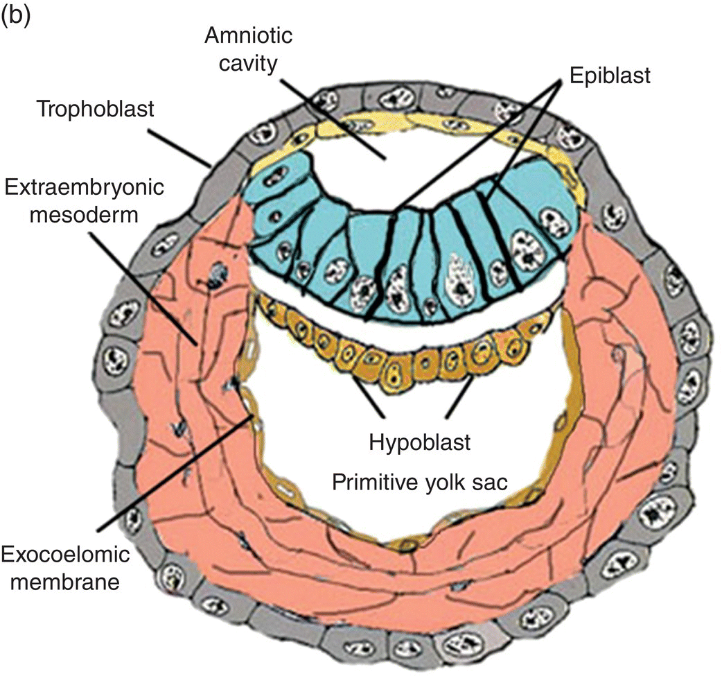
Extraembryonic cavity AKA chorionic cavity
large space between extraembryonic somatic mesoderm & extraembryonic splanchnic mesoderm
Secondary yolk sac functions [3]
supplies temporary nutrition until placenta forms fully
disappears at end of 1st trimester
Hematopoeisis
site of primordial germ cells [gametes]
*entirely lined w/ endoderm
Primary Chorionic Villi
1st stage in placenta development
Chorion forms from
extraembryonic somatic mesoderm
2 layers of trophoblast
gastrulation —> these 3 germ layers
ectoderm
mesoderm
endoderm
Gastrulation events
Primitive streak forms
Primitive node forms
Epiblast cells migrate to & thru primitive groove/streak [invagination]
influenced by growth factors
Which cell type creates all germ layers via gastrulation?
epiblast
endoderm: from displacing hypoblast
mesoderm: cells lie btwn epiblast and endoderm
ectoderm: cells remaining in epiblast
Primitive streak makes which germ layer?
mesoderm
until week 4, then diminishes & remains in sacrococcygeal region
Notochord formed via
invagination of prenotochordal cells thru the primitive node
*mesoderm
Mesenchymal cells
pluripotent; can become any cell/tissue
Key events at Day 21 [end of week 3] in development?
Heart begins to beat
utero-placental circulation functional
Ectoderm gives rise to
Skin epidermis
Nervous system [CNS, PNS]
CNS from neural tube
PNS from neural crest
Mesoderm gives rise to
Vascular system
Urogenital system
Muscles, bones, skin dermis
Endoderm gives rise to
epithelial lining of
GI tract
Respiratory tract
urinary bladder
glands
thyroid, parathyroid, liver, pancreas
Teratogen
known environmental, drug, or lifestyle factor that leads to birth defect
Which week of dvlpment is highly sensitive to teratogens?
Week 3
Malformation
complete or partial absence of a structure
OR
alterations of its normal configuration
Ex. Neural tube defects → anencephaly or spina bifida
Disruption
morphological alteration of an already formed structure by a destructive process
Ex. Amniotic bands produce vascular accidents → cleft lip, toe/finger amputations
Deformation
mechanical forces mold a part of the fetus over time
*often musculoskeletal
Ex. Club feet or hand from compression in amniotic cavity by oligohydramnios [too little amniotic fluid]
Syndrome
group of anomalies occuring together that have a specific common cause
risk of recurrence known
Ex. Down Syndrome
Teratogenic Exposures
Infectious agents [TORCH]
Hyperthermia
Radiation
Chemical agents
Thalomide, Vitamin A, Alcohol
Hormones
Maternal disease
Nutritional deficiences
Obesity
Heavy metals
Thalidomide
sedative linked to limb defects in 1961
Ex. Phocomelia - loss of limb’s long bones
Zika virus
infectious agent -→ microcephaly/small cranium in infants at early gestation
Fetal Alcohol Syndrome
reduced growth
small cranium
indistinct philtrum
thin upper lip
depressed nasal bridge
short nose
flat midface
Neural Tube Deficiency [NTD]
Folate deficiency
partial failure or reversal of neural tube closure
→ severe spina bifida, or anencephaly if in cranial region
Hydatidiform Moles
fertilization of an oocyte lacking a nucleus
followed by duplication of male chromosomes to restore diploid number
trophoblastic development can still occur and lead to placental tissue formation
secrete high levels of hCG
may produce benign or malignant tumors
pregnancy must be terminated
Ectopic pregnancy
blastocyst implants outside of body of uterine cavity
*ampulla most common site of ectopic pregnancy
frequently predisposed by chronic pelvic inflammation [PID]
IVF, smoking, and tubal damage/surgery also risk factors