Final Exam Study Guide
1/237
There's no tags or description
Looks like no tags are added yet.
Name | Mastery | Learn | Test | Matching | Spaced |
|---|
No study sessions yet.
238 Terms
Some microbial activity results in the production of oxygen
Bioremediation
Gut health and vitamin production
Production of food products: cheese, yogurt, alcoholic drinks, and fermented vegetables like kimchi
Play a role in nutrient cycling and nitrogen fixation; makes important nutrients available to plants
Found in root nodules of legumes
Used to produce products such as antibiotics
How are microbes beneficial?
Pathogenic microorganisms can cause disease
Can form biofilms on surfaces like pipes, damaging infrastructure
Biofilm can form on implants, posing a health risk to patients
Involved in food spoilage
How are microbes harmful?
Bright-field microscopy
Differential Interference Contrast (DIC) Microscopy
Confocal Scanning Laser Microscopy
Phase-Constract Microscopy
Dark-Field Microscopy
Fluorescence microscopy
Transmission Electron Microscopy (TEM)
Scanning Electron Microscopy (SEM)
What are the major types of microscopy?
Bright Field Microscopy
What type of microscopy is shown?
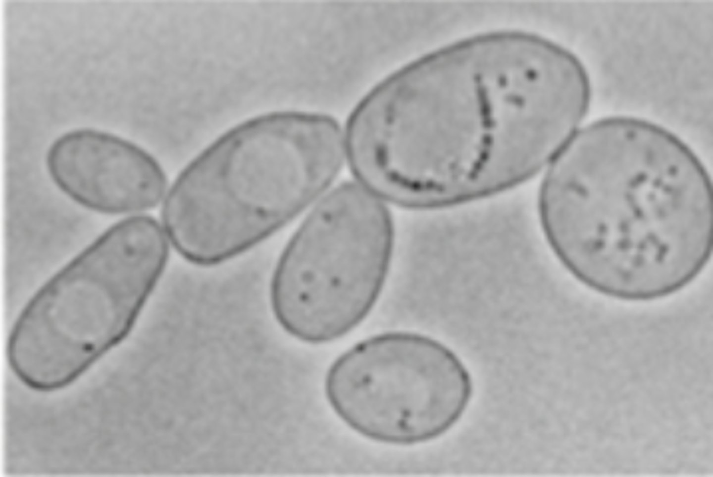
Differentail Interference Constrast (DIC) Microscopy
What type of microscopy is shown?
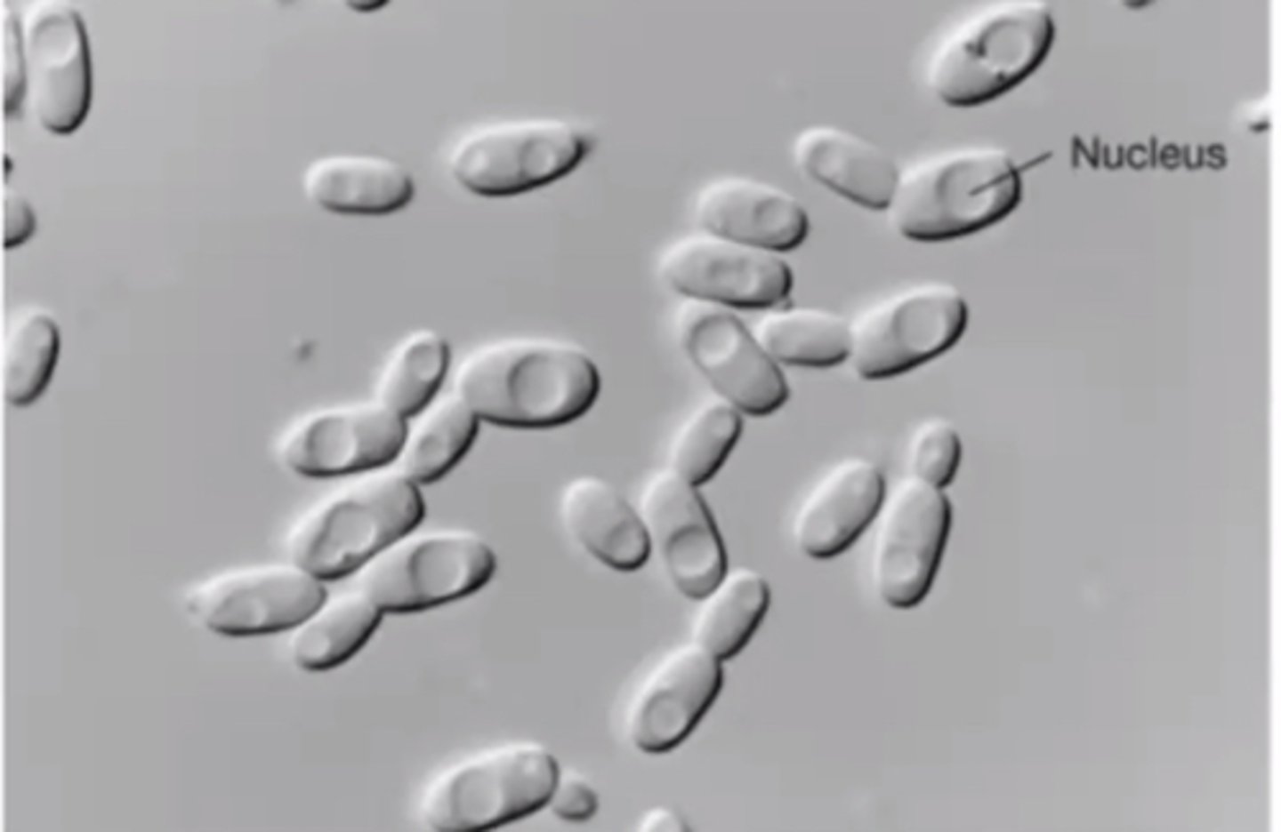
Confocal Scanning Laser Microscopy
What type of microscopy is shown?
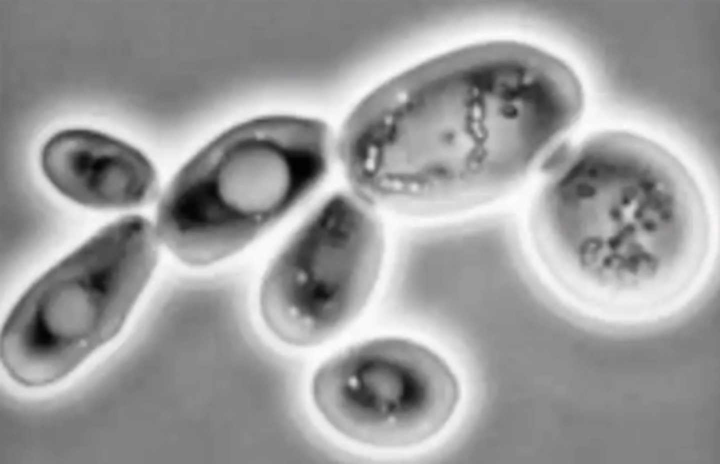
Phase-Contrast Microscopy
What type of microscopy is shown?
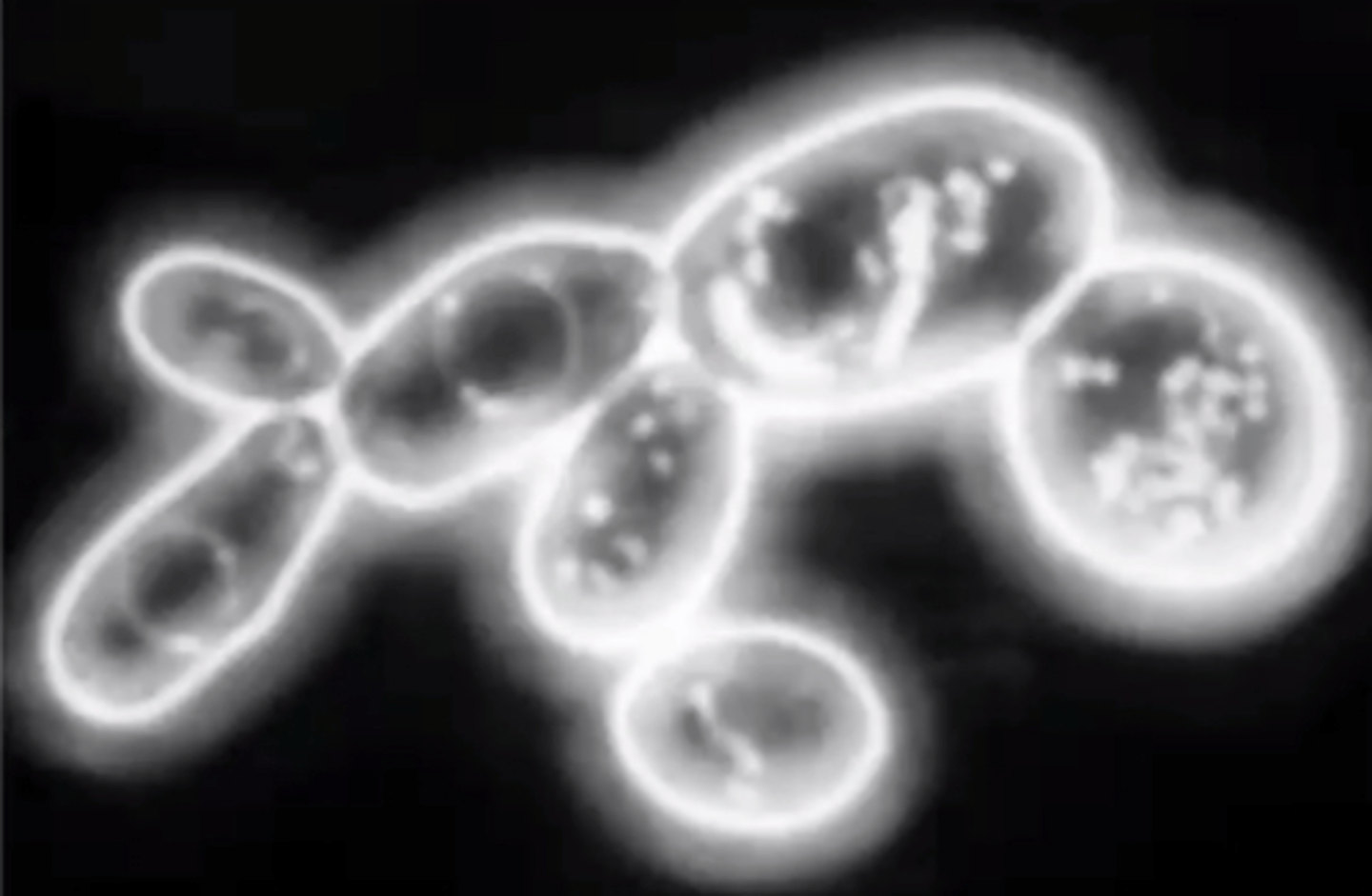
Dark-Field Microscopy
What type of microscopy is shown?
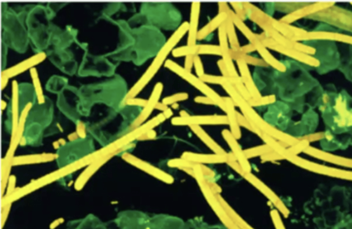
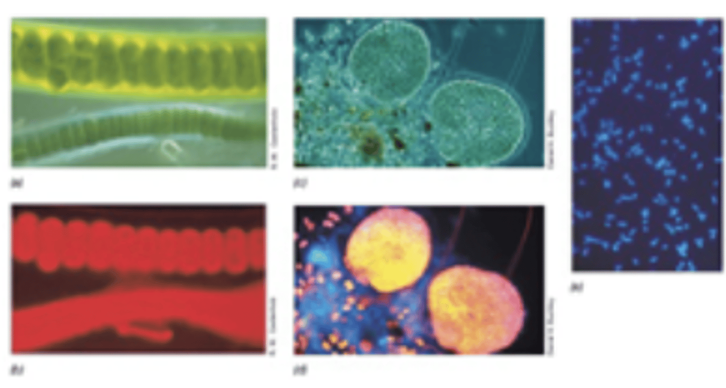
Fluorescence Microscopy
What type of microscopy is shown?
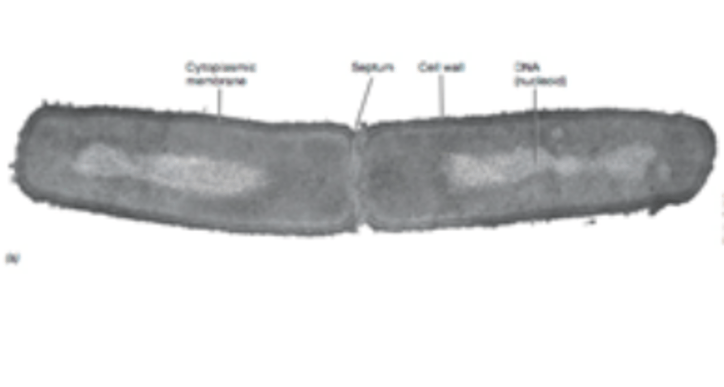
Transmission Electron Microscopy (TEM)
What type of microscopy is shown?
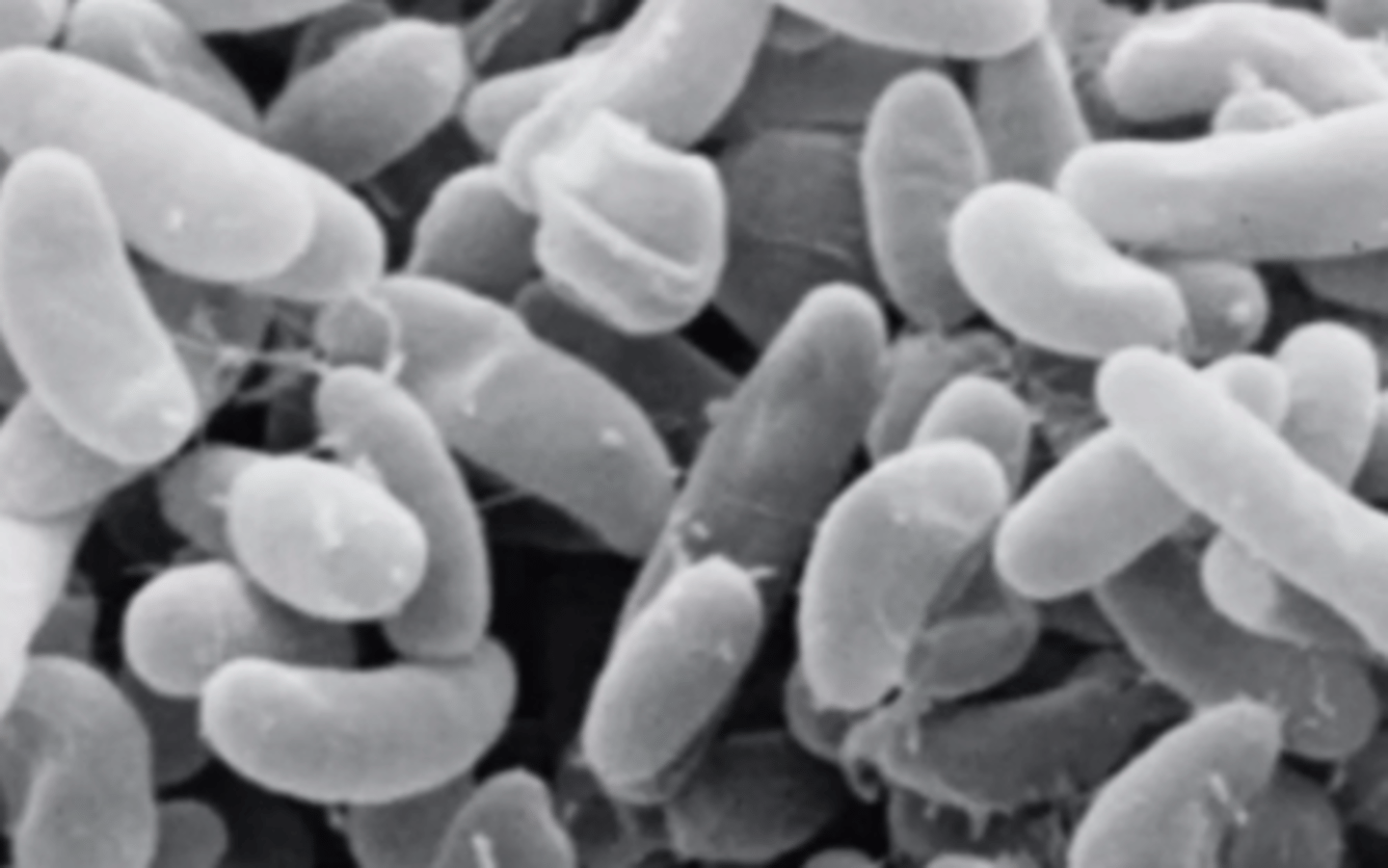
Scanning Electron Microscopy (SEM)
What type of microscopy is shown?
The most common type of light microscope often requires dyes to improve contrast.
What is Bright-Field Microscopy?
Creates a relatively 3-D image of internal cell structures, helping one to view them without stains, by using a polarizer within the microscope condenser.
What is Differential Interference Contrast (DIC) Microscopy?
Uses a laser and a fluorescence microscope to create computer-generated, high-contrast images.
What is Confocal Scanning Laser Microscopy?
A phase ring is used to create increased contrast in the specimen against a dark background.
What is Phase-Contrast Microscopy?
A stop is used to block light from hitting the specimen directly, creating a dark background and allowing living specimens to be viewed with greater contrast.
What is Dark-Field Microscopy?
Used to view natural fluorescence or with fluorescent dyes such as DAPI.
What is Fluorescence Microscopy?
Blue Fluorescent dye that complexes with cell DNA
What is DAPI?
Allows internal cell structures to be viewed at very high magnifications (to the molecular level).
What is Transmission Electron Microscopy (TEM)?
Very effective for viewing 3-D external surface structures at high magnifications.
What is Scanning Electron Microscopy (SEM)?
Coccus
Coccobacillus
Bacillus
Vibrio
Spirillium
Spirochete
Pleomorphic
What are the major bacterial morphologies?
Diplococci
Streptococci
Tetrads
Sarcinae
Staphylococci
What are the major bacterial arrangements?
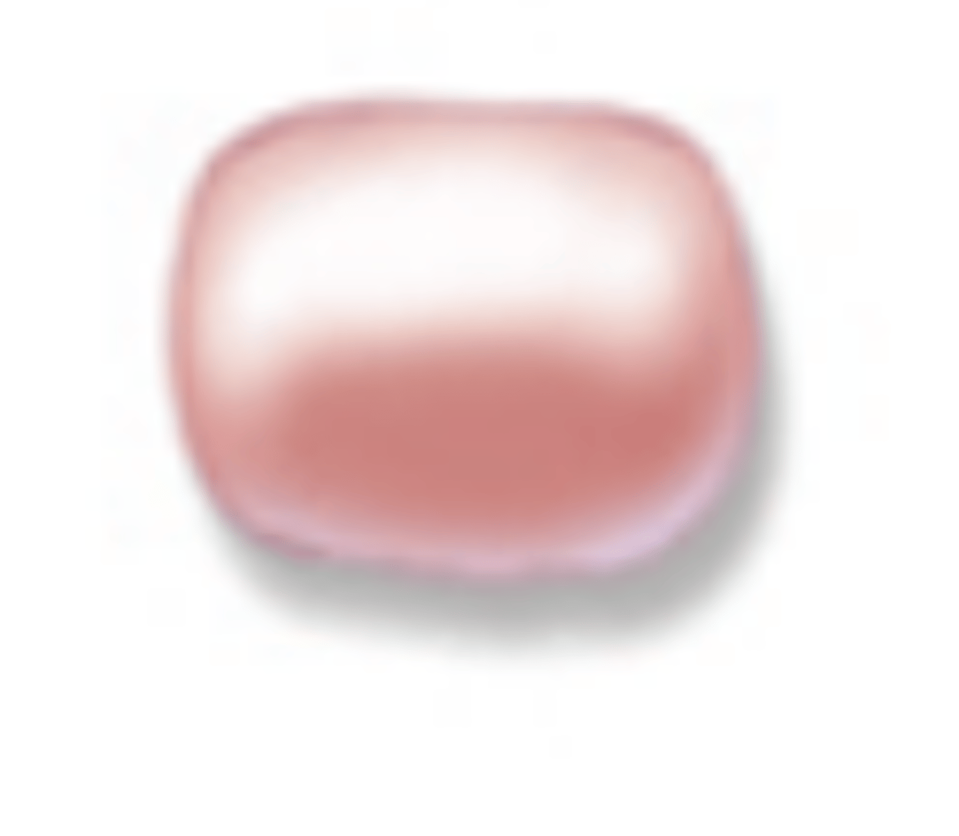
Coccus
What is the morphology of the bacteria shown?

Coccobacillus
What is the morphology of the bacteria shown?
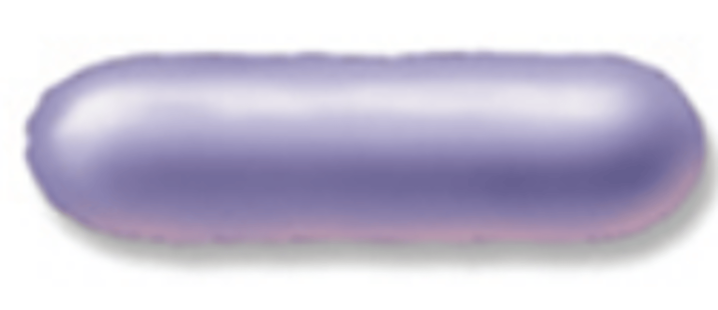
Bacillus
What is the morphology of the bacteria shown?
Vibrio
What is the morphology of the bacteria shown?

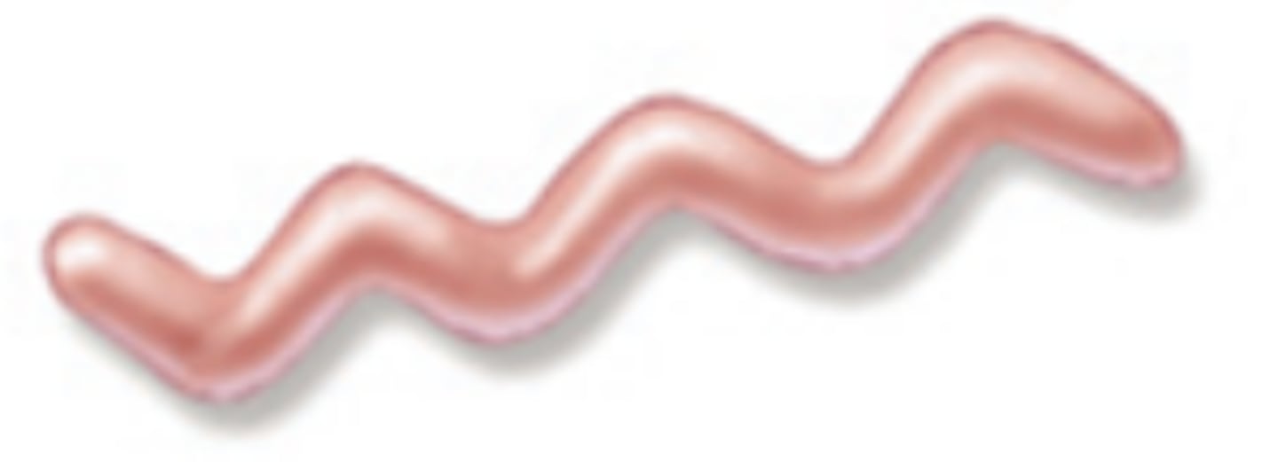
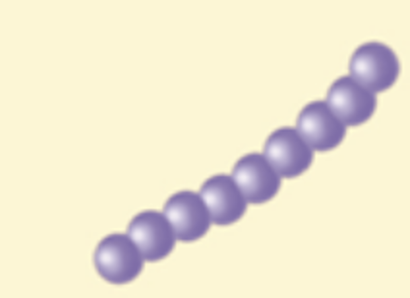
Spirillium
What is the morphology of the bacteria shown?
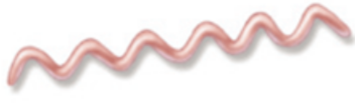
Spirochete
What is the morphology of the bacteria shown?
Pleomorphic
What is the morphology of the bacteria shown?

Diplococci
What is the arrangement of the bacteria shown?

Streptococci
What is the arrangement of the bacteria shown?
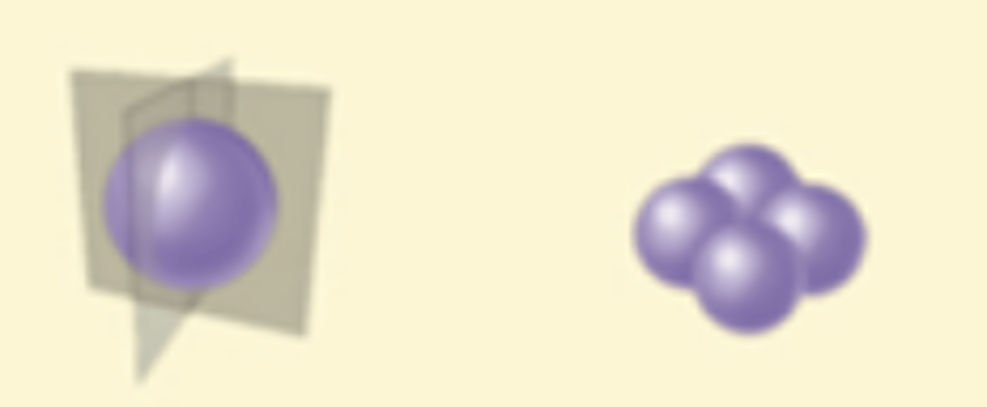
Tetrads
What is the arrangement of the bacteria shown?
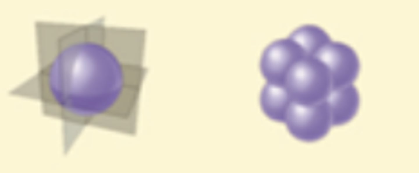
Sarcinae
What is the arrangement of the bacteria shown?
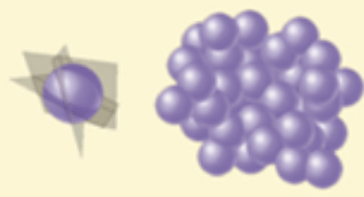
Staphylococci
What is the arrangement of the bacteria shown?
No membrane-bound organelles (no mitochondria or nucleus)
Unicellular
Circular DNA
Asexual Reproduction
Has a capsule
Has ribosomes
Has a cell membrane
Contains Cytoplasm
What is a prokaryotic microbe made up of?
Membrane-bound organelles
Both asexual and sexual reproduction
Can be unicellular or multicellular
Linear DNA
Has ribosomes
Has a cell membrane
Contains Cytoplasm
Has a nucleus
Has a Golgi Complex
Has a mitochondria
Has a rough endoplasmic reticulum
What is a eukaryotic microbe made up of?
Has ribosomes
Has a cell membrane
Contains Cytoplasm
Have a Flagellum
Have a plasma membrane
What are the similarities between eukaryotic and prokaryotic microbes?
Eukaryotes have:
- Membrane-bound organelles
- Multicellular
- Asexual and sexual reproduction
- Linear DNA
Prokaryotes have:
- NO membrane-bound organelles
- Unicellular
- Asexula reproduction
- Circular DNA
What are the differences between eukaryotic and prokaryotic microbes?
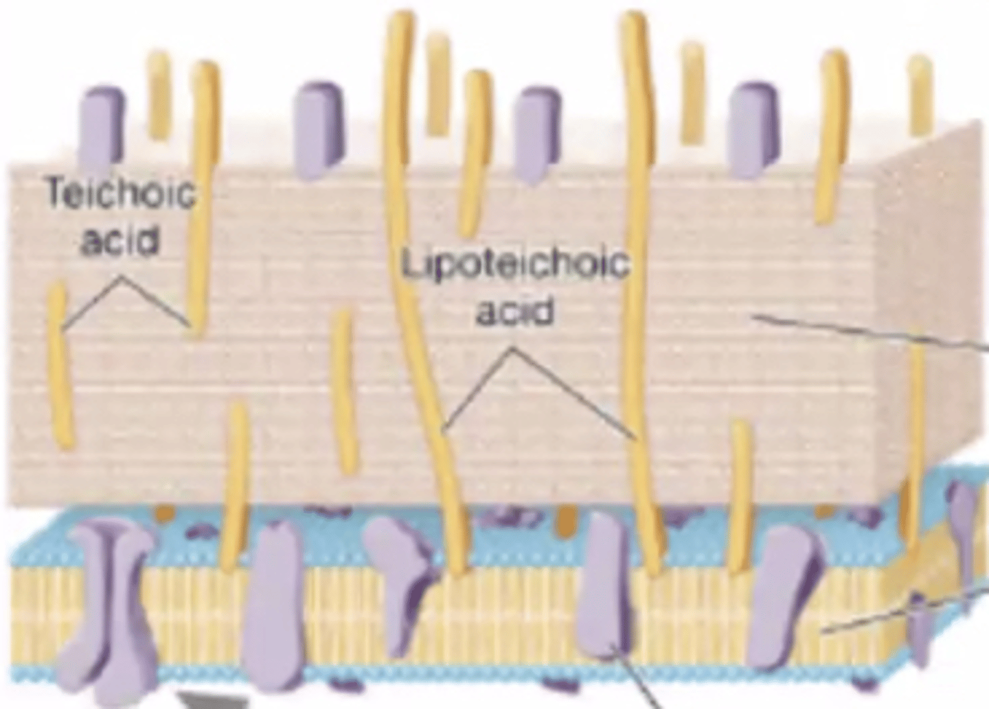
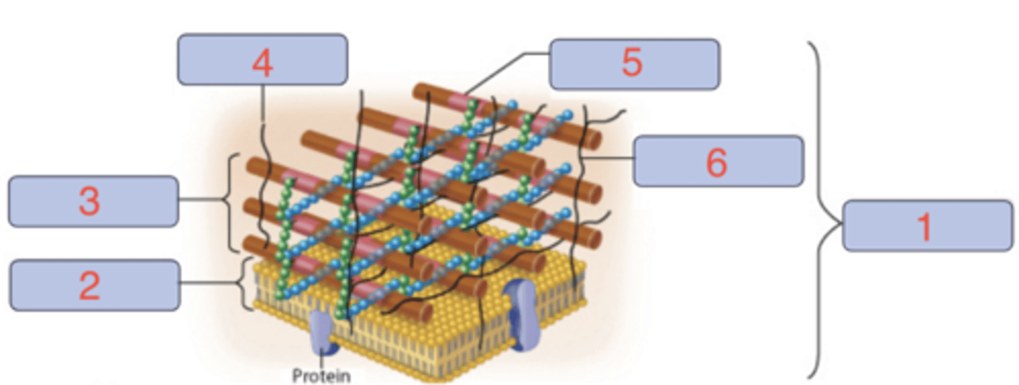
Gram-positive
Based on the structure of the cell wall, is this a gram-negative or a gram-positive bacterium?
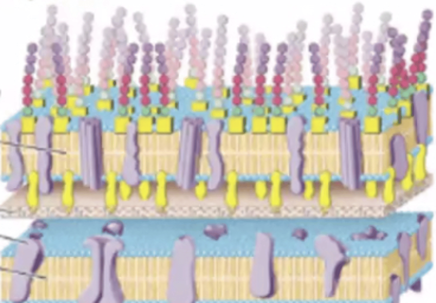
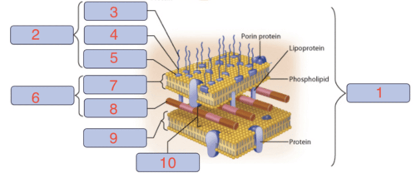
Gram-neagative
Based on the structure of the cell wall, is this a gram-negative or a gram-positive bacterium?
1. Gram-positive cell wall
2. Plasma membrane
3. Cell wall
4. Teichoic acid
5. Peptidoglycan
6. Lipoteichoic acid
Fill out the structure of this bacterium.
1. Gram-negative cell wall
2. LPS
3. O polysaccharide
4. Core polysaccharide
5. Lipid A
6. Cell wall
7. Outer membrane
8. Peptidoglycan
9. Plasma membrane
10. Periplasm
Gram-positive bacteria have a thick layer.
Gram-negative bacteria have a thin layer.
What is the difference between a gram-negative and a gram-positive peptidoglycan?
Gram-positive bacteria have teichoic acids present.
Gram-negative bacteria do not have teichoic acids present.
What is the difference between a gram-negative and a gram-positive teichoic acid?
Gram-positive bacteria do not have an outer membrane.
Gram-negative bacteria do have an outer membrane.
What is the difference between a gram-negative and a gram-positive outer membrane?
Gram-positive bacteria stain purple (retain crystal violet).
Gram-negative bacteria stain pink (safranin counterstain).
What is the difference between a gram-negative and a gram-positive stain color?
Gram-positive bacteria are more sensitive to antibiotics.
Gram-negative bacteria are less sensitive/more resistant due to their outer membrane.
What is the difference between gram-negative and gram-positive antibiotic sensitivity?
1. Flood the heat-fixed smear with crystal violet for 1 minute
2. Add iodine solution for 1 minute
3. Decolorize with alcohol for 20 seconds
4. Counterstain with safranin for 1 - 2 minutes
Describe the technique used to distinguish gram-negative from gram-positive bacteria in the laboratory.
THe gram-positive cells are purple and the gram-negative cells are colorless.
After doing the third step of staining (decolorizer), what will happen to the gram-negative and gram-positive bacteria?
The gram-positive cells are purple and the gram-negative cells are pink to red.
After doing the fourth step of staining (counterstain), what will happen to the gram-negative and gram-positive bacteria?
Has a phospholipid bilayer with embedded proteins. Made up of hydrophobic tails and hydrophilic heads.
Is semi-permeable
Has anchor proteins that play inportant roles in cell functions.
What is the general structure of a cytoplasmic membrane (plasma membrane)?
Prevents passive leakage of solutes in or out of the cell.
Plays a role in energy conservation (proton motive force) for bacteria and archaea.
Waste removal
Communication with environment
What is the general function of a cytoplasmic membrane (plasma membrane)?
Swimming (flagellar) motility
Twitching motility
Gliding motility
What are the types of motility present in prokaryotes?
Swimming (flagellar) motility
What are the types of motility present in eukaryotes?
Driven by rigid, helical flagella made of flagellin protein; rotating motion
Describe the swimming motility of prokaryotes.
Requires type IV pili (retract and pull movement)
Describe the swimming motility of prokaryotes.
No external propulsive structures; instead, an intracellular tract with "gliding motors" driven by the proton motive force.
Describe the gliding motility of prokaryotes.
Cilia
Flagella
What are the types of flagella that Eukaryotes have?
Short flagella that move in Synchrony to move the cell
Describe the cilia of a eukaryote.
Whiplike motion, contains a cytoskeleton (tubulin), and is surrounded by the cell membrane
Describe the flagella of a eukaryote.
Prokaryotic flagellum:
- Made of flagellin protein (filament, hook, basal body)
- Powered by proton motive force
- About ~20 nm
Eukaryotic flagellum:
- Made of microtubules (9+2 arrangement) and dynein motors
- Powered by ATP
- About ~200 nm
How do prokaryotic flagella differ from eukaryotic flagella?
Only some gram-positive bacteria can produce endospores.
Bacteria that produce endospores are dormant cells that can tolerate extreme environmental conditions (heat, chemicals, radiation, and desiccation).
Bacteria can transition, after very long periods of time, into vegetative cells when the conditions are favorable.
What is the significance of endospore formation?
A theory that proposes that mitochondria and chloroplasts are descendants of respiratory and phototrophic bacterial cells that were engulfed by early eukaryotes; forming a symbiotic relationship with their hosts
Evidence:Both have their own circular DNA and ribosomes,Reproduce by binary fission, & Double membranes.
What is the endosymbiotic theory?
Both have their own circular DNA and ribosomes.
Reproduce by binary fission.
Have double membranes.
Explain the relevance of the endosymbiotic theory and chloroplast/mitochondria?
Both oxidation and reduction reactions occur together.
Oxidation = Loss of electrons OIL = Oxidation Is Loss)
Reduction: Gain of electrons (RIG = Reduction Is Gain)
In redox, how are oxidation and reduction related to electrons?
** in progress**
What are the major metabolic pathways?
Fermentative organisms:
- Reduce the pyruvate produced during glycolysis
- Pyruvate is the final electron acceptor
- NADH is oxidized back into NAD+ by reducing pyruvate
- Low ATP yield
Respiratory Organisms:
- Use external electron acceptors
- O2 is the final electron acceptor
- NADH donates electrons to ETC, regenerating NAD+
- High ATP yield
How is redox balance achieved by fermentative organisms vs. respiratory organisms?
Electrochemical gradient of protons (H⁺) across a membrane, storing potential energy.
What is the proton motive force in prokaryotes and eukaryotes?
It is generated by the electron transport chain (ETC) in both prokaryotes and eukaryotes.
Pumps protons across:
- cytoplasmic membrane for prokaryotes
- inner mitochondrial membrane for eukaryotes
How is proton motive generated by prokaryotes and eukaryotes?
Prokaryotes:
- utilize the proton motive force across the cytoplasmic membrane to generate ATP and power other cellular mechanisms (motility)
Eukaryotes:
- utilize the proton motive force at the site of cellular respiration, the mitochondria, to make ATP and activate transport of molecules
How is proton motive force utilized by prokaryotes and eukaryotes?
Substrate-level phosphorylation
Oxidative phosphorylation
Photophosphorylation
What are the 3 major mechanisms by which ATP is generated?
Substrate-level phosphorylation
Direct transfer of a phosphate group from a high-energy substrate to ADP (ex., Glycolysis, TCA cycle)
Oxidative phosphorylation
Energy from electron transport & PMF drives ATP synthase (ex., ETC in respiration)
Photophosphorylation
Light energy drives electron flow to create PMF and ATP (ex., photosynthesis)
How do the 3 major mechanisms by which ATP is generated differ?
Microscopic Cell Counts
Viable Cell Counts
Turbidmetric Method
What are the techniques used to measure microbial growth?
Quick and easy
Provides accurate counts of live and dead cells
What are the benefits of Microscopic Cell Counts?
Counts Viable cells in Colony-Forming Units (CFUs)
Counts only living cells
What are the benefits of Viable Cell Counts?
Quick, easy, and can be done without destroying samples
Continuous monitoring of growth
Optical density is porportional to cell number
What are the benefits of the turbidimetric method?
Dead cells cannot be distinguished from live cells (without special stains)
Small cells are difficult to see
Dibris may be mistaken for cells
Mobile cells have to be killed
What are the drawbacks of Microscopic Cell Counts?
Highly sensitive
Provides unreliable results from natural samples (soil or water)
Targets only known species, not unknown
Time-consuming
Not all microbes grow on media
What are the drawbacks of Viable Cell Counts?
Relies on a prepared standard curve
Clumping and biofilms can interfere with accurate readings
Measures total biomass (live + dead cells)
Less accurate
What are the drawbacks of the turbidimetric method?
1. Attachment: cell adheres to the surface
2. Colonization/Microcolony Formation: cells multiply, secrete EPS (Extracellular Polymeric Substances)
3. Development/Maturation: 3-D biofilm stucture develops
4. Dispersal: Cells release to colonize new surfaces
What are the major steps in biofilm formation?
Cause infections
Contaminate surfaces
Biofilm formation at implants, human joints, and indwelling devices can be very difficult to treat
Form dental caries
Corrosion and plugging of pipes
Contaminate fuel storage tanks where they form
Dimish the speed of ships by growing in their hulls
Protect microbes from antibiotics & immune defenses (ex, on catheters, teeth plaques, infections)
How do biofilms impact human health?
Formula: N_t = N_0 × 2^n
N_t = cell number at time "t"
N_0 = initial/starting number of cells
n = number of generations
How do you calculate exponential growth?
N_5 hours = (5) x 2^(300 minutes/10)
= 5 x 2^(30)
= 5 x 900
= 5,368,709,120 cells
After eating chicken salad for lunch on a very warm afternoon in South Florida, you place your leftovers in your backpack and forget about them until you get home 5 hours later. As you contemplate whether to finish off those leftovers, you think back to your Microbiology Lecture earlier in the day, where you learned how to calculate bacterial growth. If your chicken salad was contaminated with just 5 cells of a pathogenic bacterium that has a doubling time of 10 minutes, how would you calculate the current number of pathogenic cells?
Temperature
pH
Salinity
Oxygen
What are the environmental effects on growth?
Psychrophiles: Cold-loving (<15°C)
Mesophiles: Moderate temp (20-45°C; includes pathogens)
Thermophiles: Heat-loving (45-70°C).
Hyperthermophiles: Extreme heat (>80°C).
Classify organisms based on their ability to cope with temperature.
Acidophiles: Grow best <5.5; acidic conditions
Neutrophiles: Grow around pH 5.5-8 (most bacteria)
Alkaliphiles: Grow >_ 8; basic conditions
Classify organisms based on their ability to cope with pH.
Halotolerant: Can tolerate salt (e.g., S. aureus).
Halophiles: Require high salt.
Extreme Halophiles: Thrive in saturated salt (e.g., H. salinarum).
Osmophiles: can grow in high-sugar environments
Xenophiles: can grow in very dry envrionments
Classify organisms based on their ability to cope with salinity
Aerobes:
- Obligate aerobes: Require O₂.
- Facultative anaerobes: Grow with or without O₂ (prefer O₂).
- Microaerophilic: need oxygen ut at low levels
Anaerobes:
- Obligate anaerobes: Killed by O₂.
- Aerotolerant anaerobes: Don't use O₂ but tolerate it.
Classify organisms based on their ability to cope with oxygen.
They require a host cell to replicate and need host machinery (ribosomes and enzymes).
Cannot carry out metabolic processes on their own; lack metabolic pathways for ATP and biosynthesis.
Exist as inert virions outside a host.
Why are viruses considered obligate intracellular parasites?
Naked viruses:
- nucleic acid core (DNA or RNA) surrounded by a protein coat called a capsid.
Enveloped viruses have an additional lipid membrane surrounding the capsid.
Enveloped virus:
- nucleic acid core (DNA or RNA) surrounded by a protein coat called a capsid, AND have an additional lipid membrane (envelope; host-derived). surrounding the capsid with viral glycoproteins for host attachment
What is the typical structure of a virion in a naked virus or an enveloped virus?
DNA or RNA (never both)
ss (single-stranded) or ds (double-stranded); linear or circular
Segmented or non-segmented
What do viral genomes consist of (DNA, RNA, ss, ds)?
Plaque assays (counting Plaque Forming Units, PFUs) for bacteriophages
Tissue cultures for animal viruses
How do we measure the amount of virus in a sample?
1. The. ell-phafe mixture is poured onto a solidified nutrient agar plate
2. The mixture is left to solidify
3. Incubation allows for bacterial growth and phage replication
Describe the process of plaque assays.
1. Attachment
2. Penetration
3. Synthesis
4. Assembly
5. Release
What are the 5 steps in the viral replication cycle of T4 phage?
T4 recognizes carbohydrates specifci to the LPS outer membrane of E. coli and attch using tail fibers
What happens a the attachment stage in the viral replication cycle of T4 phage?
Tail pins make contact with cell surface, T4 lyzozyme makes a pore in the cell peptidoglycan layer, and viral genome enter the cytoplasm
What happens a the penetration stage in the viral replication cycle of T4 phage?
T4 genome enters the cell and host-sepcific protein produces ceases, turning into producing viral proteins instead (early, middle, and late proteins)
What happens a the synthesis stage in the viral replication cycle of T4 phage?
T4 virions are assembles, the DNA is packed into a phage head and other steps ensue
What happens a the assembly (maturation) stage in the viral replication cycle of T4 phage?
The phage genome encodes for enzymes that break the plasma membrane and the cell wall of the host
What happens a the release (lysis) stage in the viral replication cycle of T4 phage?
Temperate phages:
- Establish a long-term relationship with their host cells, entering a state called lysogeny
Virulent (lytic) phages:
- Kill their host once infection/replication begins through lysis
How does a virulent (lytic) phage differ from a temperate phage?