Lecture 3- From LGN to cortex
1/37
There's no tags or description
Looks like no tags are added yet.
Name | Mastery | Learn | Test | Matching | Spaced |
|---|
No study sessions yet.
38 Terms
What is the lateral geniculate nucleus?
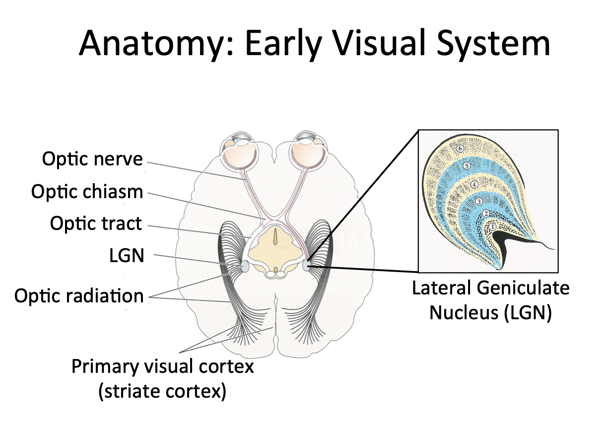
LGN = first relay station that neurons in the retina make contact with
Located in the occipital lobe
Largest of the visual areas
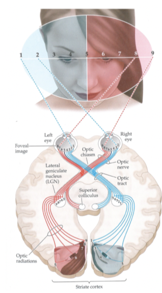
Which parts of the retina project contralaterally and which parts project ipsilaterally?
Nasal parts (parts on the retina by the nose) project onto the LGN on the other side (left onto right and vice versa)
Contralaterally
Lateral parts (away from the nose) projects onto the LGN on the same side
Ipsilaterally

The cells in the LGN are monocular. What does this mean?
Each layer gets input from only one eye
Either gets input from the left or right eye, but not both
LGN has __ layers, __ from each eye?
6, 3
Give 5 characteristics of layers 1 and 2
Large cell bodies
Magnocellular layers
Receive input from M ganglion cells (have similar properties to them)
High contrast sensitivity and low spatial resolution
Process coarse features and motion
Of the magnocellular layers, which one is the contralateral and which one is the ipsilateral layer?
Need 2 in each LGN because one receives input from each eye
Layer 1 - contralateral layer
Layer 2 - ipsilateral layer
Give 5 characteristics of layers 3-6
Small cell bodies
Parvocellular layers
Receive input from P ganglion cells (extension of the cone system to LGN)
Low contrast sensitivity and high spatial resolution
Process fine features and colour
Of the parvocellular layers, which ones are the contralateral and which ones are the ipsilateral layers?
2 contralateral (4 and 6) and 2 ipsilateral (3 and 5)
Don't know why we need four parvocellular layers
What is retinotopy in the LGN?
Neighbouring relationships that exist in the retina are retained in the LGN (retinotopic map)
So, each LGN has 6 maps of the world (one in each layer)
Each is only half of the visual world
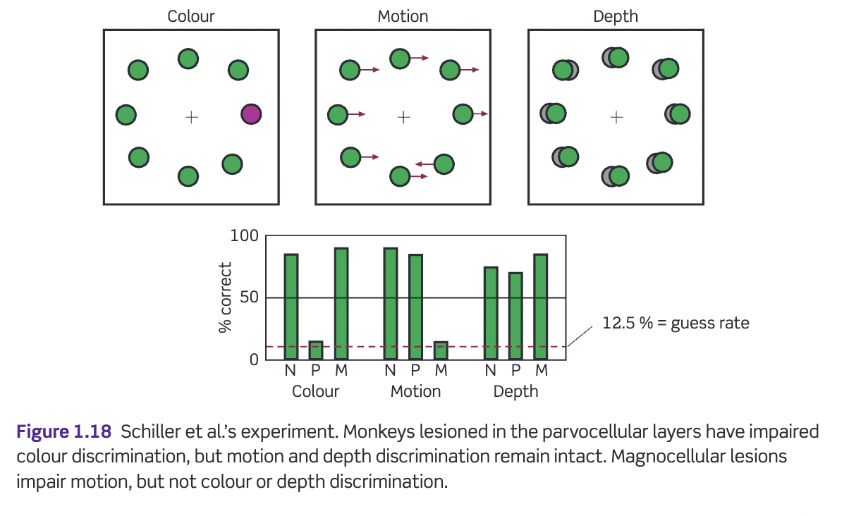
M and P cells of the LGN seem to have similar properties to the M and P cells of the retina (movement and flicker, and colour, respectively). Give a study that supports this
Schiller et al. (1990)- trained animals to fixate on a central point then move their eyes to the 'odd one out'
Small lesions in either the M or P division
What are koniocellular cells?
Mainly situated between the layers
Receive input from a special type of ganglion cell in the retina
Koniocellular cells seem to be important for…?
Seems to be important for perceiving colour (blue-yellow comparison)
This type of retinal ganglion cell (that they receive input from) has a major input from the blue cones
The function of the LGN is still a bit of a mystery. But what do we know? (3 things)
LGN is…
Richly connected to many other parts of the brain
A locus at which retinal information can be modulated by brain areas
More than just a relay station
LGN seems like a replication of the ganglion cells in the retina- important to understand it's unique function
How does LGN vary from the retina?
Due to it's extensive connections to other areas of the brain
The strongest input into the LGN is not from the retina, but from…?
The cortex itself
Biggest input to the LGN comes 'top-down'
This has led to the idea that the LGN might be important in filtering what information gets through to the cortex
Now some evidence for this from O'Conner et al. (2002)- fMRI
LGN projects to V1, the primary visual cortex. Why is the retinotopic map in V1 distorted?
Cortical magnification
Same distance between 5 and 4 and 2 and 1
However much more space in the cortex dedicated to processing the part of the stimulus indexed by 5-4
Directly focusing on the centre of the stimulus (where '5' is)
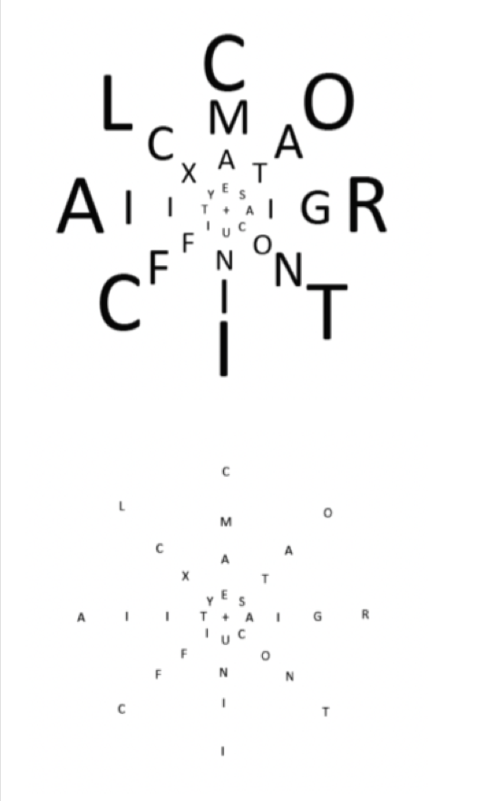
What are the consequences of cortical magnification?
Top picture; all letters equally clear
Scaled in such a way that the same number of neurons that process the small letters process the larger letters
Due to cortical magnification
A much closer to the centre of the visual field
Bottom picture; much harder to see the letters on the outside
Using less neurons to process the large letters compared to the small
Why do we not use the same amount of neurons for the whole visual field?
Trade-off between having a large visual field and having (some areas of) high acuity
If we had the same acuity in the fovea in the whole visual field, we would need eyes and brains multiple times the size of our skull
Acuity = how good you are at resolving small details
Cortical magnification highlights the importance of….?
Eye movements
Move eyes around in order to move that high resolution part of the visual system from one point to the next
Visual system patches this together to provide this high detail experience of the whole visual world
What did Hubel and Wiesel (1977) find in their experiments looking at V1 in cats?
'Accidental' firing- realised that the crucial stimulus for cells of V1 might be elongated lines rather than spots
Each cell responded vigorously to a line stimulus, but not any line
E.g. the line had to be of a particular orientation to elicit the greatest response
What did Hubel and Wiesel’s experiments tell us about the organisation of V1? (3 things)
Clear that the retinotopic mapping found in the LGN was still present (cells close to each other in V1 have receptive fields that are close to each other on the retina)
Each V1 maps half of the visual field (contralaterally)- little overlap between the two halves of the map and yet we are never aware of the join
Each cell has its own 'preferred' orientation and within a small area of the cortex there will be a cell for all possible orientations
Retinal ganglion cells/LGN neurons have centre-surround receptive fields that respond best to spots of light. However, receptive fields in V1 are…?
Elongated; respond best to lines, bars and edges
Looking at a whole array of ganglion cells
These are connected to the V1 simple cell
Looking at this, you can see the lines, bars and edges aspect of the V1 receptive field
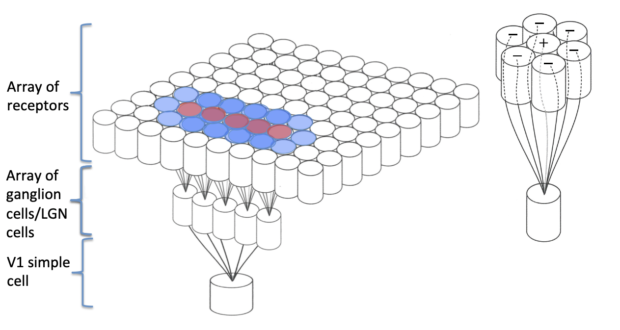
What are bar detectors?
As with the concentric receptive fields, you can have simple cell receptive fields where the middle is facilitatory or inhibitory
Bar detectors
What are edge detectors?
Some have only one excitatory and inhibitory region- will respond best to a contrast edge of the correct orientation rather than to a bar
Edge detectors
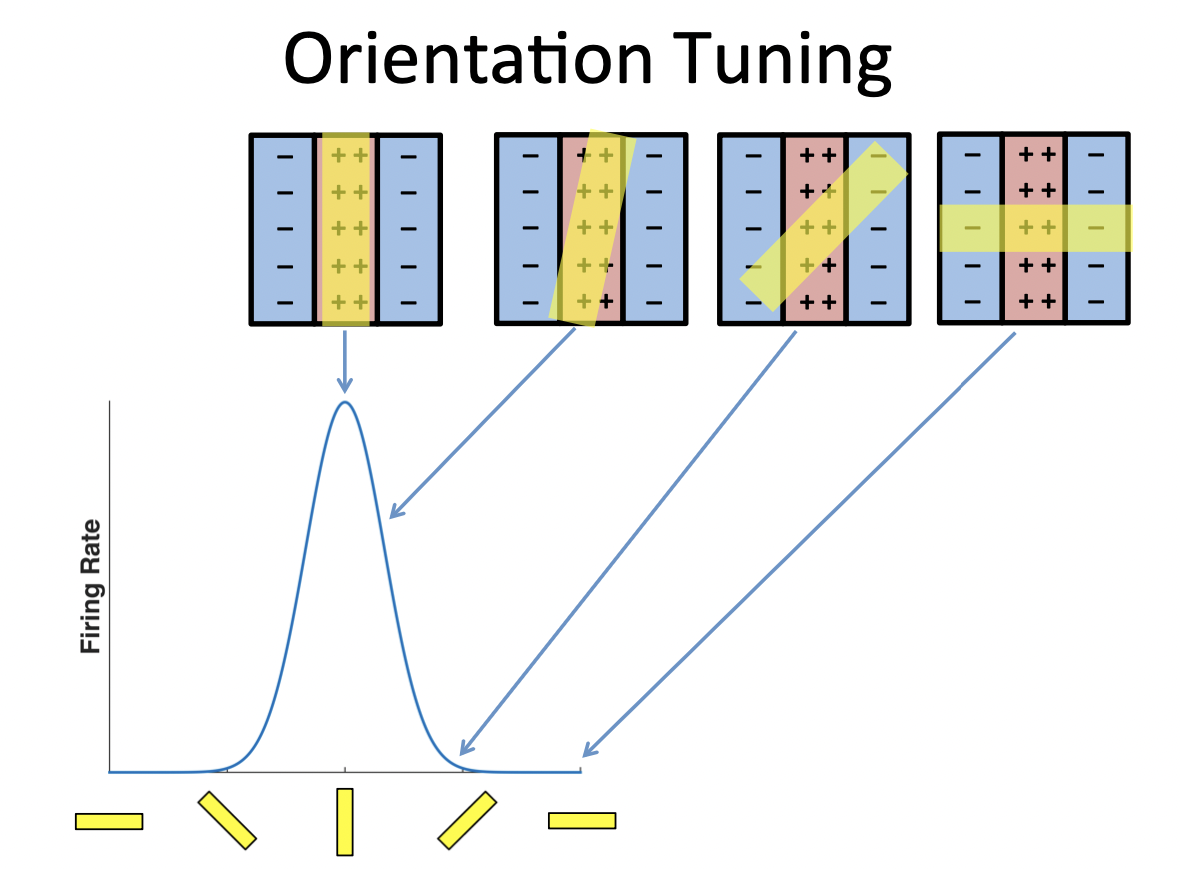
What is orientation tuning?
Neurons respond best to vertical bands of light
Not just responding to one and not other, but have an optimal stimulus which they respond best to
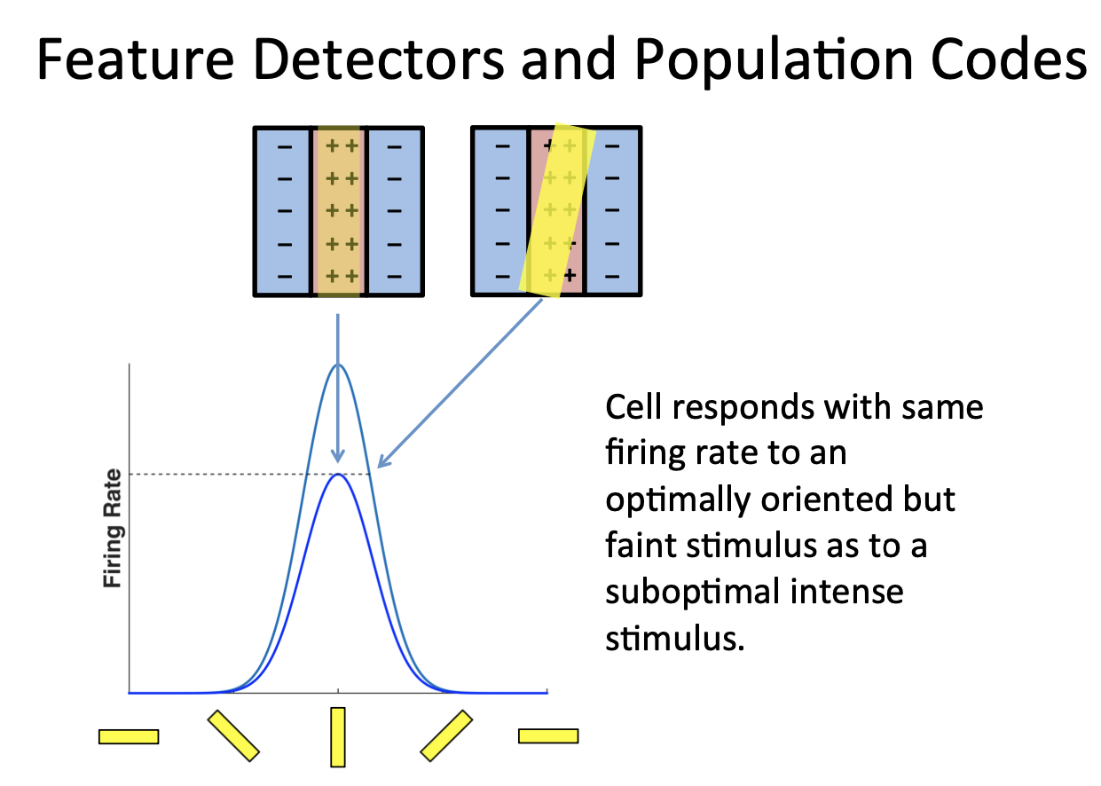
These cells don’t just respond to orientation, but intensity too. Why is this a problem?
Response of a single neuron is always ambiguous because of this
Is the response due to the certain orientation or a bright stimulus with a different orientation?
Optimal but faint stimulus activates cell equally as suboptimal intense stimulus
Have to consider activity in the context of the other neurons (associated with the same stimulus)
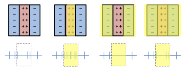
How does the brain discriminate orientation and intensity of the stimulus?
The brain looks at the relative activity of neurons that are tuned to different values of the same parameter (not just singular neurons)
Difference in relative activity is what matters, not the absolute activity
What are population codes?
Population of cells tuned to different orientations allows to disambiguate intensity and orientation- features are coded in a population code
Relative activity is what matters
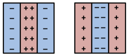
What are simple cells?
Many of the cells in area V1 have receptive fields with these discrete on and off regions
By adding up how much light falls on the ON region and how much falls on the OFF region we can accurately predict the response of the cell to any stimulus
Light on none/all of the receptive field- baseline response
Light on facilitatory part of receptive field- above baseline (and vice versa for inhibitory)
Why can’t cells in the LGN discriminate between stimuli of different orientations?
As their concentric circular receptive fields treat vertical, horizontal, and tilted lines the same
Receptive fields in V1 still have excitatory and inhibitory regions, but they are now stretched out
What are complex cells?
No spatially defined facilitatory or inhibitory regions
Just responds to that orientation no matter where the orientation shows up in the receptive field
Whole extent of the receptive field- no change in activity
If you make the bar longer than the spatial extent of the receptive field it doesn't matter
Complex cells don't care about this
How do simple and complex cells differ in phase sensitivity?
Simple cell cares where the bar is in its receptive field
This variation with the exact position (phase sensitivity) is a characteristic of simple cells but not complex cells
Complex cells give approximately the same response right across their receptive fields- said to be phase insensitive
Hubel and Wiesel proposed that one could build complex cells by…?
Appropriately 'wiring' together the output of simple cells
Simple cells that are all tuned to the same orientation, but in slightly different positions in space, could feed on to the complex cell which performs an OR operation
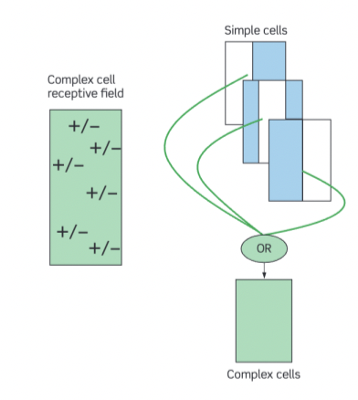
Within complex cells, what is an OR operation?
If it receives input from this cell OR this cell etc. then it will fire
When the line appears, within this small space, one of the simple cells should give a good response and therefore this is all that is needed by the 'OR gate'
Gives a response to all positions within this receptive field
However few direct tests of the validity of this theory
Complex cells seem to suggest that the brain is building more….?
Specialist cells as the information moves back from the eyes and deeper into the brain
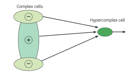
What are hypercomplex cells?
Also don't have these clearly defined facilitatory and inhibitory regions
But if you extend the bar of light beyond the receptive field = decrease in activity
End-stopping- thought to be important in object perception and inclusion of objects

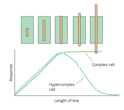
Describe what this graph is showing
For the complex cell, the response increases as the length of the bar increases until a point where the bar becomes as big as the receptive field
Larger than the receptive field = the parts outside the receptive field do not contribute to the response and hence no further increases in firing rate is seen
The hypercomplex cell initially shows the same increase in response but as the bar becomes longer still, the response begins to fall
Optimal stimulus for this cell is a bar, not only of a particular orientation, but also of a particular length
How are hypercomplex cells made?
Suggested that some complex cells, whose receptive fields are aligned with that of the hypercomplex cell, produce the excitatory response, but there are others, whose receptive fields are at either end of the excitatory cell's receptive field, that provide an inhibitory input
Therefore, as the bar becomes larger and excites these inhibitory cells, the response of the hypercomplex cell diminishes