Practical Pathology CNS
 Call Kai
 Learn
Learn Practice Test
 Spaced Repetition
 Match
Match Flashcards
 Knowt Play
1/34

There's no tags or description
Looks like no tags are added yet.
Name | Mastery | Learn | Test | Matching | Spaced | Call with Kai | Chat |
|---|
No analytics yet
Send a link to your students to track their progress
35 Terms
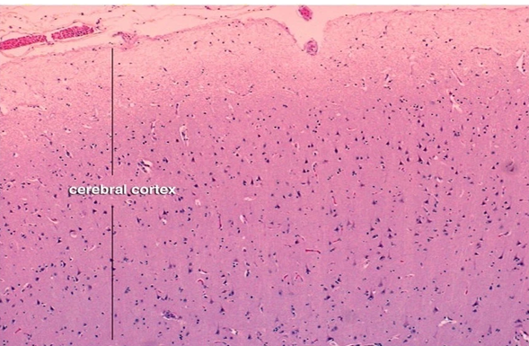
Cerebral cortex Features: Gray matter, neurons, glial cells, neuropil

Cerebral cortex Features: Gray matter, neurons, glial cells, neuropil
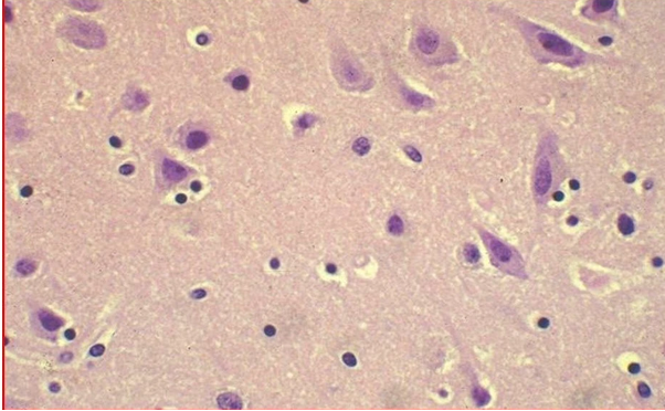
Gray matter of CNS Features: Neuron cell bodies, glial cells, neuropil
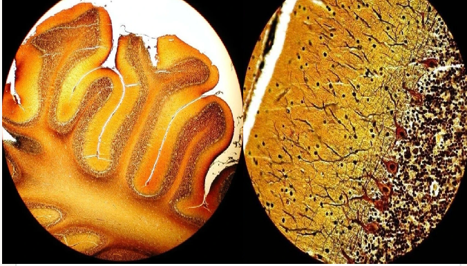
Cerebellum Features: Molecular layer, Purkinje cell layer, granular lay

Pyramidal neurons Features: Large multipolar neurons, prominent nucleolus, Nissl granules

Spinal cord Features: Gray matter, white matter, butterfly-shaped gray matter
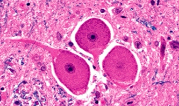
Pseudounipolar neuron Features: Large round cell body, single process, no dendrites.

Spinal ganglion Features: Large pseudounipolar neurons, satellite cells

Cerebral ischemia (Red neurons) Features: Eosinophilic cytoplasm, loss of Nissl granules, small dark nucleus.

Gray matter of CNS Features: Neuron cell bodies, glial cells, neuropil
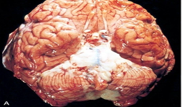
Acute pyogenic meningitis (Gross) Features: Thick suppurative exudate, thickened leptomeninges

Central canal of spinal cord Features: Ependymal cells, cuboidal/ columnar cells, cilia.

Acute pyogenic meningitis (Micro) Features: Subarachnoid neutrophils, brain edema, vasodilation
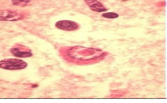
Herpes simplex virus type I Features: Necrotizing, hemorrhagic, eosinophilic nuclear inclusions

Cytomegalovirus Features: Ependymal cells, basophilic intranuclear inclusions
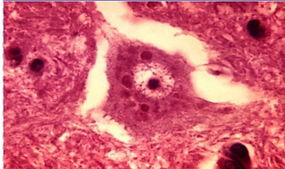
Rabies Features: Negri bodies, eosinophilic intracytoplasmic inclusions

Meningioma (Gross) Features: Well-circumscribed mass, compressing but not invading, attached to dura. P

Meningioma (Micro) Features: Cellular whorls, psammoma bodies, indistinct cytoplasmic boundaries.

Low grade astrocytoma (Gross) Features: Pale brain mass
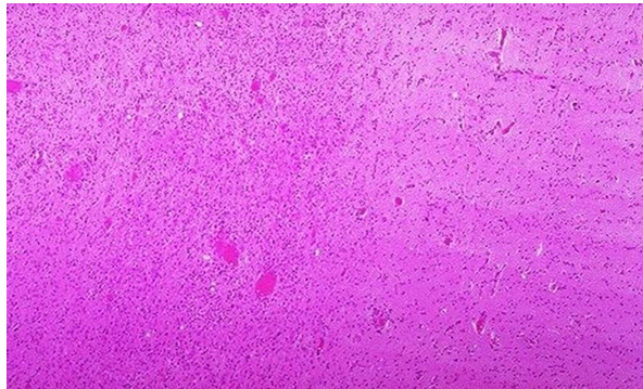
Low grade astrocytoma (Micro) Features: Greater cellularity, pleomorphism, indistinct margin.
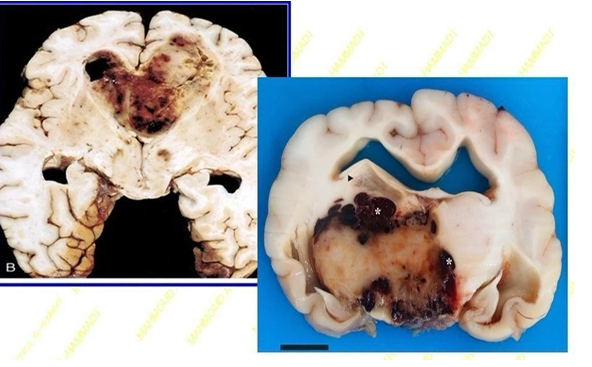
Glioblastoma (Gross) Features: Necrotic, hemorrhagic, infiltrating mass

Pilocytic astrocytoma (Micro) Features: Bipolar cells, hair-like processes, Rosenthal fibers, microcysts
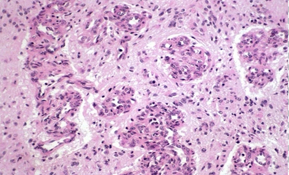
Glioblastoma (Micro) Features: Vascular endothelial proliferation, glomeruloid formation

Glioblastoma (Micro) Features: Dense cellularity, pleomorphism, pseudo-palisading necrosis

Ependymoma (Gross & Scan) Features: Arising from fourth ventricle, bulging toward cerebellum

Oligodendroglioma (Micro) Features: Uniform cells, clear cytoplasm (halo), delicate capillaries

Ependymoma (Micro) Features: Rosettes around lumens or capillaries
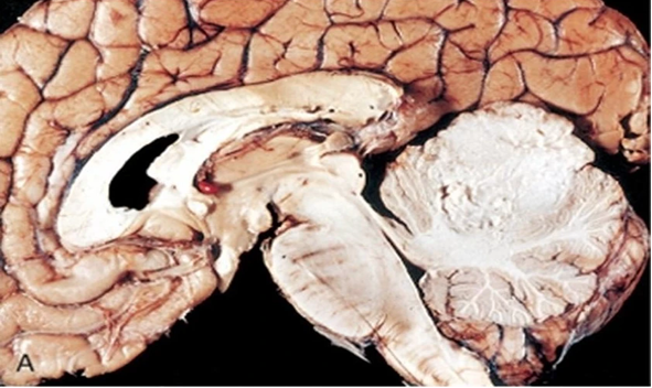
Medulloblastoma (Gross) Features: Destroying upper cerebellum.

Retinoblastoma (Gross) Features: Filling posterior eye chamber.
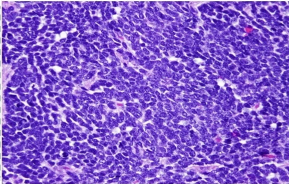
Medulloblastoma (Micro) Features: Small blue round cells, hyperchromatic nuclei, mitoses.

Retinoblastoma (Micro) Features: Small blue round cells, hyperchromatic nuclei, mitoses.

Acoustic neuroma (Gross & Scan) Features: Mass in cerebello-pontine angle

Alzheimer disease Features: Neurofibrillary tangles, plaques, silver stain

Acoustic neuroma (Micro) Features: Schwann cells, hypercellular (Antoni A), hypocellular (Antoni B), palisading (Verocay bodies).

Prion disease Features: Spongiform degeneration, vacuoles, no inflammation