Pathological Anatomy special part - 3
1/30
There's no tags or description
Looks like no tags are added yet.
Name | Mastery | Learn | Test | Matching | Spaced | Call with Kai |
|---|
No study sessions yet.
31 Terms
64. Renal failure
Renal dysfunction can be due to prerenal, renal and post renal causes.
Prerenal – reduced renal blood flow, shock, obstruction of vascular supply to kidneys, severe hypovolemia.
Renal – glomerulitis, acute tubular necrosis, chronic renal disease.
Postrenal – obstruction of uterine outflow – uroliths, inflammation, tumor.
Renal failure causes accumulation of metabolic wastes (uremia) such as nitrogenous waste (azotemia), can reduce blood pH, and alternate plasma concentration and cause imbalance in phosphorus, calcium, potassium ratios.
It also causes retention of constituents in plasma that are normally removed by kidneys.
Clinically and at necropsy, non-renal lesions of uremia in animals and are useful in recognizing renal diseases. These include pulmonary edema, fibrinous pericarditis, mediocalcinosis, atrial thrombosis, anemia, soft tissue mineralization.
65. Renal necrosis
Bilateral renal cortical necrosis – results from widespread micro thrombosis which occurs in glomerular capillaries and other arteries and afferent arterioles in DIC. Gram neg septicemia or endotoxemia. Micro thrombosis causes ischemia, hemorrhage and coagulation necrosis. Renal cortex may be pale with a zone of hyperemia. Cortex is mosaic.
Renal medullary crest necrosis – response to ischemia. NSAIDs or analgesic drugs is the cause. Drugs inhibits prostaglandin biosynthesis which is important for maintenance of normal blood flow.
Tubular necrosis – result of ischemia or toxic insult to kidneys. Tubular epithelium undergoes degeneration and necrosis and sloughing of cells. Uremia will occur as a result.
Lesions are difficult to observe grossly, but kidneys will be swollen, pale and translucent. Cut surface also pale with extensive moist.
Microscopically there is edema/swelling and coagulation necrosis. Tubular necrosis. Proximal convoluted tubules are most severely affected. Prolonged ischemia can result in necrosis of all tubular epithelium.
Nephrotoxic tubular necrosis due to heavy metals, plants, antibacterial/antifungal agents, oxalates, mycotoxins. Necrosis due to toxins.
66. Glomerulitis
Glomerulonephritis
immune mediated GN (immune complex and nephrotoxic), suppurative, viral
Glomerular damage alters filtration barrier and can lead to many things, protein in urine amongst others.
Prolonged severe proteinuria results in hypoproteinemia and generalized edema and is called nephritic syndrome.
Viral glomerulonephritis – acute systemic viral disease like in infectious canine hepatitis, causes endothelial hypertrophy and thickened mesangium, hemorrhage and cell necrosis.
Suppurative – bacteriemia with bacteria in glomeruli which form small, pale and yellow abscesses scattered around the renal cortex. Bacterial colonies, neutrophils.
Immune-mediated glomerulonephritis – antibodies to glomerular basement membrane or deposition of immune complexes within the glomeruli. Occurs in persistent infections with prolonged antigenemia. AgAb complexes starts chemotaxis and activate neutrophils which damage basement membrane. Macrophages. Kidney is swollen, glomeruli as pale greyish dots, scarring may occur.
lesions in glomeruli: Proliferative (proliferation of glomerular cells), Membranous (thickened basement membrane), or membranoproliferative (both lesions). others are hyaline, calcification.
Amyloidosis is systemic disease where amyloid accumulates as hyaline in walls of small blood vessels and glomeruli. The glomeruli then become enlarged and capillary lumen may become destroyed.
67. Acute interstitial nephritis
may be non-purulent or purulent, either focal – white spotted calf kidney, by E.coli, less severe and consists of more discrete grey areas in cortex and sometimes medulla
or diffuse – canine leptospirosis, swollen kidney and pale with random grey mottling)
hematogenously/medullar/disseminated, urinogenous, pyelonephritis
Is a result of bacterial and viral septicemias, where infectious agents enter kidney tubules and initiate inflammatory response in the kidney.
Examples – E. coli, infectious canine hepatitis. The actual pathogenesis of these lesions are unknown.
Microscopically there is aggregates of lymphocytes, plasma cells and monocytes and neutrophils.
Interstitium may be edematous.
Tubules with inflamed area may have degenerative and necrotic changes in epithelium.
68. Renal pelvic diseases, hydronephrosis
Pyelonephritis
is kidney inflammation due to bacterial infection, often starting from the lower urinary tract, reaching the kidney because of abnormal reflux. - asending form (vesicoureteral reflux) more common than ascending form
Can be acute or chronic. More common in females.
E. coli, staphylococcus, streptoc, Corynebacterium renale in cattle and C. suis in swine.
Acute pyelonephritis affects the pelvis and ureteral membrane, showing hyperemia, sometimes pus, thickening and sometimes ulcers in renal medulla.
Chronic pyelonephritis results in extensive tissue damage, necrosis, scarring and inflammatory response. Most severe lesions in the medulla. Neutrophilic infiltrate.
Hydronephrosis
is when the renal pelvis dilates due to urine outflow obstruction.
This blockage can be from various reasons like urinary stones or inflammation or tumors of ureter and bladder
Unilateral cases may cause extensive cystic enlargement.
Bilateral cases can lead to uremia and death.
Leads to kidney tubule dilation → pressure buildup → glomerular filtrate diffuse into interstitium → eventually collapse of vessels → reduced blood flow → ischemia → lobular atrophy and necrosis → intersitital fibrosis,
in severe hydronephrosis → bacterial infection → pus accumulation (pyonephrosis).
69. Chronic renal diseases
Renal fibrosis – also known as scarring.
Can be primary but more commonly occurs as chronic manifestation of the healing process of preexisting renal lesions.
Results from primary inflammation of kidney components, or following severe necrosis of renal tubules.
This causes renal dysfunction and can lead to renal failure and uremia.
Polyuria and polydipsia are common.
Hypoplastic anemia due to decreased erythropoietin.
Kidney will appear pale, shrunken, firm, with fibrous adhesions between capsule and cortex.
The fibrotic pattern may be diffuse, stripped, coarse, multifocal, patchy.
Atrophic tubules have reduced diameter and thickened membrane (hyalinized)
lymphocytes, plasma cells (these cells indicate chronic interstitial nephritis).
Dilation of Bowmans capsule. Cysts may be present.
Glomeruosclerosis, mineralization.
Familial renal disease - severe bilateral renal fibrosis in young dogs of several breeds. usually from 4months to 2 years. Doberman pinscher. Norwegian elkhounds.
70. Renal neoplasms
Not so common to have primary tumors in the kidney.
Can be epithelial, mesenchymal or embryonal in origin.
Majority of neoplasia in kidney are metastatic
Epithelial tumors: adenomas, carcinomas, transitional cell papilloma
Primary mesenchymal tumors: fibroma, fibrosarcoma, haemangiosarcoma, lymphosarcoma
Renal adenoma (Arise from epithelium of proximal tubules)
Renal carcinoma (predom in males, typically located at one pole of the kidney, causes atrophy of nearby tissue. Invades kidney tissue)
Renal cystadenocarcinoma (bilateral localization, German shepard)
Nephroblastoma (true embryonal tumor, if all three layers (blastemal, epithelial, stromal) present - triphasic).
71. Urolithiasis, cystitis
Urinary calculi (uroliths)
are concretions formed in the urinary tract, composed of salt and inorganic/organic acids and other material like cysteine.
Occurs in renal pelvis or in the lower urinary tract.
Calculi in renal pelvis predispose for pyelitis and pyelonephritis.
Bladder calculi can be single or multiple and various in size, and sometimes fine sandy material may appear as cloudy urine.
Calculi may be smooth or have irregular surface, be white, yellow, brown depending on composition.
Can obstruct, cause inflammation and painful urination and hemorrhage.
Forms due to precipitation of salts in urine – urinary pH, bacterial infection, herpes virus, nutritional and dietary factors can all lead to calculi.
Grossly the bladder wall is thin and often diffusely hemorrhagic. Mucosal ulceration and necrosis can be present.
Inflammatory changes, hemorrhage, necrosis microscopically.
Cystitis
Is the inflammation of bladder.
Results from chemical causes during therapy or from bacteria.
Occurs more commonly in females due to shorter urethra.
E. coli, Corynebacterium renale, streptococci, staphylococci.
Obstruction, neurogenic causes, trauma, faulty catheterization, and parturition can all lead to cystitis.
Acute cystitis can be hemorrhagic, fibrinopurulent, necrotizing or ulcerative.
Bladder wall may be thickened due to edema and infl cells present, can be diffuse but can also be local.
Chronic cystitis can show thickened hyperplastic mucosa and lymphocyte infiltration and fibrosis.
72. Neoplasia of lower urinary tract
Neoplasia in lower urinary tract is mainly in bladder.
More frequent in dogs, cats, and cattle.
Grow slow and late metastatic.
caused by industrial chemicals, chronic irritants, foreign bodies (sutures), viruses, bracken fern etc.
grade them accoring to TNM system to help therapy and prognosis
Epithelial tumors: adenoma, papilloma, carcinomas
Mesenchymal tumors: fibroma/fibrosarcoma, leiomyoma/leiomyosarcoma, rhabdomyosarcoma, lymphosarcoma, hemangioma/hemangiosarcoma
Adenomas (rare)
Papilloma’s (tend to be multiple, need to be differentiated from papillary hyperplasia in cattle)
Carcinomas (four histological types – TCC, SCC, adenocarcinoma and undifferentiated carcinoma. Can be solitary or multiple).
Transitional cell carcinomas – papillary, polyploid or sessile.
Squamous cell carcinoma and adenocarcinomas – shows non papillary invasive growth.
73. Anomalies, malformations and traumatic injuries of CNS
Anencephaly – absence of brain, results from abnormal closure of anterior aspect of neutral tube during development.
Hydrocephalus – increased accumulation of cerebrospinal fluid. Congenital (enlargement of cranium, prevents outflow of CSF) or acquired (obstruction of ventricular system due to infl).
Hydranencephaly – complete or almost complete absence of hemispheres, leaving only membranous sacs filled with cerebrospinal fluid.
Cranium bifidum – midline cranial defect where brain tissue may protrude.
Spina bifida – herniation of the spine, protrusion of meninges and spinal cord, or only meninges.
Syringomyelia – tubular cavitation of spinal cord that extends over several segments. A cyst in the spinal cord.
Cyclopia – fusion of both eyes which can be seen as one large orbit. All bones are absent in this area.
Cerebellar hypoplasia – reduction in size of cerebellum. Common congenital defect. Can be due to virus.
Traumatic injuries of CNS include;
Concussion (transient loss of consciousness and reflex activity after sudden injury to the head. Mildest degree with no visible damage),
Contusion (hemorrhage, but nervous tissue is retained, may be focal or diffuse),
Laceration (a traumatic injury where there is disruption to brain tissue . penetrating injury can result in secondary infection),
Fracture (can be meningeal hemorrhage, displacement of bones).
74. Oedema of CNS
Generalized cerebral edema – edema and swelling of brain in many systemic diseases.
Vasogenic edema – due to injury of vascular endothelium, which allows leakage.
Cytotoxic edema – accumulation of fluid within intracellular departments of the brain as a result of altered cellular metabolism. Cellular swelling.
Hydrostatic edema – accumulation of fluid in extracellular spaces of brain due to elevated hydrostatic pressure + hydrocephalus. Heart failure, liver disease.
Osmotic cerebral edema – water intoxication, increased body hydration. Intracellularly primarily. Renal failure, liver disease.
Sequela of cerebral edema:
Enlargement of an organ in a closed limited space → compression of cranium → brain has flattened gyri and shallow sulci, and it shifts position.
Edema can be on one side → unlilateral displacement
75. Ischemic lesions of CNS
Cerebrospinal vasculitis – affects both arteries and veins during several specific diseases. Hog cholera, malignant catarrhal fever. Parenchymal degeneration after ischemia.
Thrombus and embolisms in cerebrospinal arterioles are very seldom observed in animals. Bone marrow emboli are formed after trauma.
Infarction – necrosis of tissue after obstruction of arterial circulation in some part of brain. Can also result from other causes (cardiac arrest, hypotension by decreased cardiac output, reduced oxygen, alterted Hb fnction, toxicities, poison, nutrition)
-Affected areas will become soft, from liquefactive necrosis. Grey matter becomes hemorrhagic (hemorrhagic infarct), and white matter is often pale. (pale infarct)
76. Prion diseases
Prion diseases is characterized by:
chronic progressive fatal neurological manifestations,
both sensory and motor deficits and spongiform encephalopathy and
a result from accumulation of abnormal isoform of the host prion protein.
Pathological when there is accumulation of prion proteins in nervous tissue.
Resistance to protease. It is misfolded.
Transmissible spongiform encephalopathies (TSEs).
PrPc (cellular prion protein) is normal prion protein. Prion protein is widely distributed in tissues but concentrated in nervous tissue.
PrPSc almost never triggers immune response.
No gross abnormalities in brain.
Histologically, the spongiform changes are the identifying feature. Small rounded vacuoles in neutrophils.
Protein only hypothesis – PrPSc (prion protein scrapie) is the reason for disease, which trigger the PrP gene to produce more PrPSc.
77. Septic embolism, cerebral abscesses and viral infection of CNS
Septic embolism may be due to active endocarditis. Mostly gram-positive. Strepto. Gram neg depends on presented number of microorganism. Piece of thrombus containing MO.
Cerebral abscesses by direct implantation in wounds or by direct invasion by adjacent structure. Hypothalamus and cerebral cortex are most susceptible to abscess formation. Listeria monocytogenes has affinity for the reticular formation in the brain stem. Singular or multiple (bacterial embolism)
Viral infection of CNS:
Hematogenous route.
Virus susceptible to be removed by histiocytes in blood if they are not small or hides.
The virus spreading in nervous tissue is not known.
Rapid in CNS, Rabies. Leads to cellular degeneration – neurons (inclusion bodies), reactivity of glia, perivascular reaction. Inclusion bodies may form in neurons, neuroglia, microglia, and other mesenchymal cells.
78. Muscle degenerations
Pigmentations;
lipofuscin (wear and tear pigment which accumulate in secondary lysosomes. Rounded granules of yellow/brown pigment to the two poles of the nucleus of myofiber),
melanin (discolor the fascial sheaths of muscles at congenital melanosis in calves. Black foci or streaks. Muscle is unaffected)
myoglobin (stain muscle after muscle necrosis).

Calcification/ossification;
Calcification of necrotic muscle fibers or fibers of old age. Rarely macroscopic.
Ossification usually termed myositis ossificans, means formation of non-neoplastic bone or cartilage in extra osseous sites that may not be in muscle. Essentially a metaplasia to bone.
Steatosis - congenital, destructive muscle fiber development → lost myofibers replaced with fat
True degeneration;
earliest detectable changes start in individual myofibers as fine vacuoles, barely visible. The vacuoles will become larger. Degeneration can be reversed, but if not it continues to floccular, granular, hyaline and at the end Zenkers necrosis which is the end stage.
79. Myopathies
Myopathies include nutritional myopathies, capture myopathy, porcine stress syndrome (malignant hyperthermia), myasthenia gravis.
Nutritional myopathies
Selenium and vitamin E deficiency (white muscle disease)
lesions are areas of skeletal or cardiac muscle which is pale or white and there is coagulative necrosis with the disappearance of many muscle fibers.
Macrophages and lymphocytes and attempt of regeneration without success.
Capture myopathy
is acute myopathy which manifests as edema of muscles, with pale streaks and hemorrhagic streaks.
Pale areas with necrosis, which become mineralized and fibrosed if survival is prolonged.
PSS – malignant hyperthermia
Cellular defect in handling of Ca ions, activating myofibrillar ATP-ase and rapid intracellular glycolysis which increases body heat.
Leads to denaturation of proteins of myofibers. Cause edema.
Muscles are pale, moist, swollen (PSE pork).
Segmental hypercontraction and necrosis if the pigs live longer.
Myasthenia gravis
is a neuromuscular disease. Acquired or congenital.
Affects the neuromuscular junction, the immune system mistakenly attacks and damages receptors for acetylcholine. Affects muscular contractions.
80. Myositis
Suppurative myositis
cause abscesses in muscles.
Can be of hematogenous origin, inoculation or extension of nearby structures like bones or lymph nodes.
Arcanobacterium pyogenes is most common cause of abscesses in muscle of cattle and sheep, Corynebacterium, streptococcus equi.
Early stage – ill-defined cellulitis. Minimum scarring, may continue to formation of abscess.
Gas gangrene
caused by clostridium.
Deep penetrating wounds, can cause hemorrhagic myositis and cellulitis.
May be discolored from red to black and gas formation is observed.
Viral myositides
is rare. Causes necrosis and lymphocytes. Blue tongue virus.
Polymyositis
used for inflammatory myopathies which pathogenesis is unknown.
Canine polymyositis – necrotic areas, inflammation and lymphocytes/plasma cells.
Bovine and ovine eosinophilic myositis
eosinophils, usually detected at slaughter
green discoloration due to eosinophils.
Muscle may be atrophic, necrotic and disappeared.
81. Parasitic diseases and neoplasms of muscles
Toxoplasmosis – toxoplasma gondii.
Lesions from tachyzoites are classically necrotizing, so cause necrosis of muscle cells/myofibers.
Cysts contain bradyzoites but these cysts do not cause an inflammatory reaction.
Sarcocystis
also cause sarcocysts with zoits inside, acute inflammatory cells sometimes accompany the cyst. Masseter, diaphragm, tongue.
Trypanosoma brucei and cruzi.
Cruzi causes myocarditis and focal myositis.
Brucei cause mononuclear infiltration in almost every body tissue.
Trichinella species.
Adult worm inhabits mucosa of small intestine and produce larva which enter blood and encapsulates in muscles.
Focal myositis can be seen with presence of neutrophils, lymphocytes and eosinophils. May live for years or calcify when dead.
Cysticercosis by Taenia species.
Has cysticercus stage in animals.
Visceral larva migrans
Toxocara larva migrate through the tissue of dog.
Some are arrested in myocardium and skeletal muscle.
Focal granulomatous myositis.
Neoplasms
Primary tumors are rare.
More malignant ones than benign ones.
Rhabdomyomas or rhabdomyosarcoma.
Animals affected are younger than 2 years old.
82. Reaction of bone to injury
Necrosis
Aseptic necrosis (often head of femur) of bone has been associated in dogs with intramedullary tumors and non-neoplastic lesions.
Increased bone marrow pressure and decreased venous flow → ischemia → necrosis
Necrotic cortical bone has dry chalky appearance and periosteum may easily be removed.
Zone of hemorrhage can also be seen. Loss of osteocytes and loss of staining. Fibrous tissue and macrophages advance from the margin of lesion.
The marrow may eventually regenerate completely or form a scar
Some necrotic bone will be removed by osteoclasts if the blood supply is re-established.
Direct injury
Direct injury to periosteum leads to pain and new bone formation, may remodel and regress or persist.
Irradiation
Irradiation cause direct injury to bone cells. necrosis if dose is large
Cartilage
weakening and destruction of cartilaginous matrix may lead to premature closure of growth plate in animals with hypovitaminosis A
Vit C deficiency → arrested osteoblastic activity
Factors affecting bone
Increased oxygen tension stimulates bone resorption whereas
decreased oxygen tension causes bone to accumulate.
Exercise tends to increase bone mass.
83. Fracture repair
Fracture may be traumatic or pathologic.
After a fracture there will be swelling, heat, pain.
Hematoma will form and there is necrosis of any isolated bone fragments and the broken end of medullary elements.
Fibrous tissue (by cells in periosteum, endosteum and medullary cavity) is formed and later woven bone (soft callus, takes 2 weeks)
May be formed internally or externally.
Callus is an unorganized meshwork of woven bone that forms following fracture.
Hard callus is formed after soft callus.
Osteoclasts remove necrotic cells.
Primary callus bridges the gap, encircles fracture site and stabilizes area.
Several factors may interfere with normal repair process.
Bones are resorbed by osteoclast during remodeling, and bone is remade by osteoblasts.
Ectopic mineralization and ossification must be differentiated from each other.
Ectopic mineralization is the deposition of calcium salts of phosphate, silicate etc in abnormal locations.
Ossification is production of bone in an abnormal location.
Secondary bone healing – healing through cartilage formation.
Primary bone healing – Haversian remodeling.
84. Abnormalities of growth and development of bones
Malformation – primary structural defect due to localized errors in the embryonic period.
Deformities are alterations in shape or structure of a previously normally formed part.
Chondrodysplasia – defective cartilage development. Affected animals have short legged disproportionate dwarfism. Growth is disproportionate because the growth of bones formed in cartilage is retarded.
Osteopetrosis – hereditary disease marked by dense bone without medullary cavity, which is susceptible for fractures. Failure of osteoclasts to reabsorb primary spongiosa.
Congenital cortical hyperostosis – newborn pigs. New periosteal bone formation on major long bones of limbs. Affected limbs are visibly thickened by edema and radiating spicules of bone.
Osteogenesis imperfecta – inherited abnormally brittle bones that are prone to fracture.
85. Metabolic bone diseases
Osteoporosis
pathological reduction in bone mass.
Bones are thinner with increased porosity, related to general malnutrition.
Ca deficiency, starvation, physical inactivity.
Rickets or osteomalacia
Failure of mineralization of bone and cartilaginous matrix.
Occur in young fast-growing animals due to nutritional deficiency of P or vitamin D.
bones become bowed/bended and joints appear swollen.
Osteodystrophia fibrosa
Fibro-osseous tissue replaces resorbed bone.
Caused by hyperparathyroidism.
Demineralization of bones.
Hypervitaminosis A
cause skeletal lesions. Causes narrowing and premature closing of growth plates.
Hypertrophic osteodystrophy
young rapidly growing dogs.
Periosteal new bone is formed around metaphysis and sometimes diaphysis contributing to enlargement of bones.
Osteosis
degeneration and necrosis of osseous tissue caused by ischemia.
Caused by trauma with fracture, necrosis in areas of inflammation.
86. Benign proliferative lesions and primary bone tumors
Benign proliferative lesions;
Exostosis (benign bony projections outwards. Osteophytes are also benign outgrowths of bone),
Endostosis (bony growth development within the medullary cavity),
HPO (hypertrophic pulmonary osteopathy, is progressive bilateral thickening of distal limbs, related to chronic intrathoracic inflammation),
CMO (craniomandibular osteopathy, also known as lion jaw, there is replacement of immature bone along inner and outer surface of affected bone).
Primary bone tumors
include fibrous dysplasia and ossifying fibroma.
Fibrous dysplasia is developmental defect in young dogs. Mineralized lesions with multiple cysts with fluid inside.
Ossifying fibroma is a benign large expansive heavily mineralized tumor on maxilla/mandible of horse and cattle.
Other tumors are fibrosarcoma, chondroma, osteoma, chondrosarcoma, osteosarcoma.
87. Reaction of joints to injury
Injured joints can react in various ways.
Trauma, instability, changes in joint lubrication can trigger these reactions.
Lysosomal enzymes can break down substances in joint, increased levels of prostaglandin E2, bacteria, immune mediated, hemarthrosis, degenerative joint diseases.
The area might change color and look rough.
As cartilage wears down, the body may respond by forming new bone growths called osteophytes near the joint and mild form of synovitis.
These growths are dense and reactive, often forming from fibrous tissue and turning into bone over time.
The synovial membrane can also react to injury by thickening aka hyperplasia and hypertrophy and by forming pannus, especially in chronic conditions.
In some cases, glucocorticoids are injected into joints, and this can actually cause degenerative changes in the joint, known as steroid arthropathy.
88. Inflammations and degenerative changes of joints
Inflammations
Arthritis - inflammation of intra-articular structures.
Inflammatory cells in the synovial membrane.
Bacterial arthritis is common in farm animals. The bacteria may reach the joints by inoculation as in puncture wounds or spread by nearby tissue.
Arthropathy refers to any joint disease.
Synovitis – inflammation of synovial membrane.
Acute fibrinous arthritis
synovitis and deposition of fibrin within synovial membrane and on surfaces of intraarticular structures.
Purulent arthritis – often accompanied by progressive and often necrosis of articular cartilage, neutrophils is present.
Crystal-induced synovitis – occurs in gout as urate crystals accumulate in and around joints.
Degenerative changes of joints
Degenerative joint disease
destructive disease of articular cartilage
Focal erosion and fibrillation of articular cartilage, formation of osteophytes and bone remodeling.
Joint enlargement and deformity, pain, articular malfunction. Lesions may be diffuse but often focal.
Ring bone (horse) of interphalangeal joints.
Bone spavin of intertarsal or tarsometatarsal joints. Osteophytes and ankylosis.
Osteochondrosis
characterized by focal or multifocal failure of enchorial ossification.
Involves metaphyseal growth plate and articular epiphyseal complex.
Osteochondrosis dissecans – splitting of a piece of articular cartilage due to fissure formation in area so it forms a flap or separates completely and falls into joint space. Occurs mostly in distal condyle of humerus and femur in dogs and pigs.
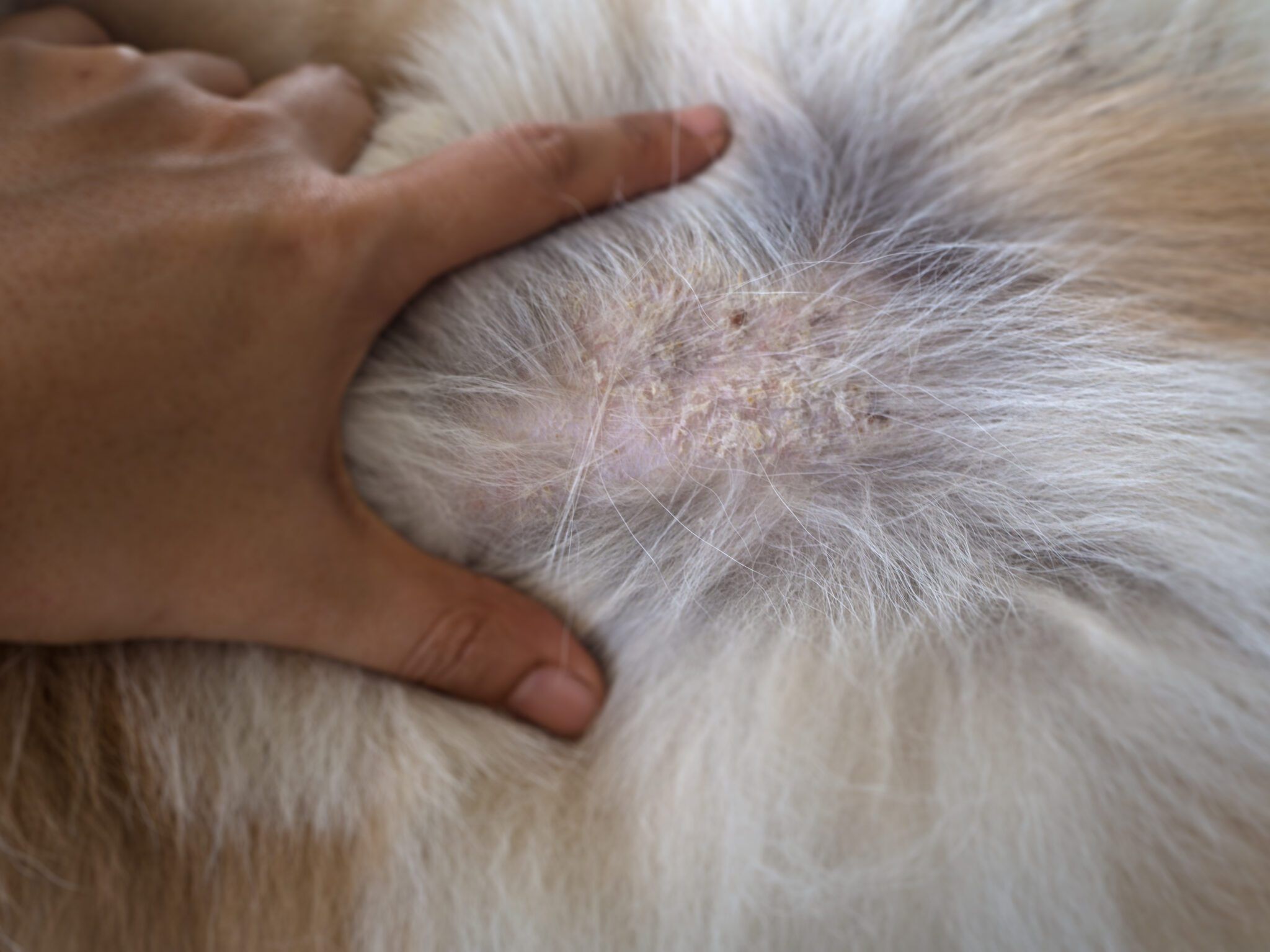
89. Skin neoplasms
One of the most common. Primary; epithelial and mesenchymal. Common types;
papilloma (epithelial origin, benign)
SCC (most common in sun damaged skin, may ulcerate),
BCC (common in cats, low grade malignancy),
hair follicle tumors,
sebaceous gland tumor (common in dogs, 3 types),
sweat gland tumors (uncommon),
perianal gland tumor (common in dogs, glandular tumors),
fibroma (uncommon, benign),
fibrosarcoma (malignant, often ulcerates)
hemangioma (more common with hemangioma),
hemangiosarcoma
mast cell tumor (older animals, 3 grades)
cutaneous lymphoma (uncommon)
histiocytic tumors
melanomas (arises from melanoblasts, melanocytes,
dendritic cells, is malignant, invasive).
90. Autoimmune dermatosis, Acral lick dermatitis
Pemphigus
group which is characterized by pustules, vesicles, bullae, erosions, ulcers.
Loss of adhesion between cells.
Immunological by development of auto antibodies directed against surface antigens of keratinocytes of epithelium in skin, mucocutaneous junctions, oral mucosa etc.

Lupus erythematosus
multisystemic disease which is characterized by presence of B-cell hyperactivity and formation of a variety of autoantibodies.
General failure of immune autoregulation due to dysregulation of t-suppressor cells or cytokines.
There is genetic predisposition for this disease, and exposure to UV light and drugs.
Acral lick dermatitis
is relatively common psychogenic dermatitis in dogs, caused by persistent chewing and licking.
Commonly in larger breeds of dogs.
Mild sensory polyneuropathy may be associated with the development of lesions.
Usually singular, occurs on carpus, metacarpus, tibial/radial area, tail and back.
Lesions are circumscribed, hairless, ulcerated.
Hyperkeratosis, acanthosis, hyperplasia, dermal fibrosis, perivasculitis, perifolliculitis.
91. Keratinization defects
Canine seborrhea
is a chronic disease of dogs, characterized by defect in keratinization with increased scale formation, excessive greasiness of skin and haircoat, and sometimes secondary inflammations.
Seborrhea sicca (dryness of skin and coat. Focal or diffuse scaling of skin)
Seborrhea oleosa (skin and hair is greasy),
Seborrheic dermatitis (scaling, greasiness, local/diffuse inflammation. Associated with folliculitis)
Primary type – inherited disorder
Secondary type – those caused by internal or external which alters the proliferation, differentiation or desquamation of surface. Like endocrine factors, nutritional, environmental factors.
Nasodigital hyperkeratosis
increased amount of horny tissue originating from and tightly adherent to the epidermis of foot pads or nasal planum.
Can be congenital, idiopathic, coexisting with other disorders. Canine distemper is very known for hyperkeratosis.

92. Mastitis
Mastitis is always attributed to invasion by Mos.
Hematogenous, and percutaneous entry is possible, the usual route is through teat canal.
3 groups; streptococcal mastitis and staphylococcus mastitis, coliform mastitis and mycoplasma mastitis.
Streptococcus agalactiae
persists in gland cistern for period waves of multiplication, increasing virulence and tissue invasion.
Edema, neutrophils, macrophages, hyperplasia.
Fibrosis takes over necrotized area.
After acute phase, there is periductal fibrosis and granulation tissue which replaces the original parts.
May lead to obstruction of milk flow. Groups of alveoli which secretion is retained may resemble small abscesses.
Grossly; hyperemia, alteration of milk quality.
Staphylococcus aureus
mastitis in younger cattle.
Most severe, forms gangrenous form, seen shortly after parturition.
Acute infl – hyperemia, edema, pain, heat, neutrophils, macrophages, erosion, necrosis. Fluid exudation, abscess formation, fistula may develop.
Granulation and fibrous tissue.
Less acute form is like streptococcus.
Coliform mastitis
sometimes associates with metritis and hematogenous entry.
E. coli, Enterobacter, and klebsiella.
Hyperemia, hemorrhage, edema, necrosis.
Endotoxins on vasculature is the coliform bacteria’s main target.
Mycoplasma mastitis
occurs in herds where bacterial mastitis have been suppressed.
Mycoplasma bovis - purulent exudate in early stages.
Nodular parenchyma – abscesses. Neutrophils, fibrosis, atrophy.
93. Granulomatous mastitis
Granulomatous mastitis can be a result of
environmental contamination or
an infusion of contaminated intra mammary preparations.
Mycobacteria and candida species for example.
Grossly the glands are hot, swollen with multiple abscesses or granulomas, surrounded by fibrous tissue, and parenchyma is replaced by fibrous tissue.
Tuberculosis;
disseminated miliary tuberculosis (early generalization and is not common. Tubercles tends to be in groups in deeper tissue, often very thick capsules),
chronic organ tuberculosis (most common. Mammary tissue is very firm and cuts readily),
caseous tuberculosis mastitis (enlargement of affected glands. Caseous areas, hyperemic, ischemic infarction, exudation of fibrin and leukocytes. Granulation tissue and hemorrhages.
94. Mammary gland tumors
Mammary gland tumors are very common in dogs.
Dysplasia, mixed tumors, benign, carcinomas and sarcomas.
Dysplasia – papillary hyperplasia and nodular myxodysplasia, both occurring with equal frequency.
Mammary mixed tumor – most common type in dogs.
Benign mixed tumors are slow and gradual for several months, can be removed but can also stay if it does not alter the individual’s health.
Malignant mixed tumors – variations in size and shape, decreased amount of stroma.
Benign tumors – usually show gradual enlargements. Freely movable. Multiple occurring in several mammae. Can be of epithelial, connective tissue and endothelial origin.
Mammary carcinomas – constitute half of all canine mammary growths, most being adenocarcinomas. The neoplastic cells have retained ability to form ducts or acini.
Sarcomas – shows rapid enlargement over only a few weeks. Can metastasize.