ANS - Sympathetic Division
1/26
There's no tags or description
Looks like no tags are added yet.
Name | Mastery | Learn | Test | Matching | Spaced | Call with Kai |
|---|
No analytics yet
Send a link to your students to track their progress
27 Terms
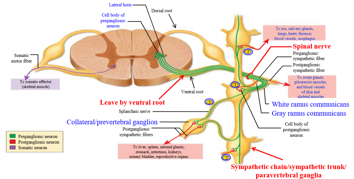
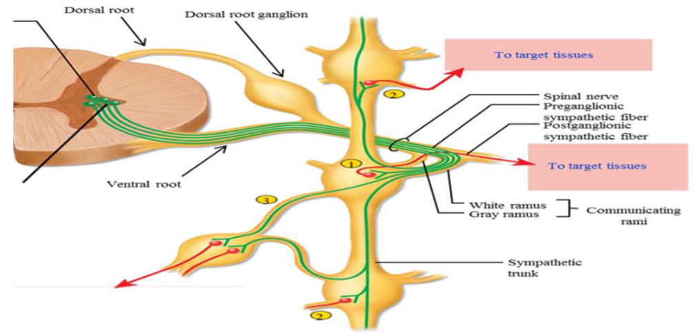
Spinal Nerve
Group of nerve axons travelling together.
Contains the axons of different neurons.
Sympathetic Pathway; First Neural Pathway - Step 1
When the dorsal root and ventral root combine, this is called a spinal nerve.
They have their cell bodies located in the lateral region of gray matter.
The preganglionic axons leaves the spinal cord via the ventral root.
They then join a spinal nerve.
Sympathetic Pathway; First Neural Pathway - Step 2
Once the preganglionic fiber leaves the spinal cord (via the ventral root) and enters into a spinal nerve, it then can enter a series of ganglia which are arranged on both sides of the spinal cord.
This series is called the sympathetic trunk/chain.
The ganglia in the circles lying in a chain & connected together.
Found on both sides of the spinal cord.
Sympathetic Pathway; First Neural Pathway - Step 3
The branches connect the spinal nerve to the ganglia.
Called “communicating rami” as the fibers of the neurons travel within them.
Sympathetic Pathway; First Neural Pathway - Step 4
The preganglionic axon, it leaves the spinal nerve and enters the rami, then into the ganglia.
This is called a white rami.
Preganglionic axon leaves spinal nerve crosses white ramus, and enters the ganglia.
It is now going to synapse with a sympathetic postganglionic fiber.
Sympathetic Pathway; First Neural Pathway - Step 5
The sympathetic postganglionic fiber will now leave the ganglion and enters back into the spinal nerve.
Enters back into the spinal nerve - grey ramas.
After, travels to a target tissue.
This is the first route that a sympathetic axon can take.
Sympathetic Pathway; Second Neural Pathway - Step 6
In another version of the pathway, the preganglionic fiber does not terminate in the ganglia and synapses with the postsynaptic neuron, but instead it goes up or down the sympathetic chain.
It will then synapse with a postganglionic neuron in a ganglia at a different level.
Sympathetic Pathway; Third Neural Pathway - Step 7
The pre-ganglionic fiber leaves the spinal cord through the ventral root, enters the spinal nerve, exits the nerve through the white ramus and enters into the ganglia.
At the same level as the spinal nerve.
Sympathetic Pathway; Third Neural Pathway - Step 8
The pre-ganglionic fiber will not synapse here.
Instead, it will pass through this ganglia and travel to the collateral ganglia in the splanchnic nerve.
The pre-ganglionic fiber will synapse with the post-ganglionic fiber in the collateral ganglia and travel to the target tissue.
Neural Pathways
Synapse immediately with a post-ganglionic neuron in the sympathetic ganglion.
Travels up or down the chain and synapses in ganglia at other levels.
Pass through chain without synapsing, continue to collateral ganglion as splanchnic nerve.
Communicating Rami
Two branches which connect a paravertebral ganglion to a spinal nerve; includes white and gray rami.
Allows for communication because of the nerve fibers that travel between them.
White Rami
Branch by which a myelinated preganglionic fiber leaves spinal nerve and enters a sympathetic ganglion.

Gray Rami
Branch by which unmyelinated post-ganglionic fibers the ganglion to re-enter the spinal nerve.

FFF - Eyes
In the sympathetic fight, flight, or freeze response, particular responses are triggered across many different organs.
There is a dilation or pupil and adjustment for far vision in this organ.
Allows you to scan the area.
FFF - Cardiovascular
In the sympathetic fight, flight, or freeze response, particular responses are triggered across many different organs.
Heart rate increases and force of contraction also increases, allowing more blood to be pumped to the skeletal muscle.
FFF - Blood Vessels (Arterioles + Veins)
In the sympathetic fight, flight, or freeze response, particular responses are triggered across many different organs.
BVs are composed of smooth muscle. The smooth muscle will constrict, regulating our BP.
FFF - Blood Vessels (Skeletal Muscle)
In the sympathetic fight, flight, or freeze response, particular responses are triggered across many different organs.
The smooth muscle will relax; it will dilate for more blood flow.
For oxygen increase.
FFF - Lungs
In the sympathetic fight, flight, or freeze response, particular responses are triggered across many different organs.
Bronchioles dilate and increase passage of air to reach lungs.
Inhibition of mucus secretion, making it easier to breathe.
FFF - Digestion
In the sympathetic fight, flight, or freeze response, particular responses are triggered across many different organs.
Even if there is food in the stomach, there is decreased GI motility and inhibition of digestive secretions.
FFF - Exocrine
In the sympathetic fight, flight, or freeze response, particular responses are triggered across many different organs.
Glands which release their secretions into ducts.
i.e. the sweat glands are stimulated, saliva is produced, but much thicker and rich.
FFF - Bladder
In the sympathetic fight, flight, or freeze response, particular responses are triggered across many different organs.
The bladder does not empty during sympathetic conditions (prevents voiding).
Urination during situations of extreme fear is not the sympathetic NS, but occurs due to the activation of both the sympathetic NS and the parasympathetic NS.
FFF - Endocrine
In the sympathetic fight, flight, or freeze response, particular responses are triggered across many different organs.
Releases epinephrine and norepinephrine.
FFF - Genitals
In the sympathetic fight, flight, or freeze response, particular responses are triggered across many different organs.
Males ejaculate and females contract their uterus.
Adrenal Gland
Composed of a outer cortex and inner medulla.
The adrenal medulla and cortex secrete different substances.
Receives only sympathetic innervation, not parasympathetic innervation.
Innervated by sympathetic preganglionic neurons.
Preganglionic Synapse at the Adrenal Medulla
Cell body in the CNS.
Short axon which innervates the adrenal medulla.
Neurotransmitter released from the pre-ganglionic neuron binds to receptors on the adrenal medulla, causing it to release its secretions into the bloodstream.
Releases epinephrine and norepinephrine.
Bloodstream to Adrenal Medulla
There is no postganglionic neuron.
The adrenal medulla itself acts like a modified sympathetic post-ganglionic neuron, but it does not have a axon or cell body.
The pre-ganglionic neuron innervates the adrenal medulla.
Effects of E and NE
By travelling in the blood, epinephrine and norepinephrine can each organs that do not receive nervous innervation.
They must first travel to the liver to be broken down, their effects on body tissues last longer.
Until the liver breaks them down, they remain active in the bloodstream, continuing to act on tissues.