Anatomy Exam 1 Flashcards
 Call Kai
 Learn
Learn Practice Test
 Spaced Repetition
 Match
Match Flashcards
 Knowt Play
1/134

There's no tags or description
Looks like no tags are added yet.
Name | Mastery | Learn | Test | Matching | Spaced | Call with Kai | Chat |
|---|
No analytics yet
Send a link to your students to track their progress
135 Terms
Cytology
The microscopic study of cells.
CELL TYPES
Connective Tissue are called
Integumentary system are called
Muscle Cells are called
CELL TYPES
Chondrocyte
Keratinocyte
Myocytes
BONE CELL TYPES
Osteocytes: ______________
Helps maintain the ________________ & monitor its composition
Housed in the ___________
Osteoblasts: __________________
______________: Process of creating new bones
Osteoclasts: ______________________
_____________: Process of breaking bone down
Osteoprogenitor cells: ___________________________________________
Osteocytes: Mature Bone Cells
Helps maintain the cell matrix & monitor its composition
Housed in the lacunae
Osteoblasts: Secretes new bone
Osteogenesis: Process of creating new bones
Osteoclasts: Breaks bone down
Osteolysis: Process of breaking bone down
Osteoprogenitor cells: Differentiates into other cells to help with bone generation or breakdown
Histology
Microscopic/Macroscopic?
Study of:
The microscopic study of tissues.
Cytology
Microscopic/Macroscopic?
Study of:
The microscopic study of cells.
Surface Anatomy
Microscopic/Macroscopic?
Study of:
The macroscopic/gross study of external and visible anatomical features and landmarks.
Ex. Look at this outline of the hip that tells you where not to cut in surgery
Regional approach
Microscopic/Macroscopic?
Study of:
The macroscopic/gross study of everything within one region
Ex. Brain
Systemic approach
Microscopic/Macroscopic?
Study of:
The macroscopic/gross study of a system
Ex. Digestive System
Approaches to Anatomy
Developmental: ____________________
Embryology: ____________________
Comparative: ____________________
Medical or Surgical: ____________________
Pathological: ____________________
Radiographic: ____________________
Approaches to Anatomy
Developmental: Egg to adult
Embryology: Fertilization to 8 weeks in utero
Comparative: Compare across organisms
Medical or Surgical: Anatomical landmarks used to see deeper structures
Like surface anatomy
Pathological: Structure relating to disease
Radiographic: – X-rays, CAT scan, ultrasound, MRI
7 Levels of Organization (Microscopic → Gross)
7 Levels of Organization (Microscopic → Gross)
Chemical
Molecules
Cells
Tissues
Organ
Organ System
Organism Level
__________: Organs working together to form a system
An organ (can/can’t) be in more than one organ system
__________ is when all organ systems are working together
__________: Basic functional/structural unit of a living organism
__________: Two or more different tissue types
Fills a specific __________ and has a defined __________
__________: Proteins, fats, vitamins
__________: All (#) body systems combine and work to sustain the life of the entire living being
__________: Group of cells combined
Organ System: Organs working together to form a system
An organ (can/can’t) be in more than one organ system
Homeostasis is when all organ systems are working together
Cells: Basic functional/structural unit of a living organism
Organ: Two or more different tissue types
Fills a specific function and has a defined shape
Molecules: Proteins, fats, vitamins
Organism Level: All 11 body systems combine and work to sustain the life of the entire living being
Tissues: Group of cells combined
Anatomical Position
The standard reference position where the body is standing upright, feet forward, and palms facing forward.
Sagittal Plane: Anatomical plane that divides the body into _____ and _____ sections.
Sagittal Plane Movement: ________________ (parallel to plane)
3 types: ___________________
Sagittal Plane: Anatomical plane that divides the body into left and right sections.
Sagittal Plane Movement: Forward and backward (parallel to plane)
Flexion: decrease in the reference angle (like flexing)
Extension: increase in the reference angle
Hyperextension: increase in the reference angle beyond anatomical position
Frontal (Coronal) Plane: Anatomical plane that divides the body into _________ and _________ sections
Frontal Plane Movement: _________ (parallel to plane)
2 types: _____________________
Anatomical plane that divides the body into front (anterior) and back (posterior) sections.
Frontal Plane Movement: Side to side (parallel to plane)
Abduction: move away from the midline of the body (absence)
Adduction: move toward the midline of the body (advance)
Transverse Plane: Anatomical plane that divides the body into __________ and __________ sections.
Transverse Plane Movement: ___________________ (parallel to plane)
2 types: __________________________
Anatomical plane that divides the body into top (superior) and bottom (inferior) sections.
Transverse Plane Movement: Rotational around the vertical axis (parallel to plane)
Medial rotation: anterior surface moving medially.
Front surface moving towards the middle of the body
Lateral rotation: anterior surface moving laterally.
Front surface moving towards the sides of the body
Movement is always __________________
Ex. Swinging your arms by your side is ______ movement, while making the sunrise motion is ______
Movement is always parallel to the plane!!!
Ex. Swinging your arms by your side is sagittal movement, while making the sunrise motion is frontal
____________: Pulling your fingers and hand backward, like making a "stop" sign or prepping for a push-up.
____________: Bend your elbow at a 90-degree angle and move your hand inward toward your stomach or opposite hip
____________: Straightening your leg out to stand up
____________: Lifting your arms straight out to the sides to form a "T" shape
____________: Keep your leg straight and turn your toes outward so they point away from each other
____________: Lifting a dumbbell during a bicep curl (bringing your forearm closer to your upper arm)
____________: Squeezing your thighs together or crossing one leg over the other
Hyperextension: Pulling your fingers and hand backward, like making a "stop" sign or prepping for a push-up.
Medial Rotation: Bend your elbow at a 90-degree angle and move your hand inward toward your stomach or opposite hip
Extension: Straightening your leg out to stand up
Abduction: Lifting your arms straight out to the sides to form a "T" shape
Lateral Rotation: Keep your leg straight and turn your toes outward so they point away from each other
Flexion: Lifting a dumbbell during a bicep curl (bringing your forearm closer to your upper arm)
Adduction: Squeezing your thighs together or crossing one leg over the other
Terms of Direction:
______________: Top & Bottom
______________: Middle & Outer Side
______________: Towards & Away from the body
Elbow is ________ and hand is ________
______________: Towards the head & Towards the tailbone
Ex. The chest is ________ to the stomach.
______________: Front & Back
______________: Front & Back
______________: Parts of the foot
________ is the sole, ________ is what you look down and see
______________: Parts of the hand
________ is the palms, ________ is the back with the fingernails
Terms of Direction:
Superior & Inferior: Top & Bottom
Medial & Lateral: Middle & Outer Side
Proximal & Distal: Towards & Away from the body
Elbow is proximal and hand is distal
Cranial & Caudal: Towards the head & Towards the tailbone
Ex. The chest is cranial to the stomach.
Anterior & Posterior: Front & Back
Ventral & Dorsal: Front & Back
Plantar & Dorsal: Parts of the foot
Plantar is the sole, dorsal is what you look down and see
Palmar & Dorsal: Parts of the hand
Palmar is the palms, dorsal is the back with the fingernails

Fill in the levels
Fill in the levels
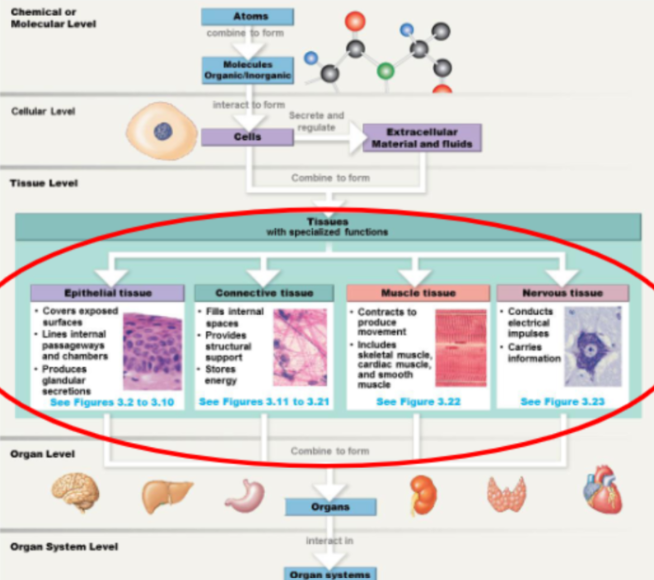
2/4 Classes of Tissues
Epithelia
Functions: (3)
Example:
Connective Tissue
Function: (3)
Example:
Epithelia (Covers, lines, and GLANDS)
Function:
Cover exposed internal and external surfaces
(Protect against physical/chemical/biological agents)
Line internal vessels, passageways, and cavities
(Control permeability)
Forms gland tissue and produce secretion in glands
Example: Skin
Connective Tissue: (Fills, structure, energy storage)
Functions:
Fill in void spaces
Provide structural support to cells, tissues, and organs
Provide a location to store energy
Example: Bone, Blood, cartilage
2/4 Classes of Tissues
Muscle Tissue
Functions: (3)
Neural Tissue
Functions: (3)
Muscle Tissue Functions: (smooth, skeletal, cardiac)
Smooth muscle tissue: Contracts to produce movement or to propel liquids
Skeletal muscle tissue: Maintains body’s correct posture
Cardiac muscle tissue: Circulates blood & blood pressure / assists in maintaining body temperature
Neural Tissue Functions: (carries info, electrical impulses, sensory motor)
Carries information to organ / tissue systems
Conducts electrical impulses
Responsible for sensory and motor function (Electrical impulse sends sensory and motor information)
Main tissue type in glands?
Epithelia
Characteristics of Epithelia
Cellularity: _______________
Polarity: _______________
Avascularity: ______________
Characteristics of Epithelia
Cellularity: Made of cells
High degree of interlocking occurring between adjacent cells, which ensures it’s not too permeable
Polarity: Uneven distribution of organelles inside cells
Avascularity: No blood vessels going to or from it (So it removes waste and gets nutrients through secretion & absorption)
Attachment Characteristics of Epithelia
Apical surface: ___________________
Doesn’t need to be the top- for example it’s the part of the blood vessel _________________
Basolateral: Where it’s anchored to the _______________
Lamina propria: A thin layer of loose connective tissue that sits directly ______________________
Attachment Characteristics of Epithelia
Apical surface: The exposed surface of the epithelia
Doesn’t need to be the top- for example it’s the part of the blood vessel touching the blood
Basolateral: Where it’s anchored to the basement membrane
Lamina propria: A thin layer of loose connective tissue that sits directly beneath the surface lining (epithelia)
Characteristics of Epithelia
Regeneration: _________________________________
Microvilli: ____________________________
Adjacent Connections: ____________________________
Characteristics of Epithelia
Regeneration: Replacement of epithelial cells at the apical suffice through the division of stem cells located near the basement membrane
Microvilli: Significantly increase the cell’s apical surface area to maximize efficiency in absorption or secretion
Adjacent Connections: Intercellular connections that maintain the epithelium's structural integrity & regulate permeability
Neuroepithelium
A highly specialized type of epithelia located in sense organs that provides sensory perception.
Simple Epithelium
Epithelial tissue consisting of a single layer of cells attached to the basement membrane.

Stratified Epithelium
Epithelial tissue consisting of two or more layers of cells (stronger than simple)
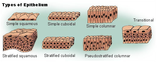
Pseudostratified Epithelium
Has nuclei of varying heights (which implies it’s stratified) but every cell has a connection to the basement membrane which means it’s simple.

Squamous Cells
Looks squashed, and looking at the top (apical surface) looks like fried eggs side by side
Look at the apical surface (top) to determine shape. It could still look cuboidal at the bottom
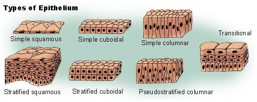
Cuboidal Cells
Box-shaped or hexagonal epithelial cells with centrally located nuclei
Looking at the apical surface, it looks mostly even and the nucleus is central

Columnar Cells
Epithelial cells whose height is consistently greater than their width.
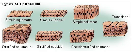
Components of Epithelium
Villi:
Definition:
Location:
Function:
Cilia:
Definition:
Location:
Function:
Components of Epithelium
Villi:
Folds of tissue composed of entire sheets of epithelial cells
Located in the intestinal walls
Increases total tissue surface area (apical surface) for nutrient absorption
Cilia:
Hair-like projections on the apical surface of specific epithelial cells
Located mainly in respiratory tract and fallopian tubes
Beats to propel fluids, mucus, or particles across the cell surface
Transitional Epithelium
Specialized epithelium capable of significant stretching, located in organs of the urinary system like the bladder.
Gland: ________________________________________
Glands are made out of __________
Relation to organs: ________________________
Example: _______________________
Gland: A cell, group of cells, or organ specialized to synthesize and release a product
Glands made out of epithelia
Relation to organs: Can be entire organ or a portion of an organ
Example: Salivary gland – produces saliva, which is functionally important for breaking down sugars and packaging food for digestion
Secretion vs. Excretion:
Secretion is ________________________, versus excretion which is ________________________
Exocytosis: ________________________
Secretion vs. Excretion:
Secretion is synthesis and release of a product for further use, versus excretion which is elimination of a waste product
Exocytosis: Transport of product outside of the cell
Exocrine Glands
Method of release:
Polarized/Unpolarized:
Further classifications as to the type of exocrine gland:
Exocrine Glands
Glands that release their secretory products onto an epithelial surface directly or through a duct system.
Cells polarized: Organelles placed in a functional location
Further classifications as to the type of exocrine gland: Single cell / Multiple Cells, Simple duct / Complex Duct
Endocrine Glands
Method of release:
Polarized/Unpolarized:
Produces:
NO DUCTS (Use diffusion into capillaries instead)
NO polarization within the cell (No need to “aim” product onto surface or into duct)
Produces HORMONES (Small, easy to diffuse)
Mode of Product Release:
Merocrine Secretion
Ex.
Apocrine Secretion
Ex.
Merocrine Secretion: Releases the product via exocytosis without losing any cell material
Ex. Saliva, Sweat
Apocrine Secretion: Releases the product along with a little bit of cytoplasm
Ex. Breast Milk, Sweat
Mode of Product Release:
Holocrine:
Ex.
Endocrine:
Ex.
Holocrine: Product packs the cell until the cell explodes
Ex. Oil gland
Endocrine: Released into a vessel for transport
Ex. Hormones
Connective Tissue: ___________________________
Function of Connective Tissues: (6)
Connective Tissue: Supportive tissue consisting mainly of extracellular matrix, ground substance, and highly specialized cells
Function of Connective Tissues (SPITED)
Store energy (lipids)
Protecting and supporting surrounding tissues
Interconnecting tissues
Transporting liquids throughout circulatory / lymph systems
Establish structural framework
Defending body from infections or other harmful substances
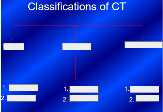
Fluid Ct: Cells and fibers supported in a ________ matrix
CT Proper: Cells and fibers supported in a ________ matrix
Supporting CT: Cells and fibers supported in a ________ matrix
Bones are ________
Fluid Ct: Cells and fibers supported in a watery matrix
CT Proper: Cells and fibers supported in a syrupy ground matrix
Supporting CT: Cells and fibers supported in a gel like matrix
Bones are calcified
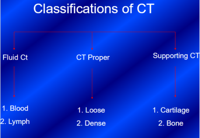
Connective Tissue (CT) Proper: Refers to CT with ______________________
Fiber Types: (3)
Connective Tissue (CT) Proper: Refers to CT with many types of cells and extracellular fibers in a syrupy ground substance
Fiber Types: Collagen, Elastic, Reticular
Connective Tissue Proper FIBERS
Collagen:
Appearance:
Attributes:
Force Type:
Elastic:
Appearance:
Attributes:
Force Type:
Reticular:
Appearance:
Attributes:
Force Type:
Collagen:
Appearance: Very long, straight, and unbranched (Moving in one direction)
Attributes: Strongest and most abundant
Force Type: Very good at handling a tensile force (moving in one direction)
Elastic:
Appearance: Branching & wavy
Attributes: Can stretch up to 100 times their length
Force Type: Can handle force in multiple directions.
Reticular: Branched and interwoven
Appearance: Thinner than collagen fibers
Attributes: Forms a delicate, net-like supportive mesh for soft organs
Force Type: Can handle force in multiple directions.
Connective Tissue Proper FIXED CELLS
Fibrocytes: ______________
Fibroblasts: ______________
Mesenchymal: ______________
Macrophages: ______________
Melanocytes: ______________
Adipocytes: ______________
Connective Tissue Proper FIXED CELLS
Fibrocytes: Maintains connective tissue fibers
Fibroblasts: Produce connective tissue fibers
Mesenchymal: Can differentiate into different cell types
Macrophages: Engulfs damaged cells or pathogens (trash collectors)
Melanocytes: Contains melanin (responsible for pigmentation)
Adipocytes: Fat cells
Connective Tissue Proper WANDERING CELLS (Able to move around / not fixed)
Macrophages:
Microphages:
Lymphocytes:
All immune system white blood cells
Macrophages: Engulf and digest pathogens and cellular debris
Microphages: Act as first responders to destroy bacteria
Lymphocytes: Responsible for targeted, long-term immunity
CT Proper:
Mast cells: _______________________________
Histamine: _____________
Heparin: _____________
Ground Substance: _____________
Loose vs Dense
Loose splits into ________, ________, and ________ (Don’t need to identify in image)
Dense splits into ________ and ________ (Should identify)
CT Proper:
Mast cells: Near blood vessels and aid in the inflammatory response
Histamine: Causes swelling, itching
Heparin: Prevents blood clotting
Ground Substance: Clear, syrupy consistency
Loose vs Dense
Loose splits into areolar, adipose, and reticular (Don’t need to identify in image)
Dense splits into regular and irregular (Should identify)

Loose or dense CT Proper?
IF DENSE: Regular or Irregular
Loose CT Proper: Light shines through loose connective tissue
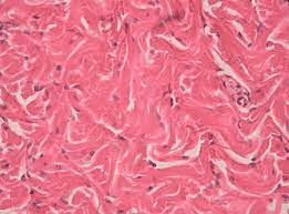
Loose or dense CT Proper?
IF DENSE: Regular or Irregular
Dense Irregular CT: Fibers & bundles in multiple directions

Loose or dense CT Proper?
IF DENSE: Regular or Irregular
Loose + Adipocytes (fat cells): Looks like white blobs
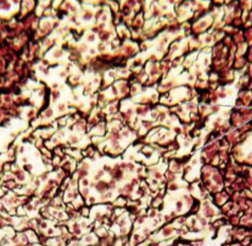
Loose or dense CT Proper?
IF DENSE: Regular or Irregular
Loose + Reticular fibers: Lots of little dots and fibers
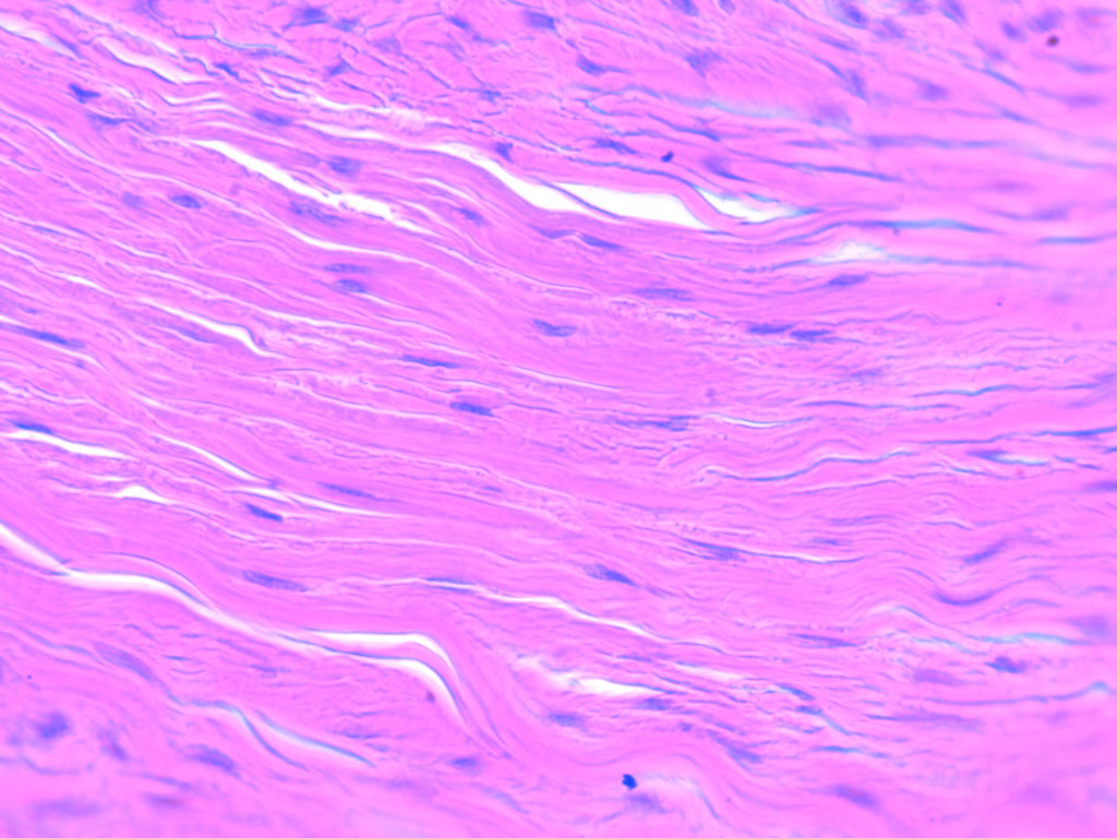
Loose or dense CT Proper?
IF DENSE: Regular or Irregular
Dense Regular CT: Fibers flow in one direction (Makes up tendons and ligaments)
Fluid Connective Tissue
Fluid Connective Tissue: Refers to distinct population of cells suspended in watery matrix containing dissolved proteins (blood & lymph)
Supporting Connective Tissue: (Less/More) diverse cell population than other types of CT.
Examples: _______ & ________
Cell type: _____________________
Lacunae: _____________________
(Vascular/Avascular) & (Loose/Dense) (Regular/Irregular)
Supporting Connective Tissue: Less diverse cell population than other types of CT.
Examples: Bone & Cartilage
Cell type: Chondrocyte (Chondro = cartilage)
Lacunae: Space where chondrocytes exists
Avascular & Dense Irregular
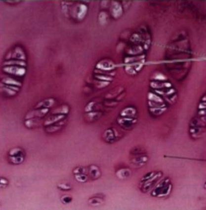
Supporting Connective Tissue: Is this Fibrocartilage, Hyaline Cartilage, or Elastic Cartilage?
Location:
Attributes:
Hyaline Cartilage (White color in the body)
Located in areas of high friction (Ex. reduces friction where bones meet)
Very tough but flexible
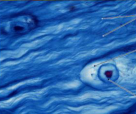
Supporting Connective Tissue: Is this Fibrocartilage, Hyaline Cartilage, or Elastic Cartilage?
Location:
Attributes:
Fibrocartilage
Located in areas of high stress (Ex. Knee joints)
Very good with one directional forces like compression

Supporting Connective Tissue: Is this Fibrocartilage, Hyaline Cartilage, or Elastic Cartilage?
Location:
Attributes:
Elastic Cartilage
Found in structures like the external ear
Highly flexible cartilage containing abundant elastic fibers
3 Divisions of the Integumentary System
Epidermis: The superficial outer layer of skin composed of stratified squamous epithelium.
Dermis: The middle layer of skin containing blood vessels, nerves, and glands.
Hypodermis: The deep subcutaneous fat layer separated from dermis by a layer called fascia
Epidermis Layers (Deep to Superficial)
_________________ (where’s it located)
_________________
_________________
_________________
_________________ (where’s it located)
Why do we sometimes say there’s 4 layers?
Epidermis Layers (Germs spin grand lucid corn)
Stratum germinativum (Stem cells attached to the basal surface)
Stratum spinosum
Stratum granulosum
Stratum lucidum
Stratum corneum (Apical surface with high levels of interlocking so things don’t get through)
The stratum lucidum is present in thick skin only (palms and bottom of feet), so we say the epidermis has 4 or 5 layers depending on the area.
Glassy-like appearance if present
2 Layers of the Dermis
__________________ (superficial layer)
Type of CT
Contains
__________________ (deeper layer)
Type of CT
Contains
Papillary layer (under epidermis- superficial layer)
Loose CT proper
Contains capillaries and nerve endings or sensory apparatus (merkel cells and dendrites)
Reticular layer (deeper layer)
Dense irregular CT proper
Contains blood vessels, hair follicles, nerves, sweat glands, sebaceous glands, collagen fibers
Hypodermis
Otherwise known as:
Type of CT:
Contains:
Distribution/accumulation of adipose tissue in hypodermis is _____ dependent
Hypodermis:
Subcutaneous layer
Loose CT proper
Contains abundance of fat cells (adipose cells) – energy store
Distribution/accumulation of adipose tissue in hypodermis is sex dependent
Main cell type in Integumentary system?
Forms what type of epithelium?
Keratinocyte is the predominant cell type of the integumentary system forming stratified squamous epithelium.
Melanocyte:
Produces:
UV exposure causes melanocytes to:
Skin color is due to:
Produces: melanin which prevent skin damage by absorbing UV radiation
UV exposure causes melanocytes to: increase melanin synthesis and transfer
Skin color is due to: the amount of melanin and skin vascularization
Accessory Structures of the Integumentary System → Hair Follicle
Root Hair Plexus: ______________________
Sebaceous glands: ______________________
Arrector pili muscle: ______________________
Accessory Structures of the Integumentary System → Hair Follicle
Root Hair Plexus: Nerve ending to sense movement of hair
Sebaceous glands: Secretes sebum (oily substance)
Arrector pili muscle: Makes your hair stand on end

Integumentary System

Accessory Structures of the Integumentary System → Nail
Lunula: Pale because ________________________
______________ can change the strength and texture of the nail
Color can indicate _________ or _________
Pressing on nail demonstrates _______________________
Nail
Lunula: Pale because it’s in a region where blood vessels are obscured
Nutritional deficiencies can change the strength and texture of the nail
Color can indicate hypoxia or anemia
Pressing on nail demonstrates how fast blood supply returns
Skin & Aging
Reduced ______ ______ and slower repair
______ is thin → More prone to injury
Reduced ______ ______ → worse temperature control
Skin & Aging
Reduced blood supply and slower repair
Epidermis is thin → More prone to injury
Reduced sweat glands → worse temperature control
Types of Bones
Long Bones: (visual description)
Epiphysis: ______________________
Diaphysis: ______________________
Medullary Canal: ______________________
Ex. _________
Short Bones: (visual description)
Ex. _________
Types of Bones
Long Bones: Long & Slender
Epiphysis: The part at the two ends of the bone
Diaphysis: The middle part of the bone (shaft)
Medullary Canal: The open area running through the center of the shaft (Marrow Cavity)
Ex. Humerus
Short Bones: Smaller & box-like
Ex. Wrist bones
Types of Bones
Flat Bones: _____________________
Ex. __________
Irregular: _____________________
Ex. __________
Types of Bones
Flat Bones: Thin, jagged edge where two flat bones meet
Ex. In the skull
Irregular: Complex shapes
Ex. Vertebrae
Sesamoid (definition & example)
Subset of irregular bones that are floating
Ex. Patella/knee cap
Surface Anatomy of a Bone
Projection/Process: ___________________
Articular: ___________________
Tubercle/Tuberosity: ___________________
Condyle: ___________________
Spine: ___________________
Surface Anatomy of a Bone
Projection/Process: Part of a bone that sticks out
Articular: Where two bones meet
Tubercle/Tuberosity: A projection
Condyle: Smooth & round articular process
Spine: Pointed process
Surface Anatomy of a Bone
Fossa: ___________________
Ramus: ___________________
Head: ___________________ (has to be a ____ bone & usually ____)
Neck: ___________________
Facet: ___________________
Fossa: Shallow depression (Ex. Cup for hip joint)
Ramus: Extension of a bone forming an angle
Head: An extension of Epiphysis (Has to be a long bone & usually rounded)
Neck: Connection between the epiphysis and diaphysis
Facet: Small flat articular surface
Spongy (Trabecular) bone vs. Compact (Cortical) bone
Spongy:
Type of matrix: _______________
Type of force: _______________
(Inside/Outside) of the bone
Compact:
Osteon: _______________________
(Inside/Outside) of the bone
Spongy (Trabecular) bone vs. Compact (Cortical) bone
Spongy:
Very branched matrix (Branches called trabeculae)
Handles multidirectional force
On the inside of the bone
Compact:
Osteon: Functional unit that creates a cylinder of compact bone
On the outside of the bone


Osteon: ________________________
Structure: ________________________
Arranged: ________________________
Matrix:
___% Inorganic, Calcium phosphate, Calcium Carbonate.
___% Organic, gel and collagen fibers
Osteon: Functional Unit of Compact Bone
Structure: One inside another inside another
Arranged: parallel to the long axis of the bone
Matrix:
65% Inorganic, Calcium phosphate, Calcium Carbonate.
35% Organic, gel and collagen fibers

Parts of the Osteon
Lamellae: ___________________________
Collagen Fibers: ________________________
Parts of the Osteon
Lamellae: Making up osteons (each “paper towel role”)
Collagen Fibers: Alternate directions w/ each lamellae (Strengthens the osteon)
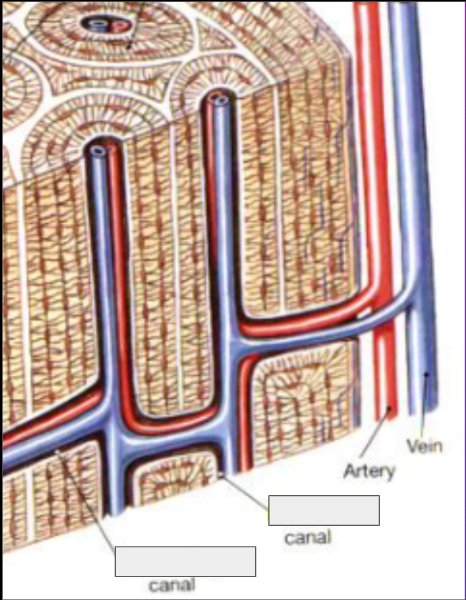
Parts of the Osteon
Lacunae: ______________________________
Canaliculi: ______________________________
Haversian Canals = _____________
Volkman’s Canals = _____________
Atomic structures in (compact/spongy) bone that contain ______ ______, including both veins and arteries, to provide _______ to the bone tissue
Parts of the Osteon
Lacunae: Where the osteocytes are located
Canaliculi: Tiny tunnels that connect lacunae to the central Haversian canals (allows for nutrient/waste transport)
Haversian Canals = central canal
Volkman’s Canals = perforating canal
Atomic structures in compact bone that contain blood vessels, including both veins and arteries, to provide nutrients to the bone tissue

Periosteum: (Location in relation to bone)
Outer Fibrous Layer:
What kind of CT: _________________
Contains: _________________
Function: _________________
Inner Osteogenic Layer:
Contains: _________________
Function: _________________
Periosteum: Surround outer surfaces of bone
Outer Fibrous Layer:
What kind of CT: Dense irregular connective tissue
Contains: Fibroblasts, collagen fibers, blood vessels, and nerves
Function: Protective barrier and an anchor point
Inner Osteogenic Layer:
Contains: Contains elastic fibers, blood vessels, and bone cells (such as osteoprogenitor cells and osteoblasts)
Function: Essential for bone growth and healing
(Periosteum surrounds outer surfaces of bone)
5 Functions: ___________________
(Periosteum surrounds outer surfaces of bone)
5 Functions: PAPIC
Participates in bone growth
Attaches bone to CT of deep fascia
Provides place for attachments
Isolates/protects bones
Circulatory and nervous supply
Endosteum
Location in relation to bone: __________________________
Active during _______ and _______
(Does/Does not) form a complete lining so that _________________________
Endosteum
Location in relation to bone: Lines inner surface of bone (lining of marrow cavity)
Active during growth and regeneration
Does not form a complete lining so that the exposed areas are accessible to osteoblasts / osteoclasts
Holes make bone accessible for bone growth/breakdown
Bone Cells (BONE = _________)
Osteocytes: _____________________
Function: ___________
Location: ___________
Osteoblasts: ________________________
Osteogenesis: _____________________
Osteoclasts: _____________________
Osteolysis: _____________________
Osteoprogenitor: _____________________
Bone Cells (BONE = OSTEO)
Osteocytes: Mature Bone Cells
Helps maintain the cell matrix & monitor its composition
Housed in the lacunae
Osteoblasts: Secretes new bone
Osteogenesis: Process of creating new bones
Osteoclasts: Breaks bone down
Osteolysis: Process of breaking bone down
Osteoprogenitor: Differentiates into other cells to help with bone generation or breakdown
4 Factors affecting Deposition and Resorption of Bone: _________________
Bone Density is (static/dynamic)
Factors affecting Deposition and Resorption of Bone (DAHM)
Diet (Vitamin D, Calcium)
Age
Hormones (Thyroid and Parathyroid gland)
Mechanical Stress (exercise)
Bone Density is dynamic = ever changing
Bone Formation and Bone Growth:
Intramembranous ossification:
Endochondral ossification:
Appositional Growth:
Bone Formation and Bone Growth:
Intramembranous ossification
(Bone formation)
Becomes spongy because it’s trapping blood vessels (parts of the bone with no bone)
Endochondral ossification
Bone develops from cartilage
(Bone formation)
Appositional Growth
(Increasing bone diameter)
Endochondral Ossification: _____________________
Step 1:
Step 2:
Endochondral Ossification: Multistep process in which bone forms out of cartilage
Step 1: The Mold Grows
The cartilage model gets bigger.
Cartilage cells in the center expand and then die off, leaving behind open spaces.
Step 2: The Bone Collar
Blood vessels wrap around the outside of the cartilage model.
Cells on the outer edge turn into bone-building cells (osteoblasts) and create a hard bone collar around the middle.
Endochondral Ossification: _____________________________
Step 3:
Step 4:
Endochondral Ossification: Multistep process in which bone forms out of cartilage
Step 3: The Primary Center
Blood vessels break into the center of the cartilage, bringing osteoblasts with them
This creates the primary center of ossification, where the dead cartilage core is replaced with spongy bone
Step 4: The Marrow Tunnel
Bone-destroying cells (osteoclasts) carve out a hollow medullary cavity (the marrow tunnel) down the center.
The bone begins growing longer at both ends, leaving just a little cartilage behind at the boundaries (metaphysis)
Endochondral Ossification: _______________________
Step 5:
Step 6:
Endochondral Ossification: Multistep process in which bone forms out of cartilage
Step 5: The Ends (Secondary Centers)
Blood vessels enter the epiphyses (the top and bottom tips of the bone).
These become secondary centers of ossification, where osteoblasts begin replacing the cartilage tips with spongy bone.
Step 6: The Final Cleanup
The tips fill up with bone, leaving cartilage in only on the outer joint surfaces (cushion) and a slice in the middle (growth plate)
endochondral ossification vs intramembranous ossification
Intramembranous ossification creates bone directly from embryonic connective tissue, while endochondral ossification replaces a hyaline cartilage template with bone
Intramembranous forms flat bones of the skull, the mandible (lower jaw), and the clavicles (collarbones), while Endochondral does most of the others
Appositional Growth: ___________________
What accelerates the process:
Osteolysis: ___________________
Appositional growth: Not a form of bone development, just makes them wider/thicker
Vessels accelerate the process
Osteolysis: At the same time as we increase in diameter, our medullary cavity is also increasing from the inside out through osteolysis, making the bones hollow so we can move around
Articulation: _____________
Joint: _____________
Continuum of joints: _____________
Articulation: Joint
Joint: Where two bones meet
Continuum of joints: From stable with good boney fit and tight ligaments (ankle) to joints that are freely moveable with loose ligaments (shoulder)
Types of Joints based on FUNCTION:
_________: No movement
Subtypes: __________________________
_________: Little movement
Subtypes: __________________________
_________: “free” movement
Subtype: _________
Types of Joints:
Synarthrosis: No movement
Subtypes: Bony fusion, Cartilaginous, Fibrous
Amphiarthrosis: Little movement
Subtypes: Fibrous, Cartilaginous, Synovial
Diarthrosis: “free” movement
Subtype: Synovial
Types of Joints based on STRUCTURE:
Bony fusion: _______________________
Fibrous joint: _______________________
Cartilaginous joint: _______________________
Synovial joint: _______________________
Types of Joints based on STRUCTURE:
Bony fusion:
Two bones fuse together
Ex. Sutures (where the skull bones fuse)
Fibrous joint:
Connective tissue holding it together
Can be synarthrotic (sutures) or amphiarthrotic (ligaments)
Cartilaginous joint:
Allows little or no movement between bones
Held together by cartilage
Synovial joint:
Diarthrotic (Ex. Knee joint)
Surrounded/held together by the joint capsule and also held together by ligaments
Synovial Joint
Surrounded by: _______________
Articular cartilage: _____________________
Joint cavity: __________________
Fibrocartilage pads & Fat Pads: __________________________
Synovial Joint
Surrounded by: Joint capsule
Articular cartilage: On either end of the bones articulating at that joint
Joint cavity: The space in the joint
Fibrocartilage pads & Fat Pads: Little sections of cartilage both used to reduce friction
Synovial Joint
Synovial membrane: ____________________________
Synovial fluid: _________________________
Ligaments: (3 Types)
Synovial Joint
Synovial membrane: Lines the cavity
Stops and never makes contact with the cartilage
Synovial fluid
Fills the joint cavity to lubricate, nourish chondrocytes, and absorb shock
Hyaluronan (glycosaminoglycans) & lubricin (protein)
Ligaments:
Extrinsic: Outside the joint capsule
Intrinsic: Housed within the joint
Capsular: Thickening of the joint capsule
Structural classification of synovial joints
Gliding joint:
Type of Movement:
Planes of Movement:
Ex.
Hinge joint:
Type of Movement:
Planes of Movement:
Ex.
Structural classification of synovial joints
Gliding joint:
Type of Movement: Linear (Slides forward/back & left/right)
Planes of Movement: 2
Ex. where clavicle meets breast bone
Hinge joint:
Type of Movement: Angular (Open & close like a door)
Planes of Movement: 1
Ex. Ankle point & flex
Structural classification of synovial joints
Pivot Joint:
Type of Movement:
Planes of Movement:
Ex.
Ellipsoidal (Condyloid) Joint:
Type of Movement
Planes of Movement:
Ex.
Structural classification of synovial joints
Pivot Joint:
Type of Movement: Uniaxial rotation
Planes of Movement: 1
Ex. Shaking head no
Ellipsoidal (Condyloid) Joint:
Type of Movement: Angular motion (Oval surface in a shallow depression)
Planes of Movement: 2
Ex. Where wrist bones meet forearm bone
Structural classification of synovial joints
Saddle Joint:
Type of Movement:
Planes of Movement:
Ex.
Ball & Socket Joint:
Type of Movement:
Planes of Movement:
Ex.
Structural classification of synovial joints
Saddle Joint:
Type of Movement: Angular (Concave on convex)
Planes of Movement: 2
Ex. Base of the thumb in the palm of the hand
Ball & Socket Joint:
Type of Movement: Multidirectional rotation (Round head in a cup)
Planes of Movement: All 3
Ex. Hip
Common Joint Diseases (“ITIS” = ___________)
Bursitis: _________________________
Synovitis: _________________________
Common Joint Diseases (“ITIS” = inflammation)
Bursitis: too much friction causes sac to become inflamed
Synovitis: Inflammation of the synovial membrane
Common Joint Diseases (“ITIS” = inflammation)
Arthritis: _______________________
Osteoarthritis: _______________________
Rheumatoid: _______________________
Common Joint Diseases (“ITIS” = inflammation)
Arthritis: inflammation of the joint
Osteoarthritis: Joint degrading from wear & tear
Aka degenerative joint disease
Wears down so it’s bone on bone contact
Rheumatoid: Swelling at the joint
Leads to inflammation of the synovial membrane
Knobby appearance
Autoimmune condition
Arthritis with degradation of hyaline cartilage
Hyaline cartilage wears away and leads to _____________ which causes _____________
Arthritis with degradation of hyaline cartilage
Hyaline cartilage wears away and leads to bone to bone contact which causes bone spurs

Options:
Medullary Cavity
Metaphysis
Synovial membrane
Compact Bone
Articular cartilages
Spongy bone
Periosteum
Joint Cavity
Fibrous Joint Capsule

Knee Joint = (also called)
Largest ________ joint in the body
Bones included: ________________
Joints included:
______________: (structure type)
______________: (structure type)
Knee Joint = GENU
Largest synovial joint in the body
Bones included: Femur, tibia, patella
Joints included:
Tibiofemoral Joint: Modified condolyte
Patellofemoral joint: Gliding