Lecture 10: Common Disorders of the Equine Hind Limb
 Call Kai
 Learn
Learn Practice Test
 Spaced Repetition
 Match
Match Flashcards
 Knowt Play
1/39

There's no tags or description
Looks like no tags are added yet.
Name | Mastery | Learn | Test | Matching | Spaced | Call with Kai | Chat |
|---|
No analytics yet
Send a link to your students to track their progress
40 Terms
cervical spine OA
caused by trauma, unnatural head carriage, OC
diagnosed by stiff neck, ± lame or neurologic, radiographs
treatment → inject facets with corticosteroid, acupuncture, mesotherapy
prognosis is fair
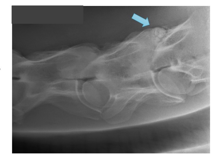
what is this?
cervical spine OA
impinging dorsal spinous processes
kissing spines
caused by trauma, poor fitting saddle
diagnosed by pain on palpation, reduced performance, radiographs
treatment → check saddle fit, inject area with corticosteroids, surgical resection
prognosis depends on severity
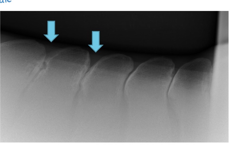
what is this?
impinging dorsal spinous processes aka kissing spines
general back muscle pain
caused by underlying hind limb lameness, poor saddle fit, poor riding, trauma
diagnosed by pain on palpation, reduced performance
treatment → check saddle fit, inject area with corticosteroids, acupuncture, rest
prognosis is good once address underlying cause
sacroiliac disease
arthritis
subluxation of SI joint = hunter’s bump
caused by trauma, inappropriate use
diagnosed by clinical signs, acute → lame, can feel crepitus by rectal palpation
treatment is rest and inject SI joint with corticosteroids
prognosis is good
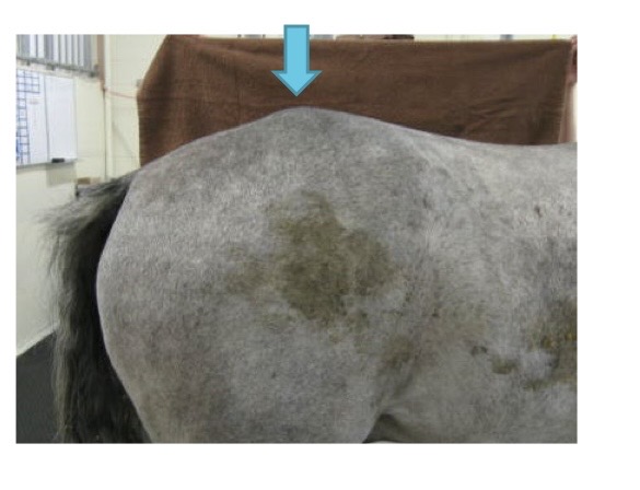
what is this?
hunter’s bump from sacroiliac disease
what are the structures involved with causing hind limb lameness in the horse ranked from most common to least common?
hock → below hock → stifle → above stifle
metatarsal 2 and 4 (splint bones) fractures
caused by kick injury
diagnosed by radiographs, observing wound
treatment → ostectomy (segmental vs complete)
prognosis is excellent
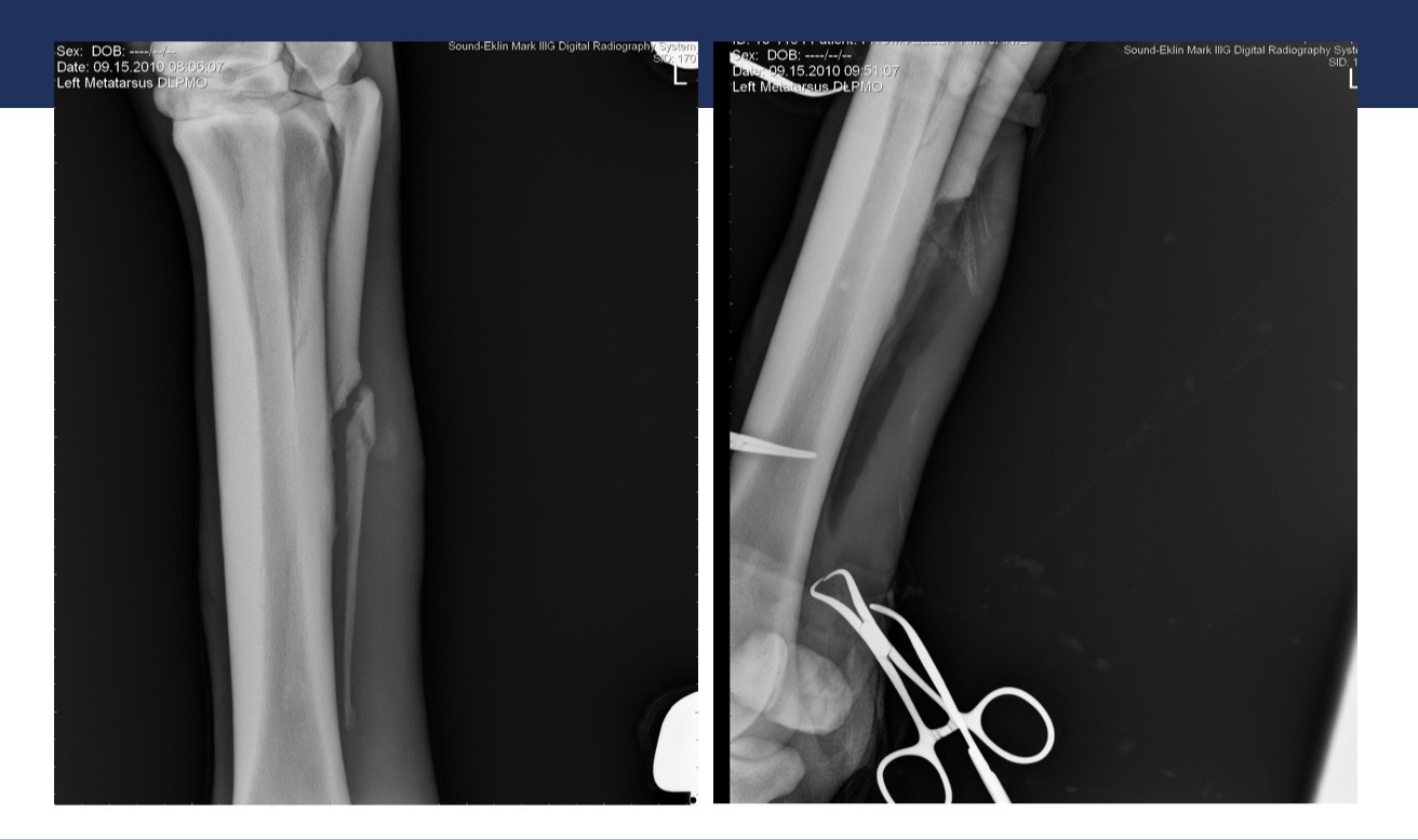
what is this?
splint bone fracture
hock lameness gait characteristics
drags toes
fails to “track up” on lame leg
lame leg adducts most
rider feels difference in their connection
reduced performance
joint palpation for hock lameness
often none
bone spavin → hard swelling
bog spavin → effusion
diagnosis of hock lameness
flexion tests
blocks
radiographs
nuclear scintigraphy
CT
what is the most common hind end lameness diagnosed in performance horses?
hock OA
what joints are affected by hock OA?
distal intertarsal and tarsometatarsal joints
hock OA
caused by athletic use or genetics
diagnosed by + upper limb flexion, blocks to distal hock joints, radiographs
treatment → intra-articular corticosteroids, shoeing, facilitated ankyloses or arthrodesis
prognosis is good
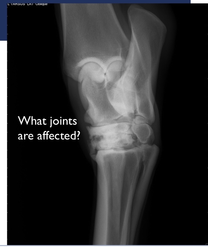
what joints are affected in this radiograph?
distal intertarsal and tarsometatarsal
tibiotarsal effusion (bog spavin)
caused by OCD, synovitis, OA (rare), blood (hemoarthrosis), infection (septic arthritis)
diagnosed by effusion in TT joint, radiographs, ultrasound, diagnostic arthroscopy
treatment → drain joint and inject corticosteroids, arthroscopy
prognosis is good
hock OC location from most to least common
distal intermediate ridge of the tibia (DIRT)
trochlear ridge (lateral > medial)
medial malleolus
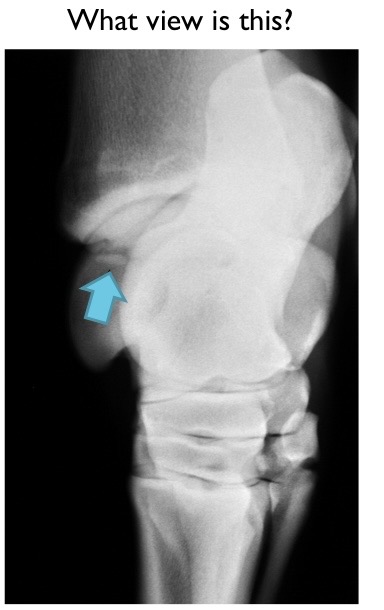
what view is this?
DLPMO
plantar ligament desmitis (curb)
caused by strain on plantar aspect of hock, sickle hocked horse
diagnosed by characteristic swelling, mild to moderate lameness, blocks, ultrasound
treatment → usually just need rest
prognosis is good, will always have a curb swelling
what breed is overrepresented for plantar ligament desmitis?
standardbreds
what is this?
plantar ligament desmitis
calcaneal bursitis (capped hock)
caused by repeated trauma → stall kicker
diagnosed by characteristic swelling, generally not lame
treatment → drain and inject corticosteroids
prognosis is excellent for function, poor for cosmesis
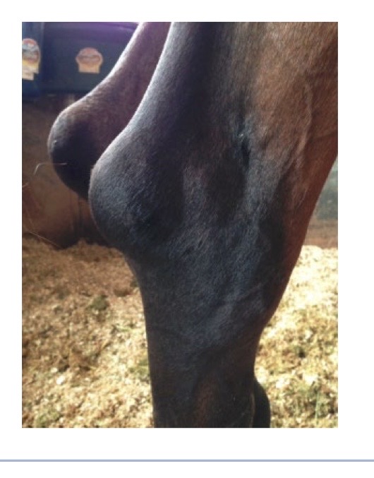
what is this?
calcaneal bursitis
septic calcaneal bursitis
caused by wound over calcaneus (common area to be kicked)
diagnosed by being very lame, pressurize bursa, contrast radiographs, ultrasound
treatment → flush bursa, regional limb perfusion, may require opening bursa and resecting part of calcaneus
prognosis is guarded (50/50)
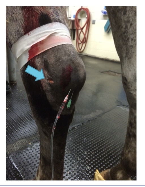
what is this?
septic calcaneal bursitis
tarsal sheath tenosynovitis (thoroughpin)
caused by wear and tear, straight hocks
diagnosed by characteristic swelling, usually not lame
treatment → usually none
prognosis is excellent for athletic use, guarded for cosmesis
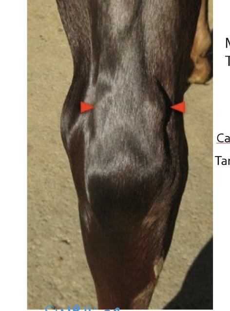
what is this?
tarsal sheath tenosynovitis
ruptured peroneus tertius
caused by trauma, full limb hind limb casts
diagnosed by ability to extend the hock with a flexed stifle, lameness and swelling, tarsus extends more than usual at the trot
treatment → avulsions near stifle, stall rest
prognosis is good, poor if it involves stifle joint
superficial digital flexor or gastrocnemius muscle/tendon rupture
caused by trauma
diagnosed by clinical signs, ultrasound
treatment → long term splinting and rest
prognosis is fair to poor, good if it heals
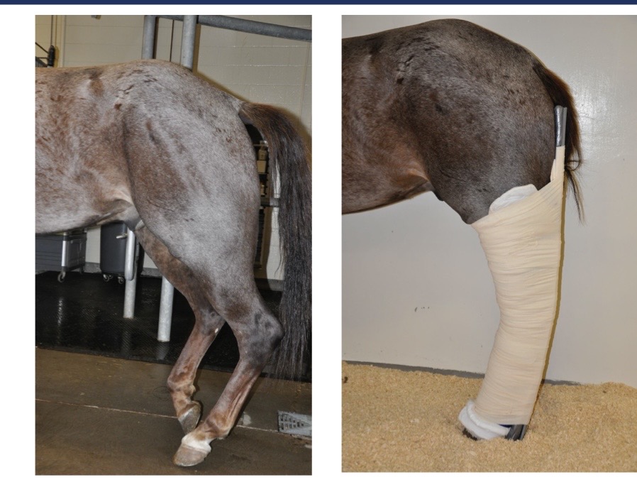
what is this?
superficial digital flexor or gastrocnemius muscle/tendon rupture
fibrotic myopathy
fibrosis ± ossification of the semitendinosus muscle
caused by trauma
diagnosed by characteristic movement (most evident at walk), palpable hardening in semitendinosus mm
treatment → semiten ± semimem tenotomy
prognosis is fair for success of treatment
gait characteristics of stifle lameness
jumps off lame leg in weight bearing phase → quicker, short step
drags toe because leaves leg out behind
increased lameness when limb is on outside of circle
stifle lameness
clinical signs → joint effusion (femoropatellar pouch easily palpable), hind end muscle atrophy with chronic problem
diagnosed by flexion test of upper limb and caudal extension, blocks, radiographs, ultrasound, arthroscopy, nuclear scintigraphy
stifle OC
most common breeds are WB, TB, QH
lesions → medial femoral cysts, trochlear ridge OCD
diagnosed by effusion, lameness when starts training, radiographs
treatments → surgery → wait until 1 yr
prognosis depends on severity and success of surgery, leads to OA
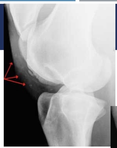
what is this?
medial trochlear ridge OCD on stifle
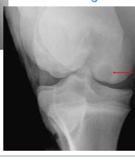
what is this?
stifle cyst lesion from stifle OC
stifle OA
caused by underlying OC, trauma, athletic use
diagnosed by blocks to the stifle
treatment → intra-articular corticosteroids
prognosis is fair - high in motion, complicated joint
upward fixation of patella
inability to release stay apparatus → patella does not disengage from the medial trochlea of femur
caused by weak quadriceps, straight legs
diagnosed by stifle being locked in extension (severe) or horse catches their stifle (mild)
treatment → immediate = back horse up, exercise strengthen quadriceps, surgery = medial patellar ligament splitting or blistering, medial patellar ligament desmotomy