BIOL B32 - Ch9 Physiology of Skeletal Muscle Fibers
1/74
Earn XP
Description and Tags
Muscular System
Name | Mastery | Learn | Test | Matching | Spaced | Call with Kai |
|---|
No analytics yet
Send a link to your students to track their progress
75 Terms
somatic motor neurons
the nerve cells that activate skeletal muscle fibers
somatic motor neurons reside where
in the brain or spinal cord
somatic is voluntary or involuntary?
voluntary
axons
long threadlike extensions of the somatic motor neuron
axons bundled with nerves do what
transmit action potentials to the muscle cells they serve to initiate contraction
action potentials
electrical impulses where it is a reversal on the membrane potential (the inside of the cell becomes positive)
when there is an electrical charge there is
potential
resting membrane potential
the electrical charge across a membrane when a cell is at rest
how much more negative is the intracellular environment of a resting membrane potential?
-70mV
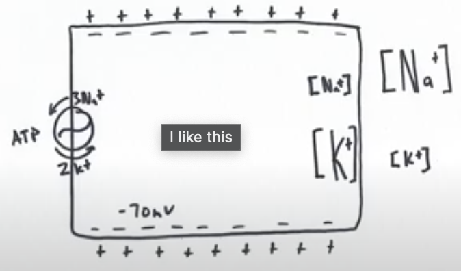
due to the selective permeability properties of the membrane the ions…
cannot freely cross and are therefore unable to reach equilibrium and active transport by the pump
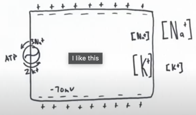
high or low concentration of sodium outside the cell?
high
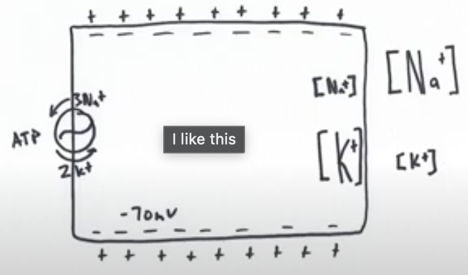
high or low concentration of potassium outside the cell?
low
ligand
a chemical messenger
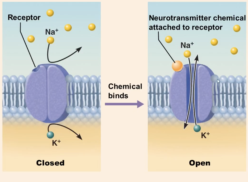
ligand-gated channels
open and close due to presence or absence of a ligand that binds to the receptor site
if a ligand binds..
the channel opens and ions move
if a ligand does not bind or is not present…
the channel closes and ions cannot move
where are ligand-gated channels found?
the dendrites and cell body of the motor neuron, and the neuromuscular junction
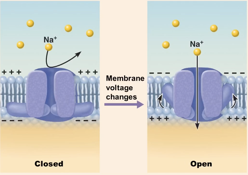
voltage gated channels
open and close in response to a change in voltage
if the membrane potential reaches a certain voltage or charge
the voltage-gated channel opens
if the membrane potential reaches a different voltage or charge
the voltage-gated channel closes
where are voltage gated channels found?
along axons of neurons and sarcolemma of muscle fiber and act much faster compared to ligand-gated channels
when a cell receives a signal, what happens to the ligand gated sodium channels?
the sodium channels open and sodium beings to leak into the cell down its concentration gradient
what is the signal the cell receives to open the ligand-gated sodium channel?
the neurotransmitter binds to its receptor
local depolarization
a step wise progression toward a more positive charge
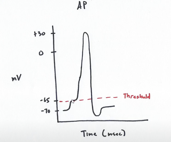
threshold
the minimum amount of depolarization required to initiate an action potential
all or none meaning for action potential
if a threshold is achieved an action potential is fired, but if it’s not then no action potential takes place and cell returns back to its resting membrane potential
what are the two phases of action potential?
depolarization and repolarization phase

-70mV
opening of ligand-gated sodium channels where sodium will leak in and produce a local depolarization
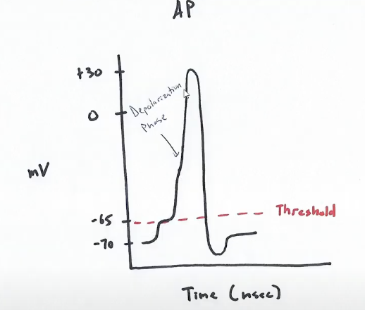
-65mV
threshold, the opening of voltage-gated sodium channels where massive amounts of sodium rush inside the cell which produces rapid depolarization
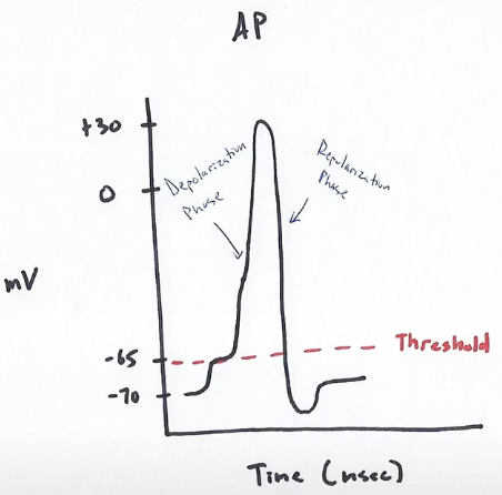
+30mV
closing of the voltage gated sodium channels and opening of the potassium channels where massive amounts of potassium rush out and produces rapid repolarization
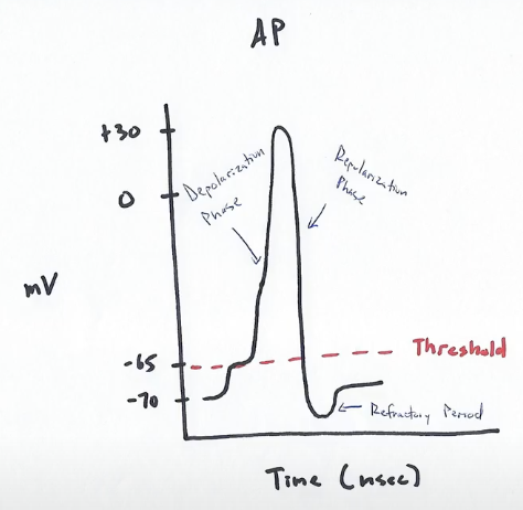
-70mV
voltage gated potassium channels close and sodium/potassium pumps reestablish the concentration gradient
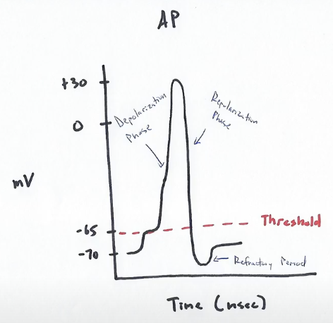
refractory period
a short period after an action potential has occurred where the cell cannot immediately fire another signal
propagation
the action potential propagates only in one direction, away from the origin of the nerve impulses, where the net result is nerve cells release neurotransmitters at their axon terminals or muscle cells contract
action potential frequency
repeated ligand release by the stimulating nerve will cause repeated action potentials that can increase the strength of a cell’s response
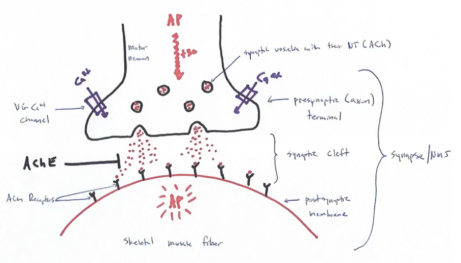
neuromuscular junction (synapse)
transfer site of a motor neuron action potential to skeletal muscle cell action potential
three major parts of a synapse
presynaptic terminal with synaptic vesicles, synaptic cleft, postsynaptic membrane
each muscle fiber is stimulated by a _____ of an axon
terminal branch
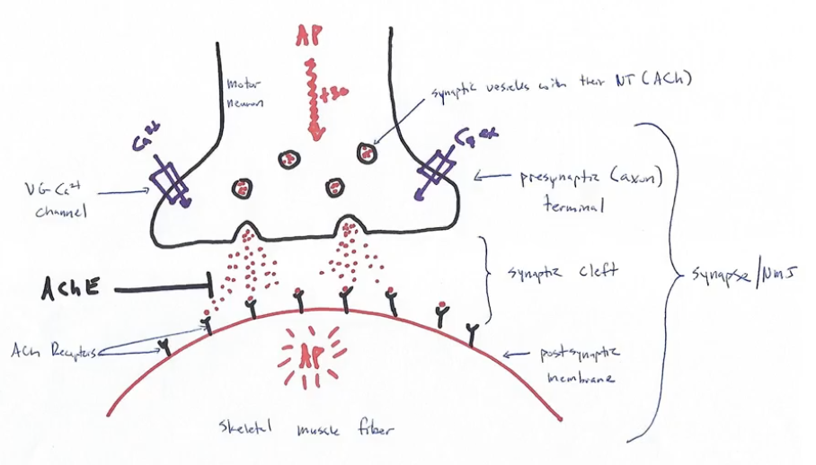
presynaptic (axon) terminal
contains the synaptic vesicles and acetylcholine
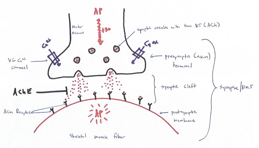
synaptic vesicles
membrane bound sacs that contain neurotransmitters
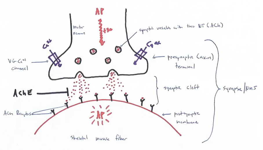
acetylcholine
the excitatory NT of the neuromuscular junction
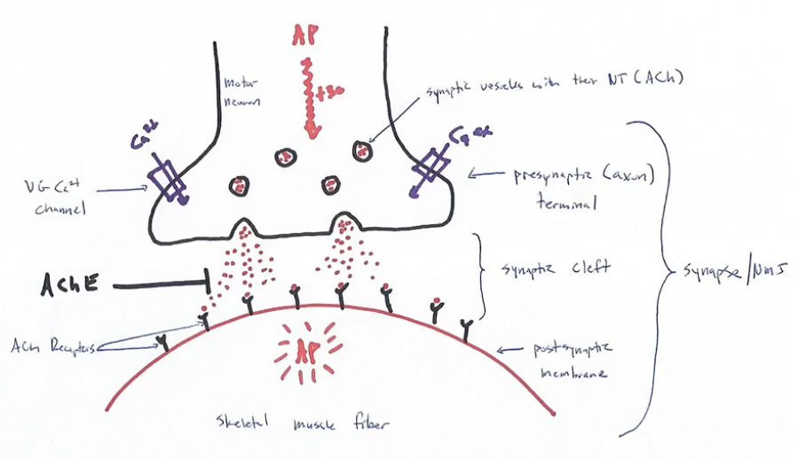
acetylcholine receptors
can only bind acetylcholine
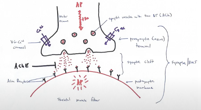
synaptic cleft
microscopic space that separates one neuron from the next cell in line
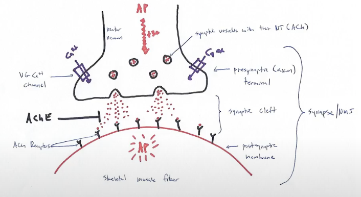
postsynaptic membrane
the motor end plate of the synapse
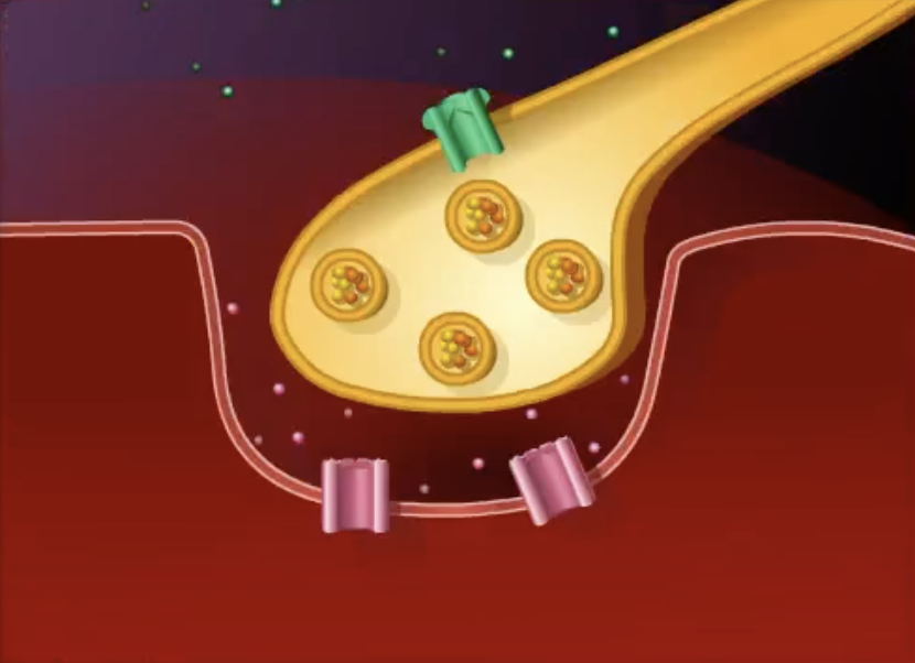
beginning of NMJ…
action potential propagates down the axon of the motor neuron and arrives at the presynaptic terminal
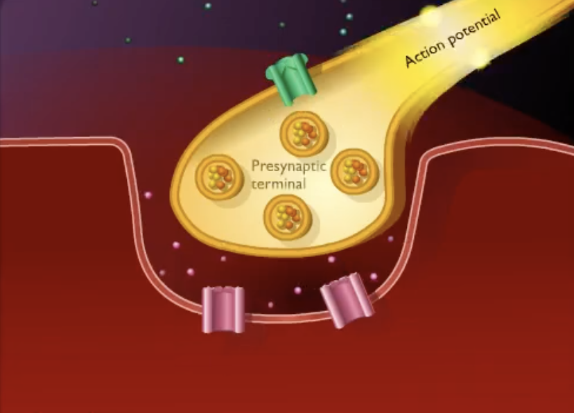
after the action potential arrives at the presynaptic terminal…
depolarization causes voltage gated calcium channels to open and calcium diffuses inward
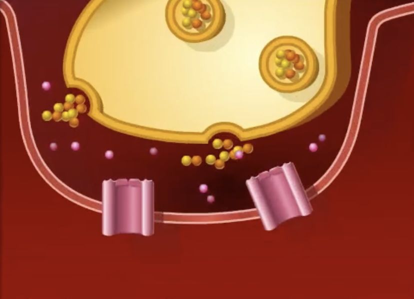
once calcium diffuses inward…
the synaptic vesicles fuse with the axon membrane and release their neurotransmitter (ACh) into the synaptic cleft by exocytosis
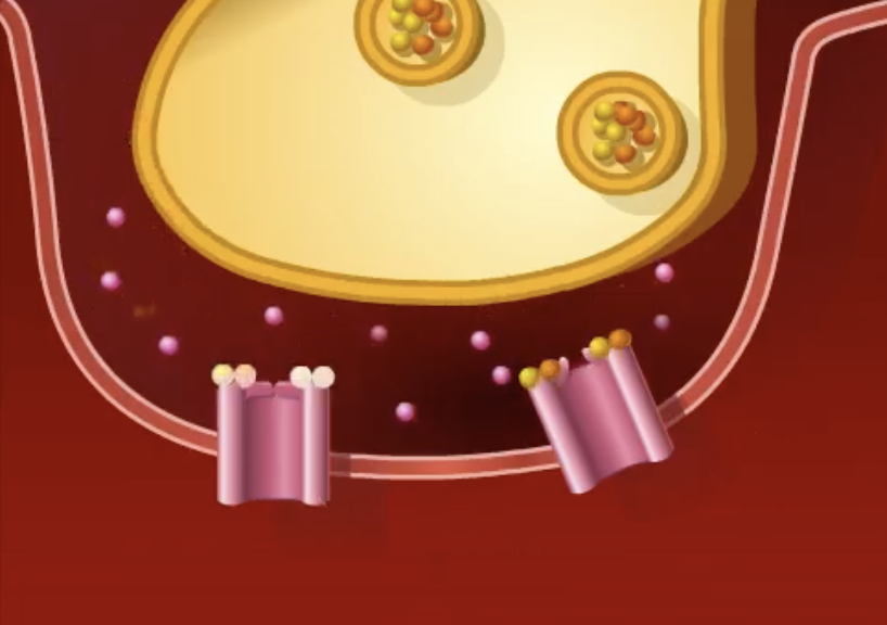
after exocytosis of the neurotransmitter…
ACh diffuses across the cleft and binds onto its receptor (a ligand gated sodium channel) on the postsynaptic cell
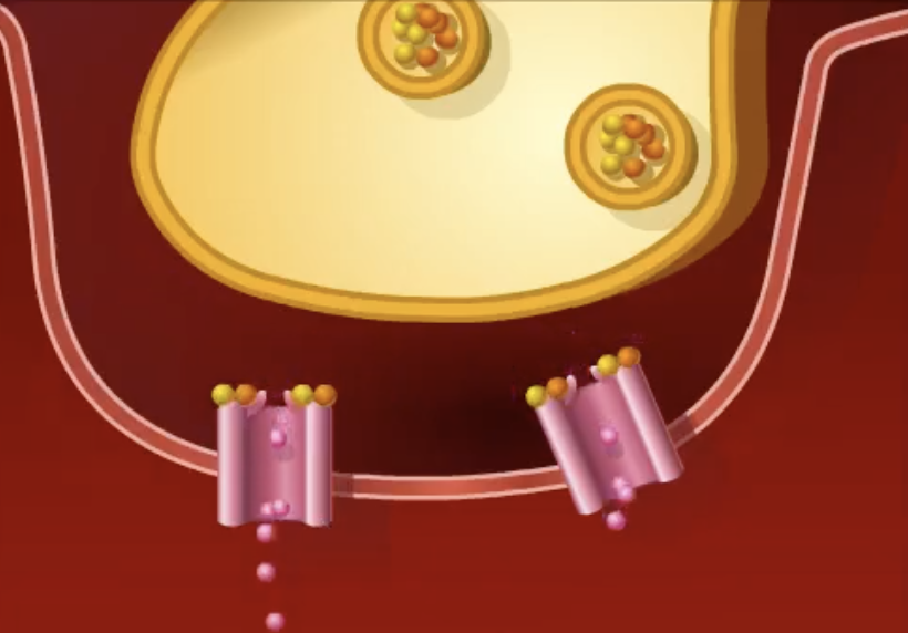
after ACh binds onto it’s receptor…
ligand gated sodium channels on the sarcolemma of the muscle fiber open and sodium diffuses inward causing a local depolarization
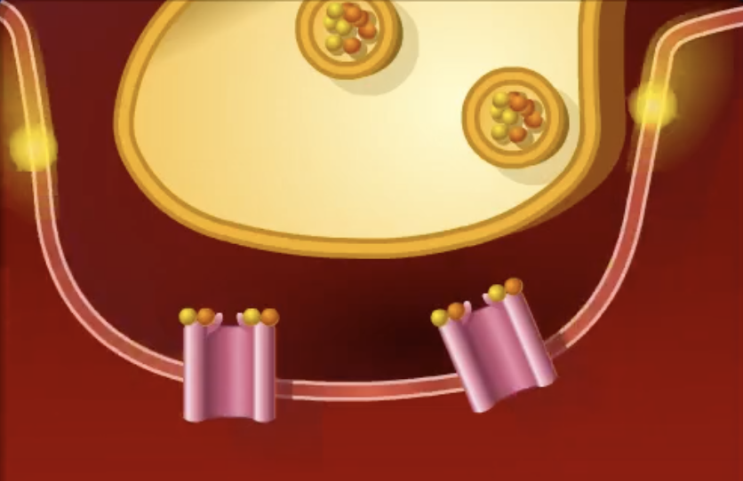
if enough local depolarizations are achieved to reach threshold…
an action potential is initiated in the skeletal muscle fiber
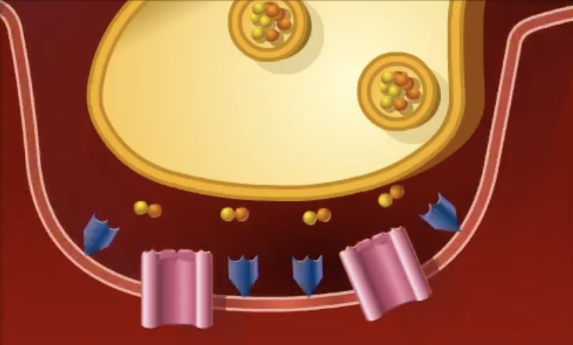
once an action potential is initiated…
neurotransmitter effects are then terminated after ACh is broken down to acetic acid and chlorine in the synaptic cleft
spastic paralysis
continued stimulation of muscle fiber and eventual muscular fatigue, even death, caused by limited AChE activity
flaccid paralysis
toxins that target ACh receptors and doesn’t allow ACh to bind so no muscle fiber stimulation
myasthenia gravis
involves a shortage of ACh receptors that are thought to be caused by an autoimmune disorder
symptoms of myasthenia gravis
drooping upper eyelids, difficulty swallowing and talking, and generalized muscle weakness
tetanus toxin
targets regulatory neurons that control release of ACh from presynaptic motor neurons and floods cleft with ACh and results in continued stimulation of muscle fiber and eventual muscular fatigue
botulism toxin
blocks release of ACh from presynaptic cell and results in flaccid paralysis
excitation-contraction coupling
sequence of events by which transmission of action potential along sarcolemma leads to sliding of actin and myosin myofilaments
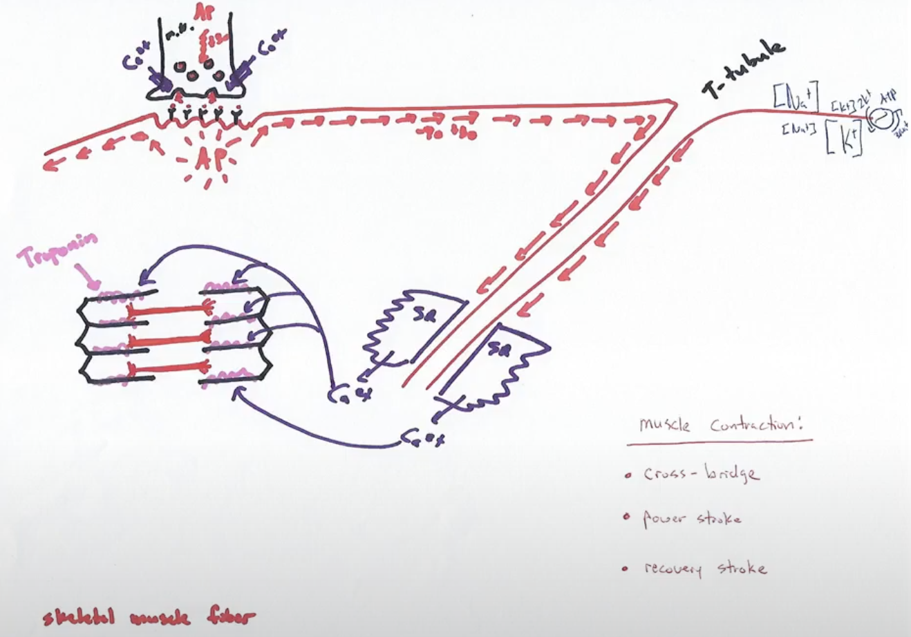
beginning of excitation-contraction coupling sequence
action potential that was initiated at the NMJ propagates along the sarcolemma until it reaches the T-tubules
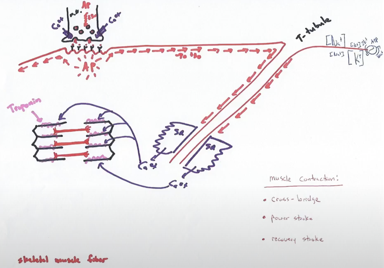
once the action potential reaches the T-tubules…
T-tubule invagination takes the sarcolemma depolarization into the sarcoplasmic reticulum
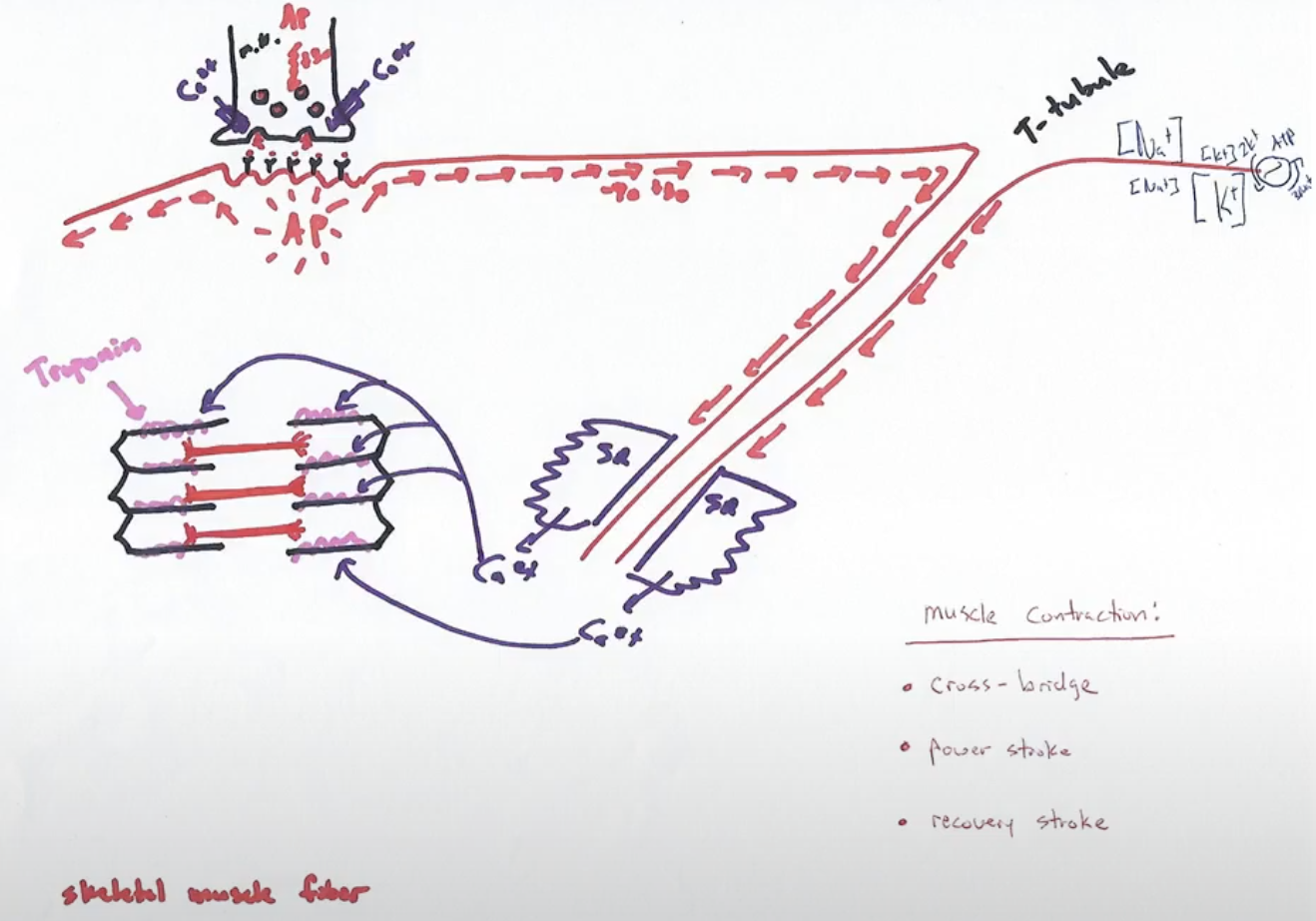
once at the sarcoplasmic reticulum…
depolarization in the T tubules causes voltage gated calcium channels to open and massive amounts of calcium diffuse into the sarcoplasm
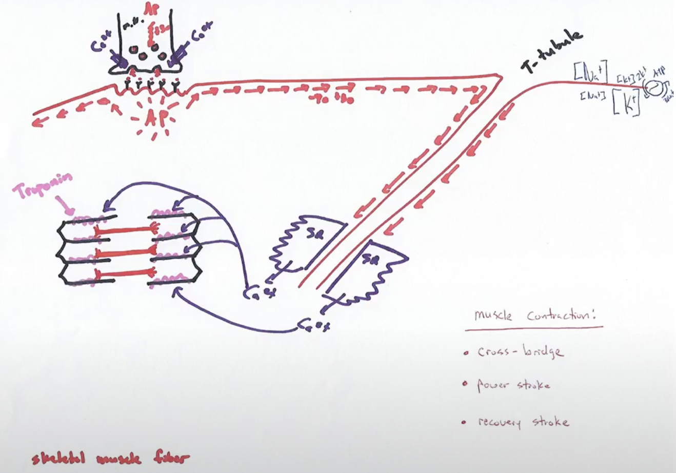
while calcium is in the sarcoplasm…
calcium binds to the regulatory protein troponin, changing its conformation and exposing the myosin binding site on actin for the first time
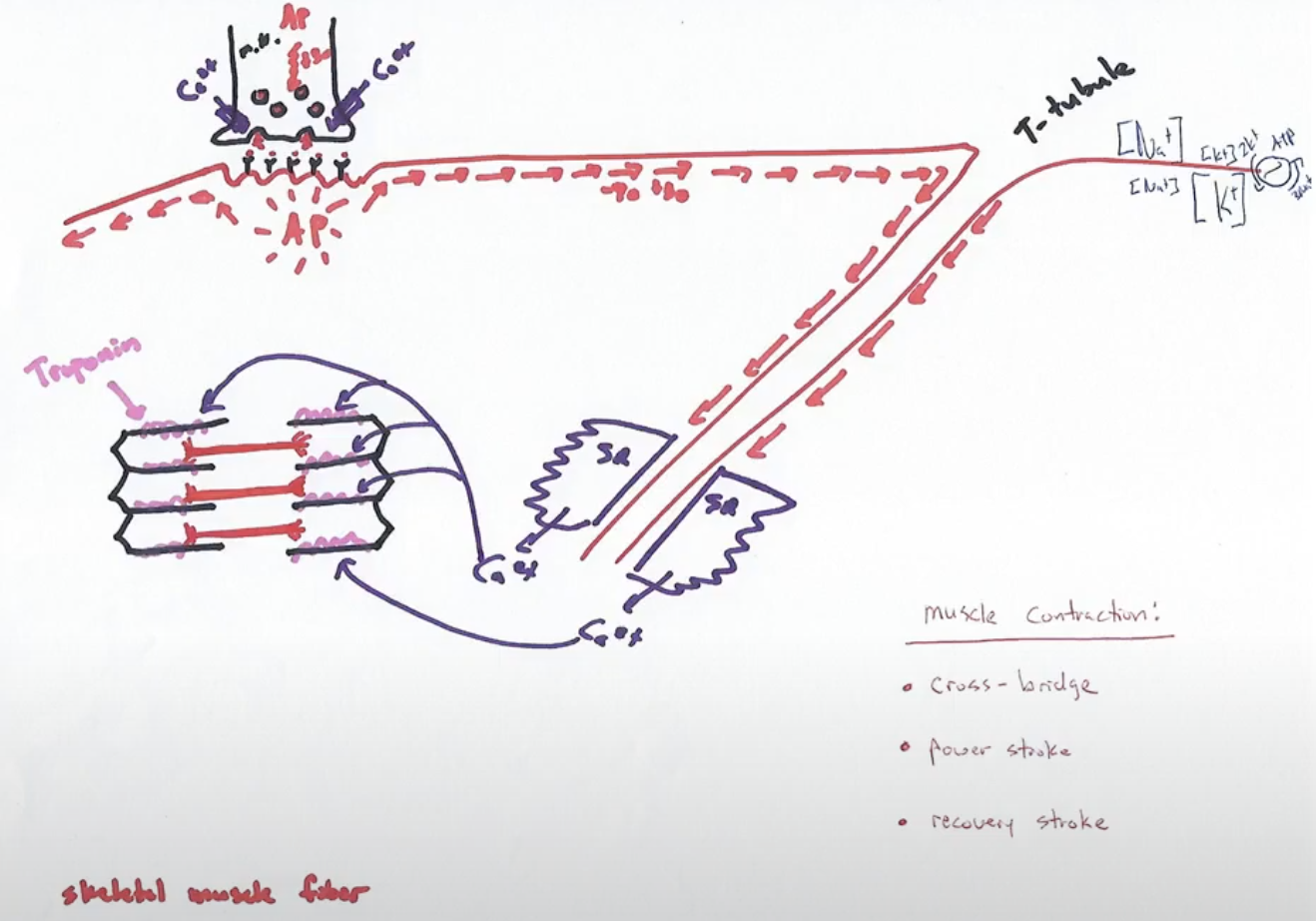
after the myosin binding site is exposed…
myosin binds onto actin through cross bridge and contraction begins
muscle fiber contraction
cross bridge activity
what are the three steps of muscle fiber contraction?
cross bridge, power stroke, and recovery stroke
cross bridge
myosin head binds actin and actin and myosin are now physically linked
power stroke
myosin head pivots and bends, pulling the actin over the top of itself and the bare zone narrows
recovery stroke
cocking of the myosin head where it is dislodged from the actin and returns to its prestroke high-energy position
if calcium is still available because of continued stimulation during recovery stroke, then
the myosin head reengages
after many quick repeats of muscle fiber contraction, what happens to the myosin heads and the sarcomere?
the myosin heads walk along the adjacent actin filaments and the sarcomere shortens and contraction is achieved in each fiber that has been stimulated by the neuron
when does relaxation of a muscle fiber occur?
when no more active potential (stimulus) is present
beginning of relaxation of muscle fiber
calcium ions are rapidly pumped back into the sarcoplasmic reticulum by active transport requiring ATP
ATP is used to do what to myosin during relaxation
it dislodges the myosin from the actin and recocks the myosin heads
what happens when free calcium is no longer present during relaxation?
troponin returns back to its original conformation and blocks the myosin binding sites on actin
what is the final step of relaxation of the muscle fiber?
sarcomeres passively lengthen
rigor mortis
actin and myosin in dying muscle cells become irreversibly cross linked after calcium and ATP synthesis ceases, so they gradually disappear as muscle proteins break down