A & P Slide 4 (Bones and Skeletal Tissue Ch 6)
 Call Kai
 Learn
Learn Practice Test
 Spaced Repetition
 Match
Match Flashcards
 Knowt Play
1/189

There's no tags or description
Looks like no tags are added yet.
Name | Mastery | Learn | Test | Matching | Spaced | Call with Kai | Chat |
|---|
No analytics yet
Send a link to your students to track their progress
190 Terms
The human skeleton initially consists of what?
just cartilage which is replaced by bone, except in areas requiring flexibility
What is skeletal cartilage?
Highly resilient cartilage tissue with HIGH content of WATER. NO BLOOD vessels or nerves.
layer of dense connective tissue surrounding cartilage like a girdle
PREVENTS outward expansion
Contains blood vessels for nutrient delivery to cartilage
perichondrium
Cartilage is made up of ________, cells encased in small cavities (______) within jelly-like _______ _____
Cartilage is made up of chondrocytes, cells encased in small cavities (lacunae) within jelly-like extracellular matrix
What are the 3 types of cartilage?
Hyaline cartilage
Elastic cartilage
Fibrocartilage
Where is hyaline cartilage found, what fiber type is in it, and what is its function?
▪Provides support, flexibility, and resilience
▪MOST ABUNDANT type; contains only collagen fibers
▪Articular (joints), costal (ribs), respiratory (larynx), nasal cartilage (nose tip)
Where is elastic cartilage found, what fiber is in it, and what is its function?
▪Similar to hyaline cartilage, but contains elastic fibers
▪External ear and epiglottis
Where is fiber cartilage found, what fiber is in it, and what is its function?
▪THICK collagen fibers: has great tensile strength
▪Menisci of knee; vertebral discs
(aids in compressive shock)
What are the 2 ways that cartilage grows in?
Appositional growth
Interstitial growth
Cartilage-forming cells in perichondrium secrete matrix against external face of existing cartilage
–New matrix laid down on surface of cartilage
appositional growth
Chondrocytes within lacunae divide and secrete new matrix, expanding cartilage from within
–New matrix made within cartilage
interstitial growth
__________ of cartilage occurs during normal bone growth in youth, but can also occur in old age. LOOK AT THE LARYNXES OF OUR CADAVERS!
Calcification of cartilage occurs during normal bone growth in youth, but can also occur in old age. LOOK AT THE LARYNXES OF OUR CADAVERS!
Is hardened cartilage the same as bone?
NO
What are the 7 important functions of bones?
1. Support
▪For body and soft organs
2. Protection
▪Protect brain, spinal cord, and vital organs
3. Movement
▪Levers for muscle action
4. Mineral and growth factor storage
▪Calcium and phosphorus, and growth factors reservoir
5.Blood cell formation
▪Hematopoiesis occurs in red marrow cavities of certain bones
6. Fat storage
▪Fat, used for an energy source, is stored in bone cavities
7. Hormone production
▪Osteocalcin secreted by bones helps to regulate insulin secretion, glucose levels, and metabolism. Does NOT affect the bone directly!!
–higher levels found in the blood when there is active bone deposition!
What are the 3 mechanical functions?
1. Support
▪For body and soft organs
2. Protection
▪Protect brain, spinal cord, and vital organs
3. Movement
Levers for muscle action
What are the warehouse functions?
1. Mineral and growth factor storage
▪Calcium and phosphorus, and growth factors reservoir
2.Blood cell formation
▪Hematopoiesis occurs in red marrow cavities of certain bones
3. Fat storage
Fat, used for an energy source, is stored in bone cavities
(Maybe hormone production ?)
During blood cell formation ______occurs in red marrow cavities of certain bones
Hematopoiesis
During hormone production ________ secreted by bones helps to regulate insulin secretion, glucose levels, and metabolism. Does NOT affect the bone directly!!
Osteocalcin
There are how many named bones in the human skeleton?
206
What are the two groups bones are divided into based on location?
Axial skeleton
Appendicular skeleton
What bones are found in the axial skeleton?
▪Long axis of body
▪Skull, vertebral column, rib cage
What bones are found in the appendicular skeleton?
▪Bones of upper and lower limbs
▪Girdles attaching limbs to axial skeleton
What are the 4 shapes that bones are also classified into? What do each look like? What are some examples in the body?
1. Long bones
▪Longer than they are wide
▪Limb bones
2. Short bones
▪Cube-shaped bones (in wrist and ankle)
▪Sesamoid bones form within tendons (example: patella). Vary in size and location between individuals!
3.Flat bones
▪Thin, flat, slightly curved
▪Sternum, scapulae, ribs, most skull bones
4. Irregular bones
▪Complicated shapes
Vertebrae and hip bones
•Bones are _____ because they contain _____ ___ __ ______
•Bones are organs because they contain different types of tissues
What tissue predominates in bone? What other types of tissue are found in bone?
Bone (osseous) tissue predominates,
but a bone also has:
▪nervous tissue,
▪cartilage,
▪fibrous connective tissue,
▪blood vessels with muscle cells and epithelial cells
What are the 3 levels of structure?
Gross
Microscopic
Chemical
What are two types of bone densities?
Compact bone
Spongy bone
What is compact bone?
dense outer layer on every bone that appears smooth and solid
What is spongy bone made up of?
a honeycomb of small, needle-like or flat pieces of bone called trabeculae
Open spaces between trabeculae are filled with ____ or ________ ____ ______
Open spaces between trabeculae are filled with red or yellow bone marrow
What is the structure of short, irregular, and flat bones?
–Consist of thin plates of spongy bone (diploe) covered by compact bone. Compact bone sandwiched between connective tissue membranes
What covers the outside of compact bone?
periosteum
What covers the inside portion of compact bone?
endosteum
What is scattered throughout spongy bone?
Bone marrow, there is no defined bone marrow cavity.
this covers area of bone that is part of a movable joint.
hyaline cartilage
What is the structure of a typical long bone?
–All long bones have a shaft (diaphysis), bone ends (epiphyses), and membranes
what is the tubular shaft that forms long axis of bone?
diaphysis
What does the diaphysis consist of?
compact bone surrounding central medullary cavity that is filled with yellow marrow in adults
these are ends of long bones that consist of compact bone externally and spongy bone internally.
–Articular cartilage covers articular (joint) surfaces
epiphyses
Between the diaphysis and epiphysis is the ____ ___
epiphyseal line
The epiphyseal line is Remnant of childhood _______ _____ where bone growth occurs.
epiphyseal plate
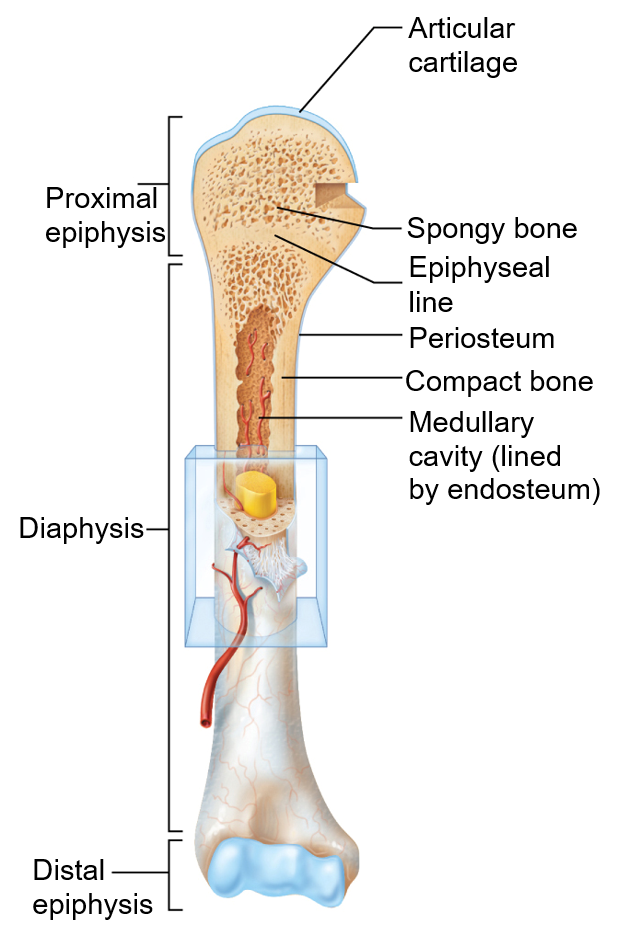
long bone
What are the two types of membranes?
Periosteum and endosteum
white, double-layered membrane that covers external surfaces except joints
periosteum
What are the two layers of the periosteum? What is found in each of these layers?
•Fibrous layer: outer layer consisting of dense IRREGULAR connective tissue and perforating fibers (Sharpey’s fibers) that secure the periosteum to the underlying bone matrix
•Osteogenic layer: inner layer abutting bone with osteogenic stem cells that give rise to most bone cells
Contains many nerve fibers and blood vessels that continue on to the shaft.
Tendons and ligaments attach here
Periosteum
Connective tissue membrane covering internal bone surface and trabeculae of spongy bone
endosteum
What cells are found in the endosteum and what does it line?
•Like periosteum, contains osteogenic cells that can differentiate into other bone cells
•Lines canals that pass through compact bone
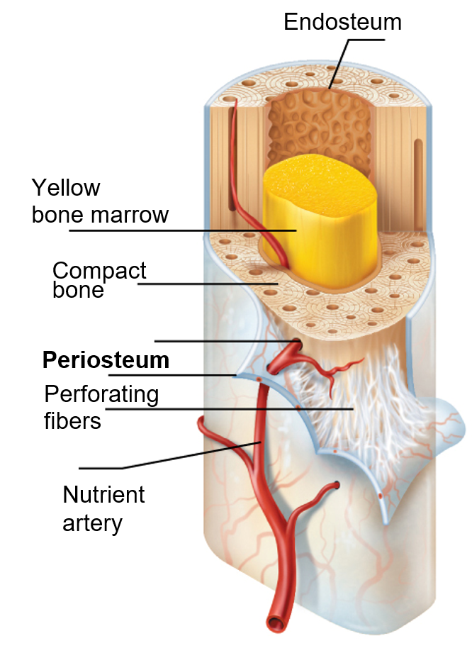
What are hematopoietic tissues in bone?
Red marrow
Yellow marrow
Where is red marrow found in newborns and in adults?
within trabecular cavities of spongy bone and diploë of flat bones, such as sternum
▪In newborns, medullary cavities and all spongy bone contain red marrow
▪In adults, red marrow is located in heads of femur and humerus, but most active areas of hematopoiesis are flat bone diploë and some irregular bones (such as the hip bone, vertebral bodies)
What can yellow marrow convert to if person becomes anemic?
red bone marrow
What tissue is found in yellow marrow?
adipose tissue
Sites of muscle, ligament, and tendon attachment on external surfaces
Areas involved in joint formation or conduits for blood vessels and nerves
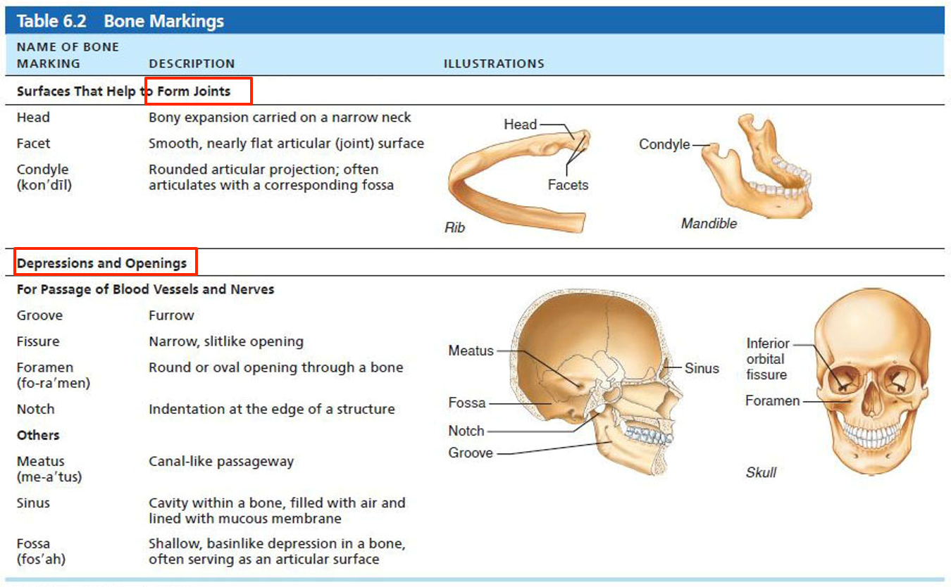
Bone markings
What are the three types of bone markings?
▪Projection: outward bulge of bone
–May be due to increased stress from muscle pull or is a modification for joints
▪Depression: bowl- or groove-like cut-out that can serve as passageways for vessels and nerves, or plays a role in joints
▪Opening: hole or canal in bone that serves as passageways for blood vessels and nerves
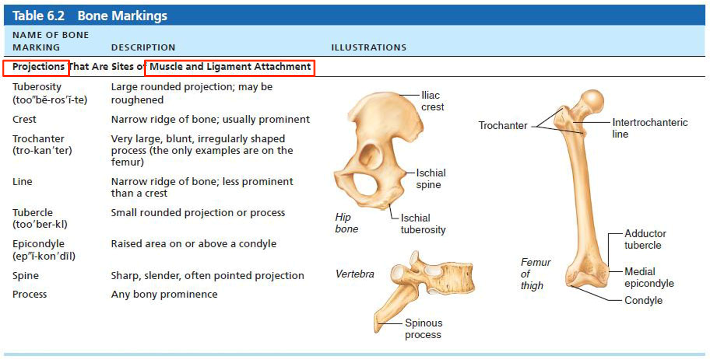
Define each of these bone markings
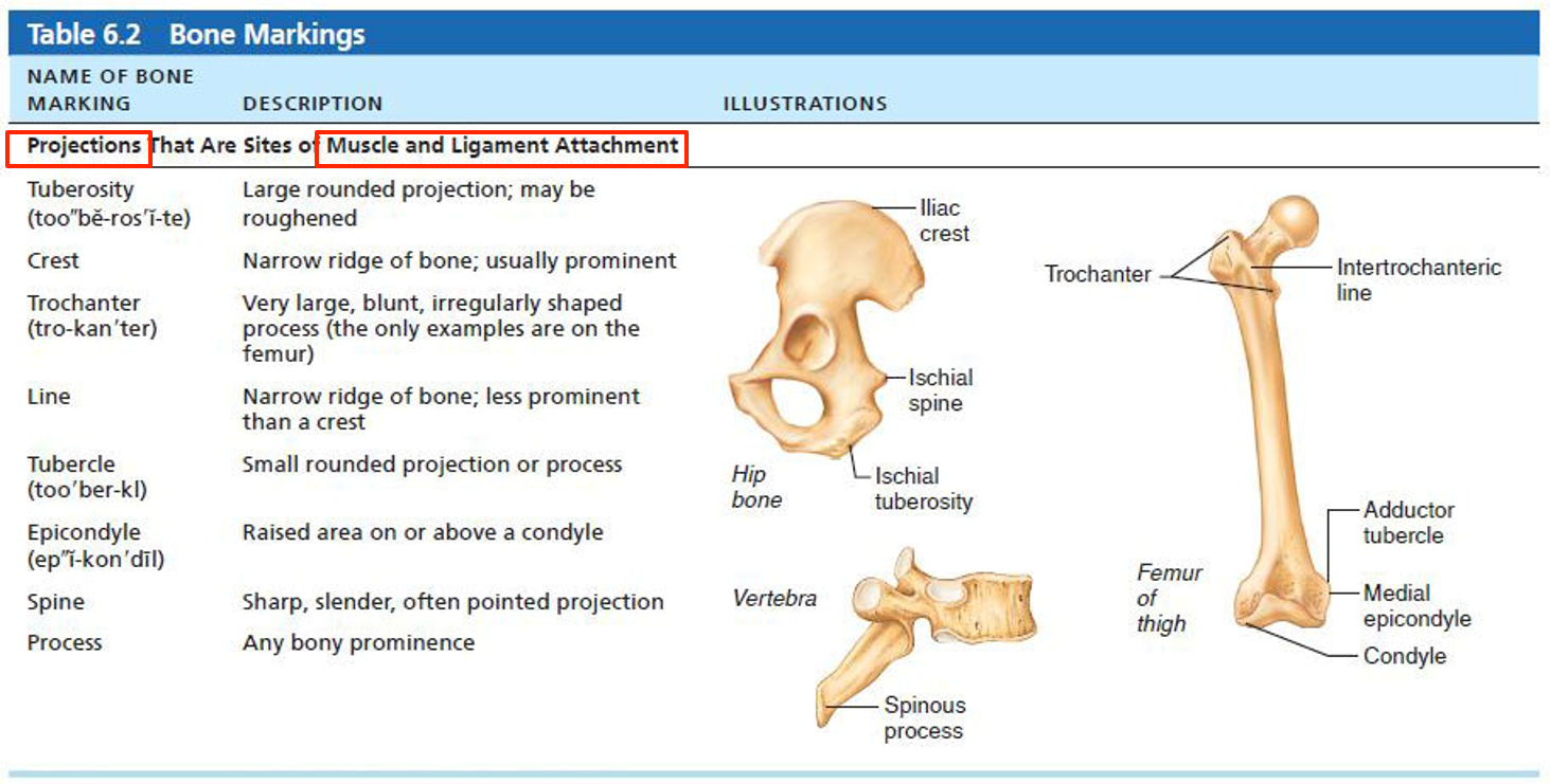
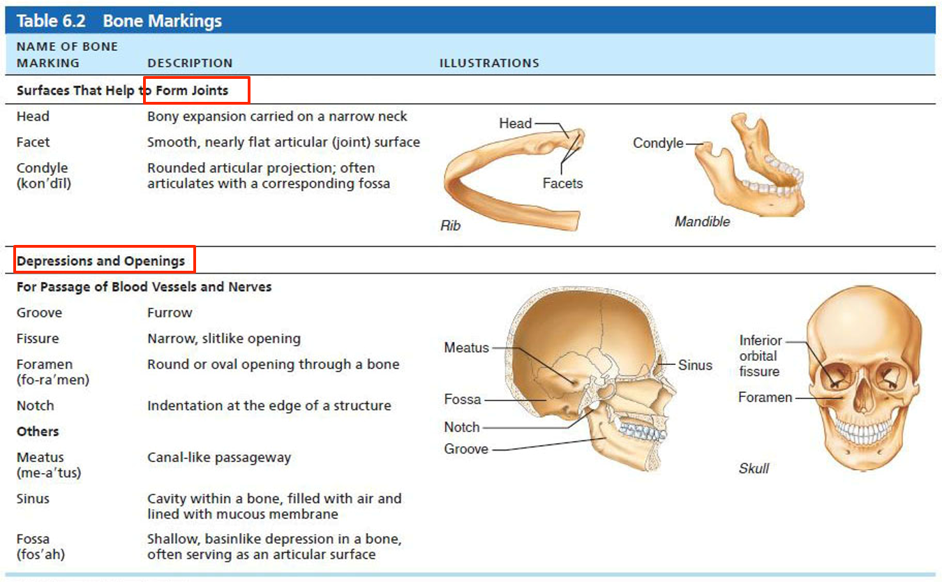
Define each of these bone markings:
What are five major cells of bone tissue?
–Five major cell types, each of which is a specialized form of the same basic cell type
1. Osteogenic cells
2. Osteoblasts
3. Osteocytes
4. Bone-lining cells
Osteoclasts
What are osteogenic cells? What is another name for them? What happens when the are stimulated?
–Also called osteoprogenitor cells
–Mitotically active (divide) STEM CELLS in periosteum and endosteum
–When stimulated, they differentiate into osteoblasts or bone-lining cells
–Some remain as osteogenic stem cells
What are osteoblasts? What do the do? What are they made up of?
–Bone-forming cells that secrete unmineralized bone matrix called osteoid
▪Osteoid is made up of collagen and calcium-binding proteins
▪Collagen makes up 90% of bone protein
–Osteoblasts are actively mitotic
What are osteocytes? What is there role? What type of sensors are they called?
Mature bone cells in lacunae that no longer divide
–Maintain bone matrix and act as stress or strain sensors:
▪Respond to mechanical stimuli such as increased force on bone or weightlessness
▪Communicate information to osteoblasts and osteoclasts to remodel the bone
What are bone lining cells? What is their role? What are they called on external vs. internal surfaces?
Flat cells on bone surfaces, help maintain matrix (along with osteocytes)
–On external bone surface, lining cells are called periosteal cells
–On internal surfaces, they are called endosteal cells
What are osteoclasts? What do they act as?
3.Derived from hematopoietic stem cells that also become macrophages
–Giant, multinucleate cells function in bone resorption (breakdown of bone)
–Cells have ruffled membrane to increase surface area for enzyme degradation of bone
What is compact bone also called? What does compact bone consist of?
–Also called lamellar bone
–Consists of:
▪Osteon (Haversian system)
▪Canals and canaliculi
▪Interstitial and circumferential lamellae
What is the structural unit of compact bone?
Osteon
Elongated cylinder parallel to the long axis of bone
▪Acts as tiny weight-bearing pillars
Osteon
The osteon is made up of several rings of bone matric called ____
Lamellae
▪Lamellae contain ________ ______ that run in different directions in adjacent rings
▪Withstands stress and resist ________ (_______)
▪Bone salts are found between collagen fibers
▪Lamellae contain collagen fibers that run in different directions in adjacent rings
▪Withstands stress and resist twisting (torsion)
▪Bone salts are found between collagen fibers
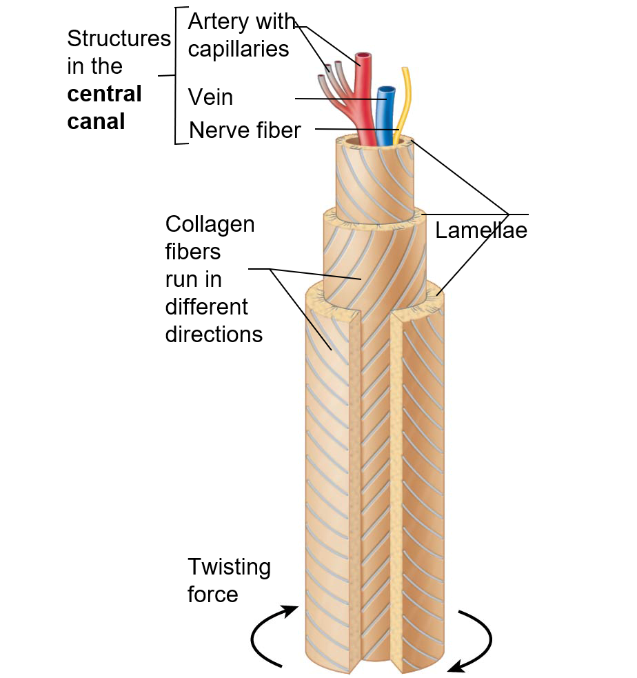
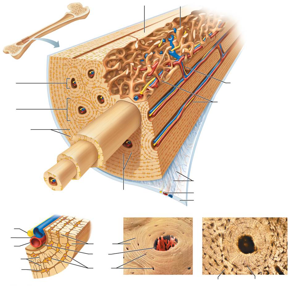
Fill out and label:

this runs –through core of osteon
▪Contains blood vessels and nerve fibers
Central (Haversian canal)
canals lined with endosteum that occur at right angles to central canal
▪Connect blood vessels and nerves of periosteum, medullary cavity, and central canal
Perforating (Volkmann’s) canals
What are lacunae?
small cavities that contain osteocytes
hairlike canals that connect lacunae to each other and to central canal
▪Allow communication between all osteocytes of osteon (nutrients and waste transport)
canaliculi
secrete bone matrix maintain contact with each other and osteocytes via cell projections with gap junctions
osteoblasts
What are the 2 types of lamellae?
Interstitial lamellae
Circumfertial lamellae
▪Lamellae that are not part of osteon
▪Some fill gaps between forming osteons; others are remnants of osteons cut by bone remodeling
Interstitial lamellae
▪Just deep to periosteum, but superficial to endosteum, these layers of lamellae extend around entire surface of diaphysis
▪Help long bone to resist twisting
Circumferential lamellae
Which type of lamellae is NOT part of the osteon?
interstial lamellae
How is spongy bone organized?
–Appears poorly organized but is actually organized along lines of stress to help bone resist any stress
______are like cables on a suspension bridge, confer strength to bone, in spongy bone.
Trabeculae
▪__ ______ are present, but trabeculae do contain irregularly arranged ______ and _______ interconnected by _______
No osteons are present, but trabeculae do contain irregularly arranged lamellae and osteocytes interconnected by canaliculi
What supplies nutrients to trabeculae?
Capillaries in endosteum supply nutrients
What are the 2 components that bone is made up of?
organic
inorganic
What cells do the organic components of bone contain?
osteogenic cells, osteoblasts, osteocytes, bone-lining cells, osteoclasts, and osteoid
is 1/3 of organic bone matrix, is secreted by osteoblasts (organic component)
Osteoid
what does the osteoid consist of? (organic component)
ground substance and collagen fibers, which contribute to high tensile strength and flexibility of bone
What is resilience of bone due to? (organic component)
•to sacrificial bonds in or between collagen molecules that stretch and break to absorb shocks and prevent fractures
•Bonds re-form
What is an inorganic component of bone?
Hydroxyapatites (mineral salts)
What do hydroxyapatites consist of?
mainly calcium phosphate crystals in and around collagen fibers
these makeup 2/3 of bone mass, and are responsible for hardness and resistance to compression. They also last long after death because of mineral composition.
Hydroxyapatites
Bone is ______________ in resisting compression and ____________ in resisting tension
–Bone is half as strong as steel in resisting compression and as strong as steel in resisting tension
what is the process of bone tissue formation?
ossification (osteogenesis)
Formation of bony skeleton begins in
month 2 of development
Postnatal bone growth occurs until ____
early adulthood
Bone remodeling and repair occur until _____
the whole lifespan
What happens at week 8 of development?
fibrous membranes and hyaline cartilage of fetal skeleton are gradually replaced with bone tissue
What is:
–Bone forms by replacing hyaline cartilage
–Bones are called _______
Form most of skeleton
endochondral ossification
cartilage (endochondral) bones
what is:
–Bone develops from fibrous membrane
–Bones are called _________
Intramembranous ossification
membrane bones
what are the 5 main steps in the process of ossification?
–Bone collar forms around diaphysis of cartilage model
–Central cartilage in diaphysis calcifies, then develops cavities
–Periosteal bud invades cavities, leading to formation of spongy bone
–Diaphysis elongates, and medullary cavity forms
▪Secondary ossification centers appear in epiphyses
–Epiphyses ossify
▪Hyaline cartilage remains only in epiphyseal plates and articular cartilages