BIO 50 LABMY 11TH AND 13TH R
1/47
Earn XP
Description and Tags
TEST PAPER IDENTIFICATION
Name | Mastery | Learn | Test | Matching | Spaced |
|---|
No study sessions yet.
48 Terms

PERICARDIUM
(7) a covering of the heart made up of three layers (an outer fibrous layer and inner serous membrane pair (parietal and visceral serous layer)


FIBROUS PERICARDIUM
(7.1) a loosely fitting superficial part of the pericardium that protects the heart and anchors in to surrounding structurest


PARIETAL LAYER of SEROUS PERICARDIUM
(7.2) layer of the serous pericardium deep to fibrous pericardium, slippery two-layered, lines he interior of the fibrous pericardium

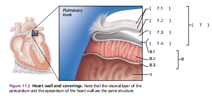
PERICARDIAL CAVITY
(7.3) the fluid-filled space between the parietal and visceral layers of the serous pericardium, which reduces friction as the heart beats.


VISCERAL LAYER of SEROUS PERICARDIUM or also EPICARDIUM
(7.4 and 8.1) the innermostlayer of the pericardium and the outermost layer of the heart wall, produces serous fluid that collects in the pericardial cavity

SEROUS FLUID
produced by the serous pericardial membranes, allows the heart to beat easily in a relatively frictionless environment
EPICARDIUM, MYOCARDIUM, ENDOCARDIUM
the three layers of the heart walls
MYOCARDIUM
(8.2) layer of the heart wall that consists thick bundles of cardiac muscle, responsible for the hearts contractions
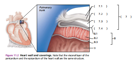
ENDOCARDIUM
(8.3) thin glistening sheet of endothelium that lines the heart chambers, continuous with the linings of the blood vessels leaving and entering the heart

HEART CHAMBER
(9) an internal cavity where the heart pumps blood and separates oxygenated and deoxygenated blood.
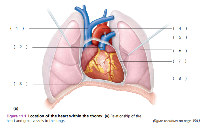
APEX
(8) directed toward the left hip and rests on the diapraghm at the level of the fifth intercostal space
BASE
POSTEROSUPERIOR aspect of the heart, from which the great vessels of the body emerge and points toward the right shoulder and lies beneath the second rib
FIFTH INTERCOSTAL SPACE
where one would place a stetoscope to count the heart rate for an APICAL pulse
INFERIOR MEDIASTINUM
where the heart is enclosed in the chest cavity, flanked on each side by the lungs
PERICARDITIS
infllamation of the pericardium, results in a decrease in the already small amount of serous fluid. causes friction betweeen the pericardial layers
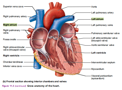
ATRIA
acts as the recieving chambers, they assist with filling the ventricles with blood that flows into them under low pressure from the veins of the body
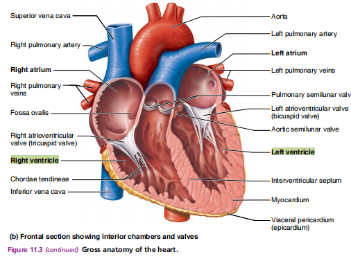
VENTRICLE
acts as the discharging chambers, they pump blood out of the heart and into circulation
INTERATRIAL SEPTUM
divides the heart into the left and right atria, preventing the mixing of oxygenated and deoxygenated blood.
INTERVENTRICULAR SEPTUM
divides the heart into the left and right ventricles, preventing the mixing of blood between them.
ARTERIES
carries blood away from the heart; thicker walls
VEINS
carries blood toward the heart; thinner walls
SUPERIOR and INFERIOR VENA CAVA
recieves oxygen-poor blood from the veins of the body
PULMONARY TRUNK
transports oxygen-poor blood to the lungs (splits into right and left arteries)
PULMONARY VEINS
returns oxygen-rich blood from the lungs to the heart.
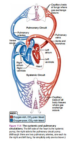
PULMONARY CIRCULATION
the circuit from the right ventricle (the pump) to the lungs and back to the left atrium (reccieving chamber); only function is to carry blood to the lungs for gas exchange (oxygen into blood - carbon dioxide inot lungs)
AORTA
the largest artery in the body that carries oxygen-rich blood from the left ventricle of the heart into smaller arteries that supply all body tissues
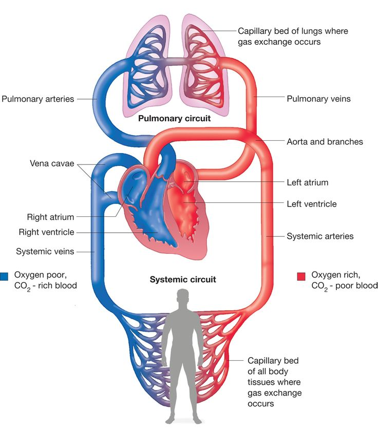
Right atrium ⟶ Tricuspid valve ⟶ Right ventricle ⟶ Pulmonary semilunar valve ⟶ Pulmonary trunk ⟶ Pulmonary arteries ⟶ Lungs (gas exchange) ⟶ Pulmonary veins ⟶ Left atrium.
the sequence of pulmonary circulation (RA - LA)
PULMONARY CIRCULATION
main role is to oxygenate the blood via the lungs, this circuit operates under low pressure and covers the shorter distance between the heart and lungs.
Left ventricle ⟶ Aortic Semilunar Valve ⟶ Aorta ⟶ Arteries ⟶ Arterioles ⟶ Capillaries ⟶ Venules ⟶ Veins ⟶ Venae cavae ⟶ Right atrium
the sequence of systemic circulation (LV - TISSUES - RA)
SYSTEMIC CIRCULATION
this circuit works under higher pressure to push blood over longer distances through arteries, capillaries, and veins throughout the entire body except the lungs.
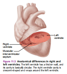
LEFT VENTRICLE
the more powerful pump between the two ventricles, has a thicker wall
ATRIOVENTRICULAR VALVES
valves located between the atria and ventricles on each side; prevents backflow into the atria when the ventricles contract
LEFT ATRIOVENTRICULAR VALVE, BICUSPID VALVE, or MITRAL VALVE
valve consisting of two cusps that allows blood to flow from the left atrium to the left ventricle, preventing backflow during ventricular contraction.
RIGHT ATRIOVENTRICULAR VALVE or TRICUSPID VALVE
valve consisting of three cusps that controls blood flow from the right atrium to the right ventricle.
CHORDAE TENDINAE
tendinous cords; achors the atrioventricular valves to the papillary muscles in the ventricle walls, and hangs limply when tthe heart is relaxed
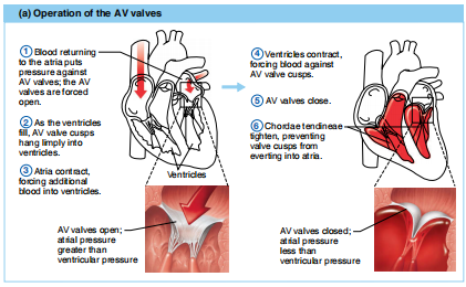
atrioventricular valves open when atrial pressure is greater than ventricular pressure, atrioventricular valves vlose when atrial pressure is less than ventricular pressure
operation of the atrioventricular valves
SEMILUNAR VALVES
valves that have three cusps that guard the bases of two large arteries leaving the ventricular chambers (PULMONARY and AORTIC)
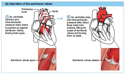
venttricle contraction and a rise in intraventricular pressure opens semilunar valves, ventricle relaxation and a fall in intraventricular pressure closes semilunar valves
operation of the semilunar valves
LEFT and RIGHT CORONARY ARTERIES
the functional blood supply to the heart muscle, branching from the aorta to provide oxygenated blood.
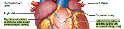
CORONARY SULCUS (ATRIOVENTRICULAR GROOVE)
encircles the heart and separates the atria from the ventricles
ANTERIOR IINTERVENTRICULAR ARTERY and CIRCUMFLEX ARTERY
coronary arteries of the left side of the heart
POSTERIOR INTERVENTRICULAR ARTERY and MARGINAL ARTERY
coronary arteries on the right side of the heart
CARDIAC VEINS
veins that drain deoxygenated blood from the heart muscle (myocardium) into anenlarged vessel on the posterior of the heart
CORONARY SINUS
enlarged vessel on the posterior of the heart that empties deoxygenated blood from cardiac veins into the right atrium.
ANGINA PECTORIS
a condition characterized by chest pain due to reduced blood flow to the heart muscle.
INFARCT
a localized area of tissue that dies due to lack of blood supply, often resulting from a blockage in the blood vessels.
MYOCARDIAL INFARCTION
heart attack or coronary