Anatomy 2 (Skeleton)
 Call Kai
 Learn
Learn Practice Test
 Spaced Repetition
 Match
Match Flashcards
 Knowt Play
1/90

There's no tags or description
Looks like no tags are added yet.
Name | Mastery | Learn | Test | Matching | Spaced | Call with Kai | Chat |
|---|
No analytics yet
Send a link to your students to track their progress
91 Terms
Axial Skeleton
Skull, vertebrae, sternum, ribs
Cranium
Frontal, parietal, temporal, occipital, sphenoid, ethnoid
Sutures
Divide pieces of the skull
Coronal
Sagittal
Lambdoid
Bregma
Where coronal and sagittal sutures meet
Lambda
Where sagittal and lambdoid sutures meet
Wormian Bones
Extra bones that form between sutures
Fontanel
Spaces due to incomplete suture lines in babies
Fossae
Interior depressions of the skull
Anterior
Middle
Posterior
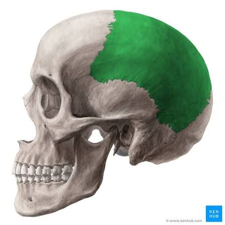
Parietal Bone
Side of the skull
Superior/Inferior Temporal Lines for muscle
Temporal Bone
Surrounds ear

Squamous Portion
Top, thin
Temporal
Petrous Portion
Bottom, thick
Temporal
Squamous Suture
Suture around temporal bone
Zygomatic Arch
Cheekbone
Temporal

Mastoid Process
Behind ear, connects to muscle
Pneumatized
Temporal

External and Internal Acoustic Meatuses
Hole for hearing and nerves
Temporal

Styloid Process
Pen shape
Temporal

Mandibular Fossa
Joint for the mandible
Temporal
Carotid Canal
Canal for carotid artery; in front on the jugular foramen
Temporal

Occipital Bone
Back of the skull
Foramen Magnum
Big hole
Occipital
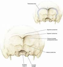
External Occipital Protuberance
Bump for muscle attachment
Occipital

Occipital Condyle
Bumps that connect to C1
Occipital
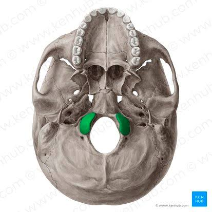
Hypoglossal Canal
Canal under occipital condyle for tongue nerve
Occipital

Inferior and Superior Nuchal Lines
Nuchal ligament attachment
Occipital
Jugular Foramen
Next to the foramen magnum for nerves and vessels
Occipital

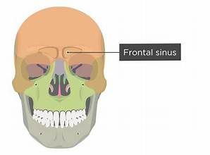
Frontal Sinus
Air pockets above eye socket
Frontal
Lacrimal Fossa
For tear glands
Frontal

Supraorbital Foramen
Opening on top of the socket for nerves
Frontal

Orbital Surface
Inner surface of eye sockets
Frontal
Glabella
Smooth surface between brows
Frontal
Ethmoid
Between frontal bones
Olfactory Foramina
Holes for olfactory nerves on cribiform plate
Ethmoid
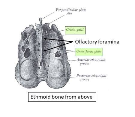
Cribiform Plate
Roof of nasal cavity
Ethmoid

Crista Galli
Middle of the cribiform plate
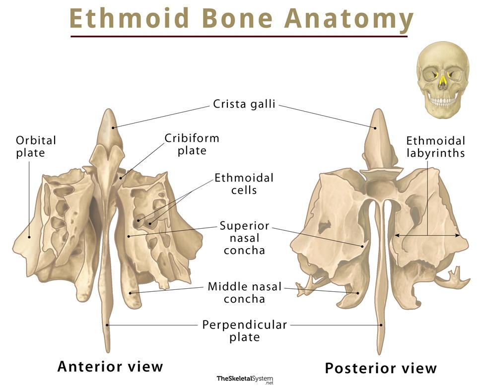
Nasal Concha (Middle, Superior)
Superior: Just below the cribiform plate
Middle: Extends down from ethmoid, next to perpendicular plate

Ethmoid Sinuses
Air space between cribiform plate and orbital surface

Sphenoid
Winged bone at the bottom of the cranium
Foramen lacerum
2 jagged holes towards the center
Sphenoid

Foramen Ovale
2 oval-shaped holes next to the foramen lacerum
Sphenoid

Foraman Rotundum
Round holes in front of foramen ovale
Sphenoid

Foramen Spinosum
Small hole behind the foramen ovale
Sphenoid

Sella Tursica
Saddle shape of sphenoid
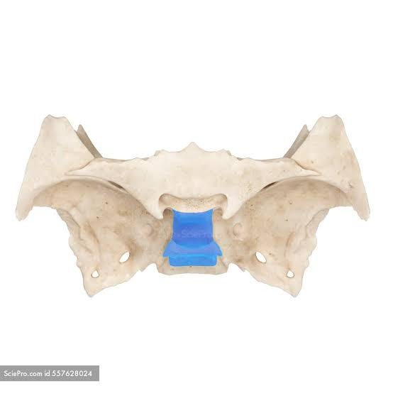
Pterygoid Plates
2 Medial: Inner plates
2 Lateral: Outer plates
Sphenoid

Sphenoid Sinuses
In front of lesser wing

Superior Orbital Fissures
Between wings
Spenoid

Inferior Orbital Fissure
Bottom of the eye socket, formed with other orbital bones
Sphenoid

Optic Canal
Hole through lesser wing
Sphenoid

Zygomatic Bones
Cheekbones
Infratemporal Fossa
Hole between cheekbone and skull
Lacrimals
2 bones between maxilla and ethmoid

Inferior Nasal Conchae
Extends from lower walls of nasal cavity

Maxilla
2 bones between nose and teeth; upper jaw

Alveolar Process
Ruffled edge for teeth
Maxilla

Incisive Fossa
Depression behind front teeth on upper palate
Maxilla

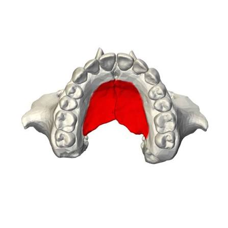
Palatal Process
Front part of hard (upper) palate
Maxilla
Infraorbital Foramen
Holes below eye sockets
Maxilla

Maxillary Sinus
Inside of the maxilla

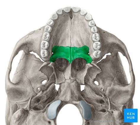
Palantine Bone
Back of the hard (upper) palate
2 Vertical parts
1 Horizontal part
Vomer
“Plow” shape; inferior nasal septum

Nasal bones
2 bones making the bidge of the nose

Mandible
Bone forming the jaw
Mandibular Notch
U Shape

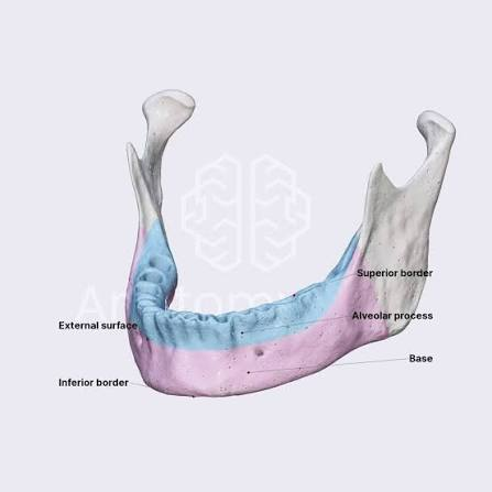
Mandibular Alveoloar Process
Jagged edge for teeth on the mandible
Mandibular Foramen
Hole in the inside angle of the mandible

Mental Foramen
Holes in the front of the mandible

Submandibular Fossa
Depression on the back of the mandible under the mylohoid line
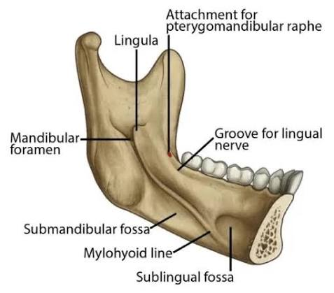
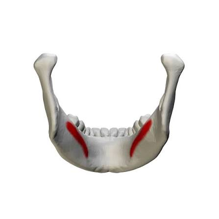
Mylohoid Line
Ridge on the inside of the mandible for muscle attachment
Mandible Ramus
Angled process of the mandible

Condyloid Process
Head of the mandibular ramus for the joint

Coronoid Process
Traingular projection of the mandibular ramus

Orbital Bones
Frontal
Maxillary
Lacrimal
Ethmoid
Palantine
SPhenoid
Zygomatic
Paranasal Sinus Function
Warm air
Lighten skull
Produce mucous
Resonate sound
Cervical Spine
7 vertebrae in the neck
Convex
Thoracic Spine
12 vertebrae in the back
Concave
Lumar Spine
5 vertebrae in the lower back
Convex
Sacral Spine
5 fused vertebrae connecting to the pelvis
Concave
Kyphosis
Extreme concavity
Breathing issues
Lumbardosis
Extreme convexity
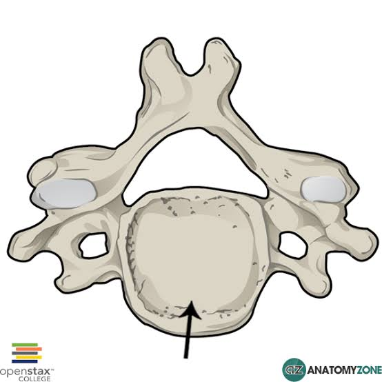
Vertebral Body
Thick cylinder
Pedicle
Connects the vertebral body to the arch/articular processes

Lamina
Connects the pedicles together to form the arch

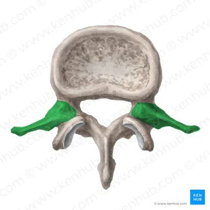
Transverse Articular Processes
Bony projections on the arch of the vertebrae extending to the side
Superior Articular Processes
Bony projections on the arch of the vertebrae extending upward

Inferior Articular Processes
Bony projections on the arch of the vertebrae extending downward

Spinous Articular Process
Bony projection on the arch of the vertebrae extending outward anterioirally

Vertebral Facets
Joints between the vertebrae on the superior and inferior articular processes
Intervertebral Disks
Fibrocartilage disks to absorb shock
Nucleus Pulposus
Jelly substance in disks
Surrounded by a fibrotic ring that can cause herniated disks if it tears
Intervertebral Foramen
Hole formed by 2 adjacent vertebrae

Vertebral Foramen
Hole formed by body, arch, and lamina of vertebrae
