6.3 THERMORECEPTION
 Call Kai
 Learn
Learn Practice Test
 Spaced Repetition
 Match
Match Flashcards
 Knowt Play
1/47

There's no tags or description
Looks like no tags are added yet.
Name | Mastery | Learn | Test | Matching | Spaced | Call with Kai | Chat |
|---|
No analytics yet
Send a link to your students to track their progress
48 Terms
THERMORECEPTION
usually free endings with small receptive fields.
THERMORECEPTION two types
Activated by cold
Activated by heat
Activated by cold
Temperatures below body temperature, no longer stimulated if the skin is under 5ºC
Activated by heat
Beyond 45ºC nociceptors take control, to avoid damage to the skin & underlying tissues.
Location of THERMORECEPTION
Widely distributed through the skin, but also in the hypothalamus & the spinal cord to detect body temperature.
Thermoreceptors
heat receptors
cold receptors
heat receptors
They begin to activate when the skin surface rises above 30°C & are most stimulated at 45°C.
Beyond this temperature, pain receptors take control, to avoid damage to the skin and underlying tissues.
cold receptors
They start having activity when the skin surface drops below 35°C & are most stimulated when the skin surface is at 25°C.
They are no longer stimulated once the skin surface drops below 5°C, explaining why the tissues stop perceiving the cold after a long time in icy temperatures.
perception of pain (pathways)
specific
pain receptors
Nociceptors
although stimuli is different →specific receptors for pain
Nociceptors
They are the sensory afferents responsible for transmitting pain, generally free nerve endings.
How is increasing temperature (pain intensity) encoded by nociceptors?
Higher temperature →
More action potentials
Higher firing frequency
👉 This is called frequency coding
After threshold (~45°C):
Pain intensity = rate of firing of nociceptors
The axons of the neurons that transmit pain neuronal information are
slower compared to sensory fibers that transmit touch & proprioception.
Perception of pain can be separated into
early perception of sharp pain (first pain)
later duller & burning sensation (second pain)
1st pain
Aδ fibers
Early perception of acute pain
Small, myelinated fibers
Phasic or rapidly adapting. Several types
Mechanosensitive: Respond to mechanical stimuli
Mechanothermal: respond to thermal & mechanical stimuli
2nd pain
C-fibers
more diffuse & burning sensation
Small & unmyelinated fibers
Tonic or slow adapting
Polymodal nociceptors: Mechanical, thermal, & chemical
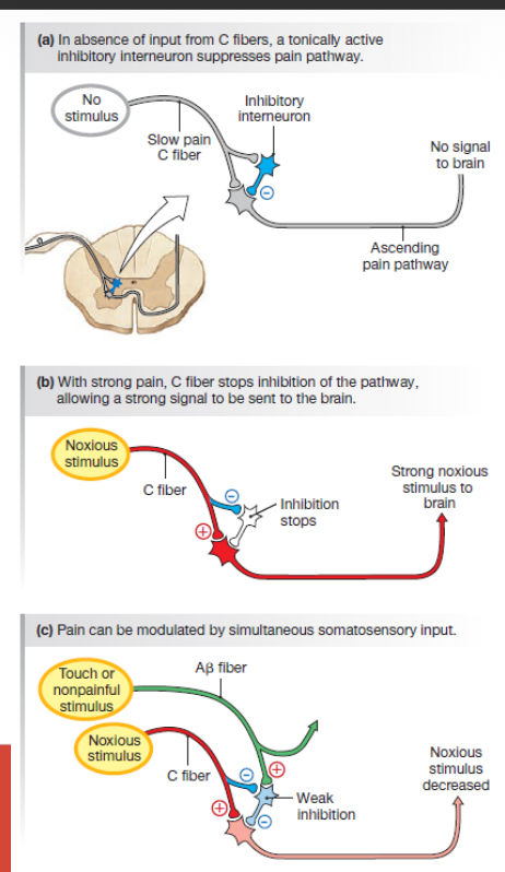
PAIN GATE THEORY
rubbing a damaged area can reduce pain
Transmission of nerve impulses from afferent fibers to 2nd order neurons in the spinal cord is modulated by a gating mechanism in the dorsal horns.
Activation of mechanoreceptors modulates the transmission of nociceptive information to higher centers.
Interactions in the local circuits of the dorsal horn, reduce the sensation of pain
PAIN MODULATION: DESCENDING PATHWAY and LOCAL CIRCUITS
descending neurons that can modulate (inhibit) active neurons in the dorsal horn of the spinal cord
These neurons activate local circuits and cause the release of inhibitory molecules that MODULATE the pain signal, decreasing it.
Pain modulators in the descending pathway
- Enkephalins
- Endorphins
- Dynorphin
Cannabinoids
Norepinephrine
Serotonin
Endogenous opioids
Enkephalins
Endorphins
Dynorphin
pain modulation is a mechanism behind
opioid analgesia — morphine mimics endogenous opioids.
Also explains why adrenaline, exercise, and placebo inhibit pain.
-Alterations in pain processing when the peripheral or central afferent fibers are damaged
generate chronic pain spontaneously or with mild stimuli.
Peripheral sensitization:
activation of peripherial nociceptor (1st order neuron) is lowered
the nociceptor itself that modifies its trigger threshold by inflammatory molecules present in the tissue
Reason for increased pain → First order neuron interaction with immune system-derived molecules
Central sensitization
It takes place due to an increase in the excitability of 2nd order neurons the dorsal horn of the spinal cord.
Hyperalgesia
exaggerated pain response to a normally painful stimulus. It is due to sensitization.
Allodynia
induction of pain by an innocuous stimulus or in the absence of a stimulus. It is often due to central sensitization.
Neuropathic pain
both peripheral & central sensitization usually disappear when an injury heals.
When this does not happen, we speak of neuropathic pain (source in the somatosensory nervous system).
Paresthesia/dysesthesia:
abnormal sensation (tingling, burning, itching) in response to touch.
It can be central or peripheral (After radiation therapy)
Phantom limb syndrome
sensory perceptions (spikes, pain...) in a limb that has been amputated.
Caused by the reorganization of the somatosensory cortex.
Activity in adjacent areas activates neurons previously representing the lost limb
Interpreted by the brain as sensations in the missing limb
CNS origin & usually disappears after the reorganization of the circuits (plasticity).
REFERRED PAIN
Pain perceived in the skin or muscle, even though the actual source is visceral
Visceral sensory neurons
from organs
Somatic sensory neurons
from skin
angina pectoris
heart
kidneys
Heart (angina pectoris)
Actual source: Heart (visceral pain)
Signals enter spinal cord
These same spinal segments also receive input from → Left arm, chest, & jaw (females)
Kidney (angina pectoris)
Actual source: Kidneys
Signals enter spinal cord at lower thoracic/lumbar levels
These correspond to:
Lower back (lumbar region)
The fibers of the visceral nociceptors share
same dorsal root ganglion & segment of the spinal cord with the fibers that come from skin nociceptors
→ brain misinterprets pain as coming from the skin.
The response to pain is multifactorial
parallel pathways of pain → Sensory-discriminative aspects & Affective-motivational aspects
Sensory-discriminative aspects
Processing of the sensory information → Location, Intensity, Duration & Type of pain
Affective-motivational aspects
Unpleasant feeling, autonomic activation fear, anxiety, (fight or flight) autonomic activation
Sensory-discriminative
aspects
Pathway: Spinothalamic tract → Thalamus (VPL) → Somatosensory cortex (SI, SII)
Function:
Location of pain
Intensity of pain
Type of stimulus
→ “Where is the pain and how strong is it?”
Affective-motivational
aspects
Pathway: Spinothalamic tract → medial thalamus → limbic system
Includes:
Anterior cingulate cortex
Insula
Amygdala
Hypothalamus
Function:
Emotional response to pain
Suffering/unpleasantness
Motivation to react
→ “How does the pain feel emotionally?”
Mechanoreceptors
provide information on the position of the limbs and other parts of the body in space
muscle spindles
detects changes in muscle length
Golgi tendon organ:
Detects changes in muscle tension
PROPIOCEPTION RECEPTOR TYPE
muscle spindle
(Tendons, joints & muscles)
TOUCH RECEPTOR TYPE
encapsulated:
• Merkel corpuscles
• Meissner corpuscles
• Pacinian corpuscles
• Ruffini's corpuscles
PAIN, TEMPERATURE RECEPTOR TYPE
Nerve endings free
PAIN TEMPERATURE PRURITUS RECEPTOR TYPE
Nerve endings free (unmyelinated)