anatomy lab Cardiac conduction system & ECG
 Call Kai
 Learn
Learn Practice Test
 Spaced Repetition
 Match
Match Flashcards
 Knowt Play
1/37

There's no tags or description
Looks like no tags are added yet.
Name | Mastery | Learn | Test | Matching | Spaced | Call with Kai | Chat |
|---|
No analytics yet
Send a link to your students to track their progress
38 Terms
Cardiac conduction system
Transmits action potentials throughout the heart to promote coordinated, sequential heart muscle contractions
• Composed of specialized, noncontractile cardiac cells
Depolarization
responsible for travel of action potentials
• Caused by influx of Ca2+ ions, which reverses membrane potential
Action potentials trigger
contractions
Conduction System
● A series of depolarization events throughout the heart
● Conducted through heart via bundles of electrically excitable cells and electrical conduction structures
Sinoatrial node (SA node)
Spreads depolarizing stimulus through both atria
-(impulse cannot pass directly into ventricles due to insulating fibrous skeleton)
Pacemaker of the hear
SA node
(sets rhythm ~75 bpm)
Atrioventricular node (AV node)
Action potential is delayed, ensuring the atria are completely contracted before ventricular contraction begins
AV bundle (bundle of His)
Connects atria to ventricles
Bundle branches (right & left)
Conduct action potential through interventricular septum
Purkinje fibers (subendocardial conducting network)
Conduct action potential through myocardium of ventricles
ECG (electrocardiogram)
Measures the electrical activity of the heart
p wave
atrial depolarization
PR segment
AV nodal delay
QRS complex
ventricular depolarization (atria repolarizing simultaneously)
ST segment
time during which ventricles are contracting and emptying
T wave
ventricular repolarization
TP interval
time during which ventricles are relaxing and filling
Baroreceptors
◦ Respond to changes in blood pressure
Chemoreceptors
◦ Respond to concentration of chemicals (e.g., O2 , CO2 , pH)
Cardioinhibitory center
Stimulated by:
• Increase in BP
• Increase in O2
• Decrease in CO2
• Increase in pH
slows heart rate
Cardioacceleratory center
Stimulated by:
• Decrease in BP
• Decrease in O2
• Increase in CO2
• Decrease in pH
accelerates heart rate

what is A?
SA node

what is B?
Atrioventricular node

what is C?
Atrioventricular bundle or bundle of HIS

what is D?
Right and left bundle branches

what is E?
Purkinje fibers (subendocardial conduction network)

what is A?
P wave

what is B?
Q peak
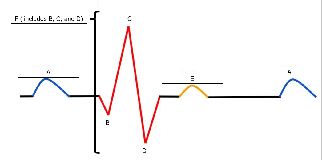
what is C?
R peak

what is D?
S peak

what is E?
T wave
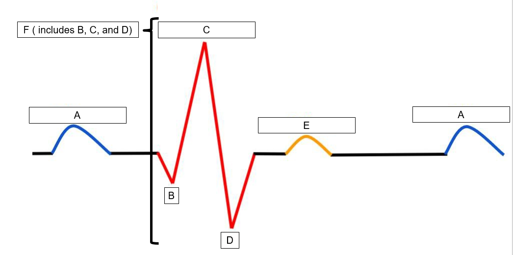
what is F?
QRS complex
The wave of atrial depolarization reaches this structure in the right atrium shortly after the SA node depolarization. |
atrioventricular node (AV)
This component contains the cardiac pacemaker cells and has an unstable resting membrane potential causing the node to undergo rhythmic depolarization that spreads throughout the right and left atria
sinoatrial node (SA)
These structures carry the action potential into the two sides of the heart's myocardium.
right and left AV bundle branches
These structures transmit the action potential to the myocardium of the ventricles to cause contraction.
Purkinje fibers
The depolarization spreads down this structure that is located in the interventricular septum
atrioventricular (AV) bundle or Bundle of HIS
Identify the scenarios in which the the cardioinhibitory (parasympathetic) center would be stimulated
Increase in blood oxygen levels
Increase in blood pH
Decrease in blood carbon dioxide levels