Biochem Comp 2
 Call Kai
 Learn
Learn Practice Test
 Spaced Repetition
 Match
Match Flashcards
 Knowt Play
1/112

There's no tags or description
Looks like no tags are added yet.
Name | Mastery | Learn | Test | Matching | Spaced | Call with Kai | Chat |
|---|
No analytics yet
Send a link to your students to track their progress
113 Terms
Almost all amino acids are chiral, meaning that the
alpha carbon is asymmetric
Biological proteins are composed almost exclusively of
L amino acids
D- alanine, one of the few D amino acids, is found in
peptidoglycan in bacterial cell walls
At neutral pH,
the carboxyl group has a negative charge
the amino group has a positive charge
Though amino acids have a positive and negative charge, it is a
neutral molecule called a zwitterion (unstable)
Uncharged hydrophilic amino acids
polar but have no net charge
Glycine doesn’t fit into any category because
it doesn’t have a real R group (R = H) . It is the only AA that doesn’t have an steroisomer
Aliphatic (hydrocarbon side chain) amino acids
valine
leucine
proline
isoleucine
alanine
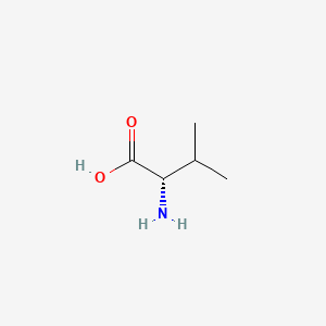
Valine
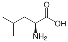
Leucine
Proline

Isoleucine

Alanine

Uncharged polar amino acids
Asparagine
Glutamine
Serine
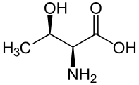
Threonine
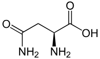
Asparagine
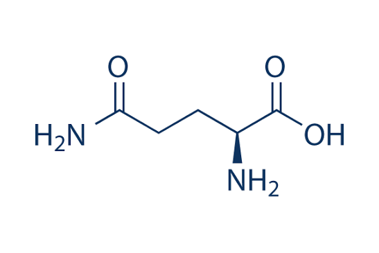
Glutamine
Serine (phosphorylation)

Threonine
Aromatic amino acids
Phenylalanine (hydrophobic)
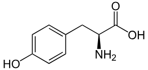
Tyrosine (hydrophilic)
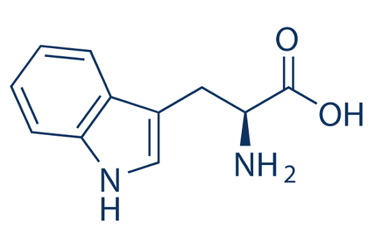
Tryptophan (uncharged hydrophilic)
Phenylalanine

Tyrosine
Tryptophan
Basic Amino acids
arginine
lysine
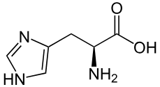
histidine
Histidine (side group is imidazole)
Arginine
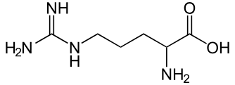
Lysine

Cysteine
can form a disulfide bond with another cysteine (called a cystine)

Methionine
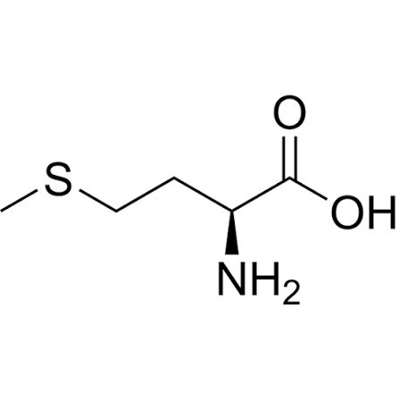
Sulfur Containing Amino Acids
cysteine
methionine
The peptide bond forms when
the
carboxyl group of one amino acid
joins the amino group of another,
releasing water
The peptide backbone is highly repetitive and
consists of the repeating pattern:
α-carbon → carbonyl carbon → amide
nitrogen
Protein function determined by amino acid sequences, but
also depends on
folding, interactions, modifications, and
cellular environment
Primary level of protein folding
amino acid sequence
Secondary
a helix and b sheet (local folding)
Tertiary
3D shape of one chain
Quaternary
arrangement of multiple chains
Anfinsen's principle
The primary
amino acid sequence contains the
information needed to determine the
protein's final three-dimensional
structure.
Homomeric (quaternary structure)
Composed of two
or more identical subunits.
Molecular chaperones
are
specialized proteins that assist in
the folding process, although the
final structure is ultimately
determined by the primary amino
acid sequence
Alpha-helix
Right-handed helix structure
R-groups project outward
β-sheets
consists of extended polypeptide strands arranged next to one another.
Can be:
• Parallel
• Antiparallel
Polyproline Helices
Proline-rich sequences
The outward-facing R-groups
are often hydrophobicimportant for protein-protein interactions
Protein Domains/motifs
an independently folding,
stable, and compact three-dimensional unit
that acts as a fundamental building block of
protein structure and function
Proteins Denaturation
loss of a protein’s normal
shape.It happens when heat, acids, bases, or
chemicals disrupt the protein.The protein unfolds and loses its normal
function.
Protein Misfolding
when a protein fails to achieve
its correct structure.
Collagen
rich in glycine, proline
left handed helix
3 chains twisted together
Collagen Requirements
Lysine and proline in collagen are
modified by hydroxylation.This helps collagen molecules form
strong cross-links.Vitamin C is needed for this process.
Cross-links give collagen strength and
stability.Without enough vitamin C, collagen
becomes weak.Severe vitamin C deficiency can cause
scurvy
Hemoglobin and myoglobin function as
carriers to bring O2 to the cell to participate in cellular
respiration to produce ATP molecules
Both myoglobin and hemoglobin are classified as
globular proteins
Myoglobin stores the oxygen whereas
hemoglobin brings the oxygen to the tissues
Myoglobin
Has higher affinity to O2 than hemoglobin
Binds and releases O2 to muscle cells
Myoglobin Structure
single polypeptide chain folded into a tertiary
structure known as globin fold (composed of 8 alpha helices)
with one O2 binding site. The helices are folded in a way to
create a hydrophobic O2 binding pocket containing heme group
with an iron atom (Fe+2) in its center
Heme structure
Composed of planner
protoporphyrin ring
composed of four pyrrole
rings the lie with their N
atoms in the center (organic
part) binding an Fe+2
(inorganic atom).
Hemoglobin
Take the O2 from the lungs to the tissues and takes CO2 from the tissues back
to the lugs to be expelled out of the body
Hemoglobin structure
tetrameric (4 polypeptide subunits, 2 alpha subunits and 2 beta subunits). Each
subunit has a heme molecule which means each is bound to Fe+2 (total 4 O2 molecules bound)
Hemoglobin has 2 ___ chains and 2 ___ chains
alpha and beta
The saturation curve of myoglobin has a
hyperbolic shape
The saturation curve for hemoglobin has a
sigmoidal shape (S shape)
Myoglobin becomes more saturated at a
lower
pressure than hemoglobin and requires very low
pressures to release O2
hemoglobin where the binding of one O2 to one subunit in
hemoglobin leads to
increasing the affinity of the other subunits for O2 (allosteric regulation)
Allosteric regulation of hemoglobin results in a conformation change also called
the change from tense (T) state to relaxed (R) state
T state happens when there is
low affinity for O2
R state happens when there i
high affinity
for O2
When O2 binds to the iron (Fe+2) group, it pulls up the Fe+2 into the heme group because it decreases
the electron density around the Fe+2, allowing it to become smaller in size, thus able to fit in the
porphyrin ring. This change in the conformation (the pull)
also pulls the proximal histidine group and
changes the overall interaction between hemoglobin subunits
T to R state
In deoxygenated hemoglobin, the quaternary structure is very constrained (T state). When O2
binds and leads to conformational change, it relieves this strain and allows the molecules to go
into the relaxed state (R state)
At low pressure (low O2), the hemoglobin binding
is slow, but as more O2 is binding,
there is a sharp rise in the curve (the affinity to O2
shoots up and we notice the increase in saturation
with O2)
There are 3 allosteric factors affecting Hb affinity, binding and dissociation to O2:
hydrogen ions
2,3 Bisphosphoglycerate
Covalent binding of CO2
Hydrogen Ion (Bohr effect)
by increasing the concentration of
CO2, the H+ ion conc also increases leading to a
decrease in blood pH thus lowering Hb affinity to
O2
In order for the cells to get rid of CO2,
it will
react with H2O to give carbonic acid by the aid of
an enzyme called carbonic anhydrase. Carbonic
acid then dissociates to H+ and bicarbonate ion (aka Bohr effect)
In the Bohr effect, the affinity and binding of Hb to
O2 is
NOT affected by the pH
How do H+ ions really affect Hb?
H+ ions bind
the Hist 146 on the β subunit and the amino group of the terminal
residue in Hb α 122 positions forming stabilized salt bridges that
leads to changing the conformation of Hb and stabilizing the Hb
in T state, making it more likely to unload O2 to the tissues that
need it
Carbon Dioxide (Haldane effect)
O2 affects the affinity of Hb to CO2
and H+increases the number of
the polar bicarbonate ions dissolved in the blood
2,3-Bisphosphoglycerate
(2,3-BPG)
Relatively small molecule that contains multiple
negative charges. These characteristics allow this
molecule to bind in the positive pocket inside Hb
2,3-Bisphosphoglycerate
(2,3-BPG) in regards to Hb and O2
2,3-BPG lowers the affinity of
Hb to O2. Therefore, more Hb will be able to release
O2 in the tissues
Red blood cells can modulate Hb affinity for O2 by
altering
the rate of synthesis or degradation of 2,3-
BPG
2,3-BPG binds to the positive pockets inside the Hb, which are
2 Histidine 143 and 2 Lysine 82 from the B subunits
Fetal Hb subunits are
two α subunits and 2 γ subunits
The difference between γ subunits and is that
γ subunits do NOT have Histidine 143. Instead,
it has serine
Serine is neutral which means the positive pocket in the
fetal Hb is
less positively charged than the maternal/adult Hb
The lower positive charge means the 2,3-BPG binds less to the fetal Hb. The physiological
significance of this is that the
fetal Hb has higher affinity to O2 than maternal/adult Hb (allows for efficiently O2 intake)
Carbon monoxide
(CO) poisoning
CO binds with Hb at the same binding site of O2
CO has higher affinity to Hb than O2
More CO binds that O2 = death
Sickle Cell Disease
a genetic error occur leading to the substitution of glutamate with valine in the B subunits
Since valine is non polar, it causes the HB molecules to form a linear shape, resulting in (sickle cell)
Thalassemia
there is unbalanced production of α subunits or ꞵ subunits (the genetic mutations that cause thalassemia affect the synthesis of either α or ꞵ subunits)
ꞵ-Thalassemia major (homozygous ꞵ-Thalassemia)
severe disorder caused by inheriting two alleles of the nutation
ꞵ-Thalassemia intermediate:
Less severe clinical phenotype. Can be the result of two different mild mutations or homozygosity for a mild mutation
ꞵ-Thalassemia minor (ꞵ-Thalassemia trait):
Heterozygous disorder that involves a single mutation.
Signal transduction
the process of converting an extracellular signal to a molecular response inside the cell
1) Release of the primary messenger
The primary messenger release occurs as a result of external or internal stimuli
2) Reception of the primary messenger
The majority of primary messengers bind to proteins in the cell membranes (receptors) that transfer this information to the cell’s interior
Signal molecules are also called
ligands
3) Delivery of the message to the interior of the cell (transduction)
The information from the receptor-ligand complex are transferred to the inside of the cell by second
messengers
Second messengers
small molecules that mediate the next steps in the transduction pathways via the
change in their concentration in response to change in environmental signals
The consequences of using second messengers:
signal may be amplified
they are free to diffuse to different sites within the cell and influence other processes
Used in multiple pathways
Activation of effectors that directly influence the physiological response
This results in the activation (or inhibition) of pumps, channels, enzymes or transcription factors
that affects cellular processes
Termination of the signal:
After the completing of the cell response to the external signal, the transduction pathway must be
terminated (to preserve cell responsiveness to new signals)
What are G protein-coupled receptors
(GPCRs)?
7-transmembrane helix proteins
transduce extracellular signals into important physiological effects
Components of GPCRs
7 transmembrane helix
functional extracellular and intracellular loops
G proteins (Ga, Gb, and Gy subunits)
G Protein A subunits contains two loops, called
switch I and Switch II
Gs
leads to the stimulation of adenylyl cyclase leading to the conversion of ATP to cAMP
Gi
leads to the inhibition of adenylyl cyclase leading to the prevention of conversion of ATP
to cAMP