BIO 201 Respiratory System

0.0(0)
Studied by 0 people
call kaiCall Kai
Locked
learnLearn
examPractice Test
spaced repetitionSpaced Repetition
heart puzzleMatch
flashcardsFlashcards
GameKnowt Play
Card Sorting

1/146

encourage image

There's no tags or description

Looks like no tags are added yet.

Last updated 9:46 AM on 7/28/26
Name
Mastery
Learn
Test
Matching
Spaced
Call with Kai
Chat

No analytics yet

Send a link to your students to track their progress

147 Terms

1
New cards

Respiration

Gas exchange of O2 and CO2

  • Occurs between the atmosphere and body cells

    • Cells need O2 for aerobic metabolism (ATP Production)

    • Cells need to dispose of CO2 produced by aerobic metabolism

2
New cards

Respiratory System

Body system that provides the means for gas exchange. Consists of…

  • Respiratory passageways in the head, neck, and trunk & the lungs

3
New cards

Functions of the Respiratory System

  • Air Passageway (= airways)

    • Moves air in and out of alveoli as we breath

  • Site for exchange of oxygen and carbon dioxide

    • Oxygen diffuses from alveoli into the blood

    • Carbon dioxide diffuses from the blood into the alveoli

  • Detection of Odors

    • Atmospheric air contains odorant molecules

    • Detectable by olfactory receptors in the superior nasal cavity

  • Sound Production

    • Air moves across vocal cords of the larynx (voice box)

    • Vocal cords vibrate, producing sound

  • Expel contents of abdominopelvic cavity

    • Assist with defecation, urination, and childbirth by increasing pressure in the thoracic cavity

  • Assist in the flow of venous blood and lymph

    • Pressure changes in thoracic cavity assist with flow of venous blood and lymph in both thoracic and abdominopelvic cavities

      • “Respiratory Pump”

  • Acid-Base Homeostasis

    • Eliminates carbon dioxide to regenerate blood’s main buffer system

  • Assists in endocrine function: Production of angiotensin II

    • Lungs produce ACE (angiotensin-converting) enzyme that converts angiotensin I → angiotensin II

    • Critical to the maintenance of blood pressure and fluid homeostasis

4
New cards

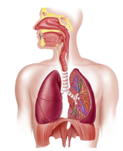
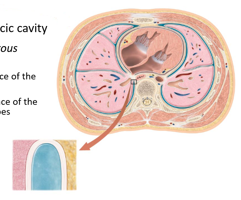
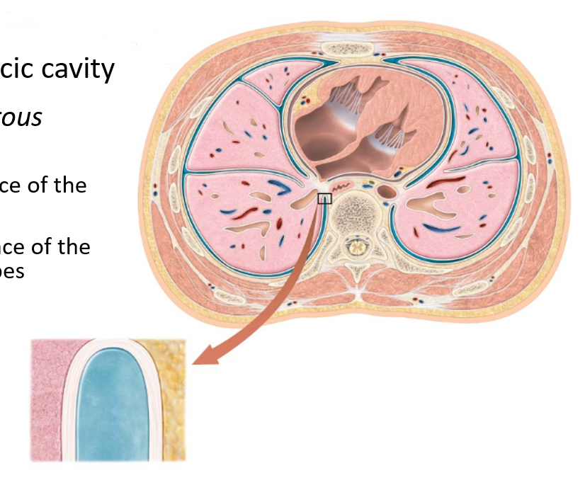
Respiratory System Anatomy

  • Organs found in the head, neck, and thoracic cavity:

    • Blood vessels = Pulmonary circuit

    • Thoracic Cage (Protection)

      • Bones and cartilage

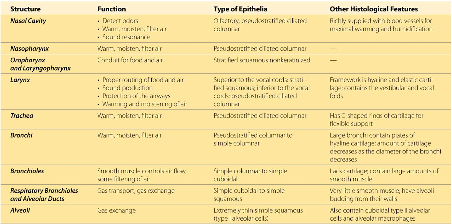
      • Respiratory muscles

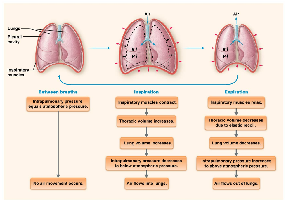
    • Pleura

      • Surface membrane of the lungs and the inside of the thoracic cage

    • Lungs

      • Spongy, paired organ

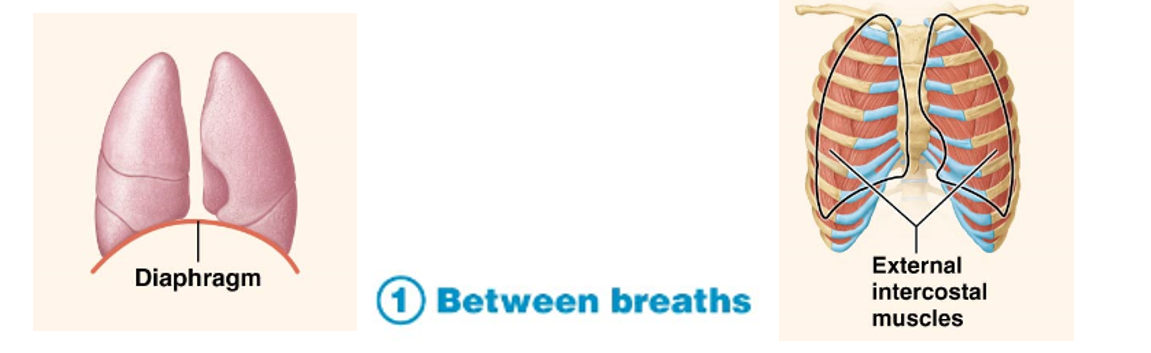
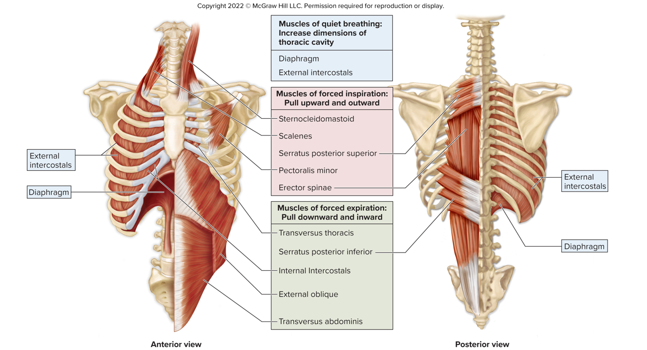
      • Within boundaries of rib cage and diaphragm

    • Airways

      • Hollow passages that collectively transport gases
<ul><li><p>Organs found in the head, neck, and thoracic cavity:</p><ul><li><p>Blood vessels = Pulmonary circuit</p></li><li><p>Thoracic Cage (Protection)</p><ul><li><p>Bones and cartilage</p></li><li><p>Respiratory muscles</p></li></ul></li><li><p>Pleura</p><ul><li><p>Surface membrane of the lungs and the inside of the thoracic cage</p></li></ul></li><li><p>Lungs</p><ul><li><p>Spongy, paired organ</p></li><li><p>Within boundaries of rib cage and diaphragm</p></li></ul></li><li><p>Airways</p><ul><li><p>Hollow passages that collectively transport gases</p></li></ul></li></ul></li></ul><p></p>
5
New cards

Structural Organization of Respiratory System

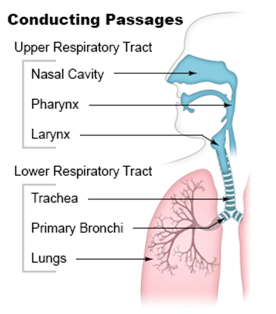
  • Upper Respiratory Tract

    • Nose, nasal cavity, pharynx, larynx

  • Lower Respiratory Tract

    • Trachea, bronchi, bronchioles, alveolar ducts, alveoli
<ul><li><p><strong>Upper Respiratory Tract</strong> </p><ul><li><p>Nose, nasal cavity, pharynx, larynx</p></li></ul></li><li><p><strong>Lower Respiratory Tract</strong></p><ul><li><p>Trachea, bronchi, bronchioles, alveolar ducts, alveoli</p></li></ul></li></ul><p></p>
6
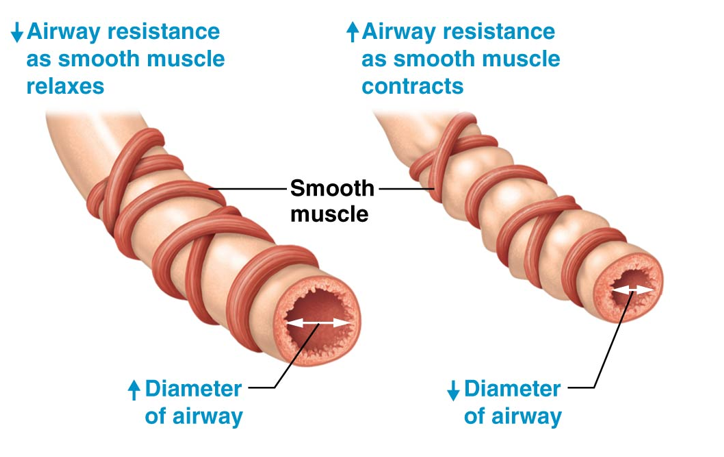
New cards

Functional Organization of Respiratory System

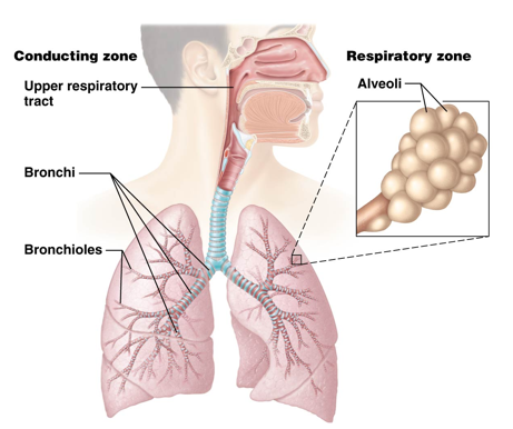
  • Conducting Zone

    • Structures in this zone transport, warm, and filter air

      • Nose to terminal bronchioles

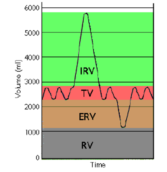
  • Respiratory Zone

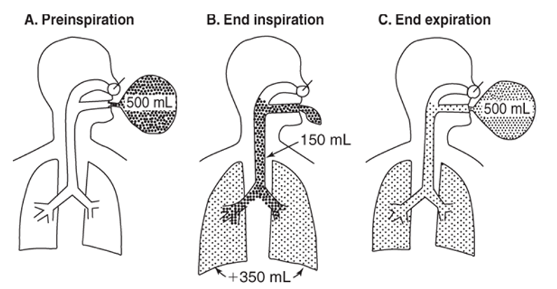
    • Structures of this zone participate in gas exchange

      • Respiratory bronchioles, alveolar ducts, alveoli
<ul><li><p><strong>Conducting Zone</strong></p><ul><li><p>Structures in this zone transport, warm, and filter air </p><ul><li><p>Nose to terminal bronchioles</p></li></ul></li></ul></li><li><p><strong>Respiratory Zone</strong></p><ul><li><p>Structures of this zone participate in gas exchange</p><ul><li><p>Respiratory bronchioles, alveolar ducts, alveoli</p></li></ul></li></ul></li></ul><p></p>
7
New cards

Structural & Functional Organization of the Respiratory System
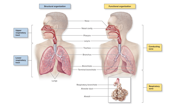
knowt flashcard image
8
New cards

Tissues of the Respiratory System

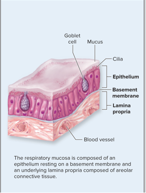
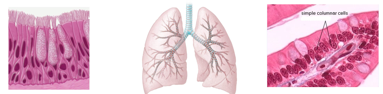
  • Mucosa = mucous membrane: respiratory lining (always moving/waving)

    • Epithelium resting on a basement membrane

    • Cilia under mucus

    • Underlying lamina propria: areolar connective tissue (loose connective tissue)

    • Secretes mucus from goblet cells

      • Contains mucin protein

      • Contains antimicrobial substances

        • Increases mucous viscosity and serves to trap dust, dirt, pollen, etc.

        • Lysozyme (antibacterial enzyme)

        • Defensins (antibacterial proteins)

        • Immunoglobulin A (antibody)

<ul><li><p><strong>Mucosa </strong>= mucous membrane: respiratory lining (always moving/waving)</p><ul><li><p><strong>Epithelium </strong>resting on a basement membrane</p></li><li><p><strong>Cilia </strong>under mucus</p></li><li><p>Underlying <strong>lamina propria</strong>: <strong>areolar connective tissue</strong> (loose connective tissue)</p></li><li><p>Secretes mucus from <strong>goblet cells</strong></p><ul><li><p>Contains <strong>mucin </strong>protein</p></li><li><p>Contains antimicrobial substances</p><ul><li><p>Increases mucous viscosity and serves to trap dust, dirt, pollen, etc.</p></li><li><p><strong>Lysozyme </strong>(antibacterial enzyme)</p></li><li><p><strong>Defensins </strong>(antibacterial proteins)</p></li><li><p><strong>Immunoglobulin A </strong>(antibody)</p></li></ul></li></ul></li></ul></li></ul><p></p>
9
New cards

Lamina Propria

Bottom layer of respiratory mucosa composed of areolar (loose) connective tissue
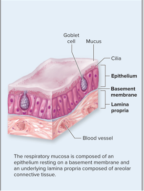
<p>Bottom layer of respiratory mucosa composed of areolar (loose) connective tissue</p>
10
New cards

Goblet Cells

Unicellular glands residing in the respiratory mucosa secrete mucus. These cells also contain the protein mucin and antimicrobial substances.
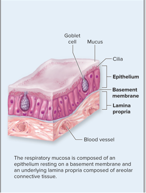
<p>Unicellular glands residing in the respiratory mucosa secrete mucus. These cells also contain the protein mucin and antimicrobial substances.</p>
11
New cards

Respiratory Epithelium

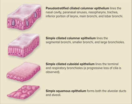
Layer of epithelial cells between the mucus lining and basement membrane in respiratory mucosa.

  • Progressively becomes thinner from the nasal cavity to the alveoli

    • Pseudostratified Ciliated Columnar Epithelium lines the

      • Nasal Cavity

      • Paranasal Sinuses

      • Nasopharynx

      • Trachea

      • Inferior Portion of Larynx

      • Main Bronchi

      • Lobar Bronchi

    • Simple Ciliated Columnar Epithelium lines the

      • Segmental Bronchi

      • Smaller Bronchi

      • Large Bronchioles

    • Simple Ciliated Cuboidal Epithelium lines the

      • Terminal Bronchioles

      • Respiratory Bronchioles

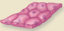
    • Simple Squamous Epithelium lines the

      • Alveolar Ducts

      • Alveoli

    • Stratified Squamous Epithelium is found in high abrasion areas

      • Osopharynx

      • Laryngopharynx

      • Vocal Cords

      • Superior Portion of the Larynx

<p>Layer of epithelial cells between the mucus lining and basement membrane in respiratory mucosa.</p><ul><li><p>Progressively becomes thinner from the nasal cavity to the alveoli</p><ul><li><p>Pseudostratified Ciliated Columnar Epithelium lines the </p><ul><li><p>Nasal Cavity</p></li><li><p>Paranasal Sinuses </p></li><li><p>Nasopharynx </p></li><li><p>Trachea </p></li><li><p>Inferior Portion of Larynx </p></li><li><p>Main Bronchi </p></li><li><p>Lobar Bronchi </p></li></ul></li><li><p>Simple Ciliated Columnar Epithelium lines the</p><ul><li><p>Segmental Bronchi</p></li><li><p>Smaller Bronchi</p></li><li><p>Large Bronchioles </p></li></ul></li><li><p>Simple Ciliated Cuboidal Epithelium lines the </p><ul><li><p>Terminal Bronchioles </p></li><li><p>Respiratory Bronchioles </p></li></ul></li><li><p>Simple Squamous Epithelium lines the</p><ul><li><p>Alveolar Ducts</p></li><li><p>Alveoli</p></li></ul></li><li><p>Stratified Squamous Epithelium is found in high abrasion areas </p><ul><li><p>Osopharynx </p></li><li><p>Laryngopharynx </p></li><li><p>Vocal Cords</p></li><li><p>Superior Portion of the Larynx </p></li></ul></li></ul></li></ul><p></p>
12
New cards

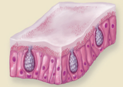
Pseudostratified Ciliated Columnar Epithelium

Epithelium that lines the

  • Nasal Cavity

  • Paranasal Sinuses

  • Nasopharynx

  • Trachea

  • Inferior Portion of Larynx

  • Main Bronchi

  • Lobar Bronchi

<p>Epithelium that lines the</p><ul><li><p>Nasal Cavity </p></li><li><p>Paranasal Sinuses </p></li><li><p>Nasopharynx </p></li><li><p>Trachea</p></li><li><p>Inferior Portion of Larynx</p></li><li><p>Main Bronchi</p></li><li><p>Lobar Bronchi </p></li></ul><p></p>
13
New cards

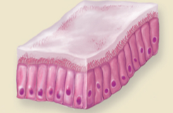
Simple Ciliated Columnar Epithelium

Epithelium that lines the

  • Segmental Bronchi

  • Smaller Bronchi

  • Large Bronchioles

<p>Epithelium that lines the </p><ul><li><p>Segmental Bronchi</p></li><li><p>Smaller Bronchi</p></li><li><p>Large Bronchioles </p></li></ul><p></p>
14
New cards

Simple Ciliated Cuboidal Epithelium

Epithelium that lines the

  • Terminal & Respiratory Bronchioles (a progressive loss of cilia is observed)
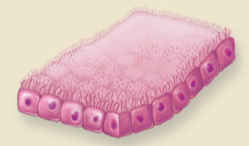
<p>Epithelium that lines the </p><ul><li><p>Terminal &amp; Respiratory Bronchioles (a progressive loss of cilia is observed) </p></li></ul><p></p>
15
New cards

Simple Squamous Epithelium

Epithelium that forms both

  • Alveolar Ducts

  • Alveoli

<p>Epithelium that forms both </p><ul><li><p>Alveolar Ducts </p></li><li><p>Alveoli </p></li></ul><p></p>
16
New cards

Stratifed Squamous Epithelium

Epithelium found in high abrasion areas, such as

  • Oropharynx

  • Laryngopharynx

  • Vocal Cords

  • Superior Portion of the Larynx

17
New cards

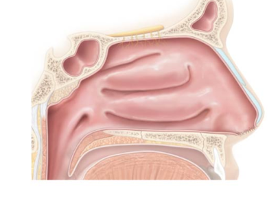
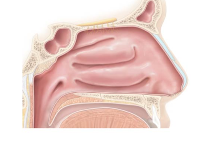
Nasal Cavity

A hollow space framed by bone and hyaline cartilage that extends anteriorly from the nostrils to the two posterior nares. Functions include

  • Being the entryway into the respiratory system

  • Warms and humidifies inhaled air

  • Filters debris from inhaled air

    • Mucosa (mucous membrane) and nose hair

  • Secrete antibacterial substance

  • House olfactory receptors

  • Enhances the resonance of voice

    • Deepness of voice

    • How you can tell if someone is congested
<p>A hollow space framed by bone and <strong>hyaline cartilage</strong> that extends anteriorly from the nostrils to the two <strong>posterior nares</strong>. Functions include</p><ul><li><p>Being the entryway into the respiratory system</p></li><li><p>Warms and humidifies inhaled air</p></li><li><p>Filters debris from inhaled air</p><ul><li><p><strong>Mucosa </strong>(mucous membrane) and nose hair</p></li></ul></li><li><p>Secrete antibacterial substance</p></li><li><p>House olfactory receptors</p></li><li><p>Enhances the resonance of voice</p><ul><li><p>Deepness of voice</p></li><li><p>How you can tell if someone is congested </p></li></ul></li></ul><p></p>
18
New cards

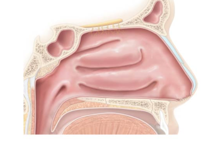
Nasal Septum

composed of bone and hyaline cartilage
<p>composed of bone and hyaline cartilage </p>
19
New cards

Vestibule

Bristle-like hairs in the nasal cavity prevent large objects from entering.

  • Lined with stratified squamous epithelium

    • Similar to external skin

    • Resists mechanical stress

<p>Bristle-like hairs in the nasal cavity prevent large objects from entering.</p><ul><li><p>Lined with <strong>stratified squamous epithelium</strong></p><ul><li><p>Similar to external skin</p></li><li><p>Resists mechanical stress</p></li></ul></li></ul><p></p>
20
New cards

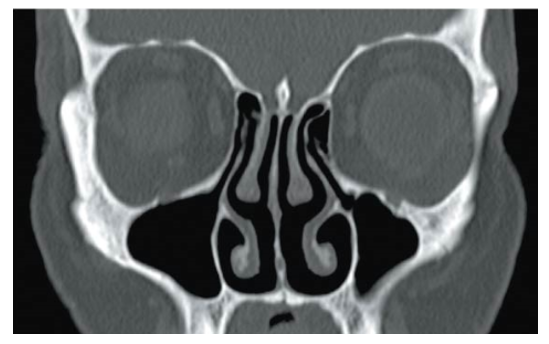
Nasal Conchae

3 bony projections of the nasal cavity that curl around three narrow passages

  • Superior conchae

    • Curl around the superior meatus

  • Middle conchae

    • Curl around the middle meatus

  • Inferior conchae

    • Curl around the inferior meatus

  • Functions:

    • Create turbulence in the airflow, slowing it down so air can be warmed and rid of dust and debris

    • Lots of hairs present to trap debris

<p>3 bony projections of the nasal cavity that curl around three narrow passages</p><ul><li><p>Superior conchae</p><ul><li><p>Curl around the superior meatus</p></li></ul></li><li><p>Middle conchae</p><ul><li><p>Curl around the middle meatus</p></li></ul></li><li><p>Inferior conchae</p><ul><li><p>Curl around the inferior meatus</p></li></ul></li><li><p>Functions:</p><ul><li><p>Create turbulence in the airflow, slowing it down so air can be warmed and rid of dust and debris</p></li><li><p>Lots of hairs present to trap debris</p></li></ul></li></ul><p></p>
21
New cards

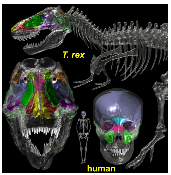
Paranasal Sinuses

Hollow cavities found within the frontal, ethmoid, sphenoid, and maxillary bones.

  • Connected to the nasal cavity by small passageways

  • Warm, filter, and humidify air

  • Also enhance voice resonance and reduce the weight of the skull
<p>Hollow cavities found within the frontal, ethmoid, sphenoid, and maxillary bones.</p><ul><li><p>Connected to the nasal cavity by small passageways</p></li><li><p>Warm, filter, and humidify air </p></li><li><p>Also enhance voice resonance and reduce the weight of the skull</p></li></ul><p></p>
22
New cards

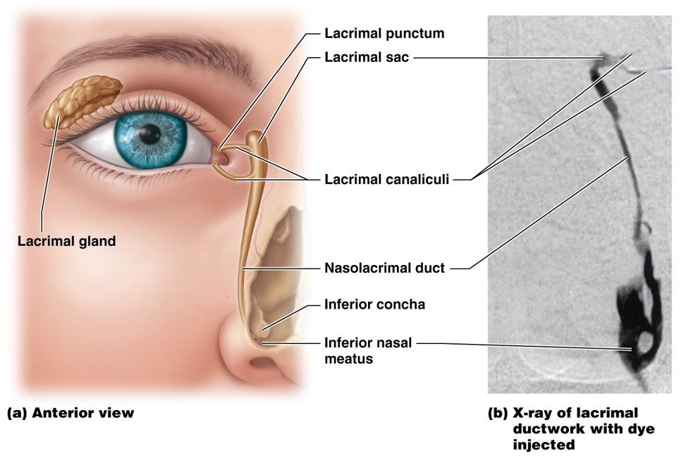
Nasolacrimal Ducts

Ducts that drain lacrimal secretions from eye surfaces into the nasal cavity
<p>Ducts that drain lacrimal secretions from eye surfaces into the nasal cavity </p>
23
New cards

Olfactory Mucosa

Mucosa located on the roof of the nasal cavity that houses special sensory receptors for smell

24
New cards

Respiratory Mucosa of the Nasal Cavity

Pseudostratified ciliated columnar epithelium & goblet cells

  • Traps foreign particles in mucus

  • Ciliated cells propel debris and mucus toward the posterior nasal cavity and pharynx

25
New cards

Runny Nose (Clinical Connection)

Also known as rhinorrhea, it occurs as a result of

  • Increased production of mucus (allergies, virus)

  • Increased secretions of lacrimal glands draining into the nasal cavity (crying)

  • Exposure to cold air (water condensation + less effective cells)

Mucus can also travel into the pharynx
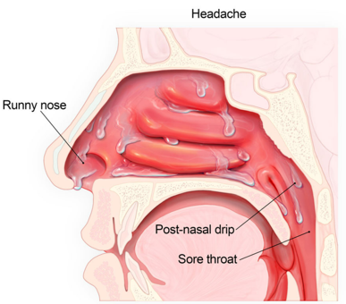
<p>Also known as <strong>rhinorrhea,</strong> it occurs as a result of</p><ul><li><p>Increased production of mucus (allergies, virus)</p></li><li><p>Increased secretions of lacrimal glands draining into the nasal cavity (crying)</p></li><li><p>Exposure to cold air (water condensation + less effective cells)</p></li></ul><p>Mucus can also travel into the pharynx</p>
26
New cards

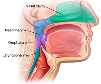
Pharynx

The throat is separated into three anatomical divisions

  • Nasopharynx

    • Behind the nose

  • Oropharynx

    • Behind the mouth

  • Laryngopharynx

    • Behind the larynx

<p>The throat is separated into three anatomical divisions</p><ul><li><p>Nasopharynx</p><ul><li><p>Behind the nose</p></li></ul></li><li><p>Oropharynx</p><ul><li><p>Behind the mouth</p></li></ul></li><li><p>Laryngopharynx</p><ul><li><p>Behind the larynx</p></li></ul></li></ul><p></p>
27
New cards

Nasopharynx

The uppermost part of the pharynx. Lined with pseudostratified ciliated columnar epithelium for warming, humidifying, and filtering inspired air

  • Internal Nares (=posterior nares) are located on the posterior uvula

  • Uvula and soft palate move posteriorly during swallowing to prevent food from entering the nasopharynx and nasal cavity
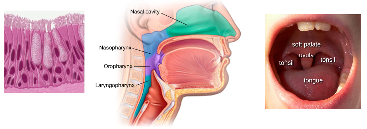
<p>The uppermost part of the pharynx. Lined with <strong>pseudostratified ciliated columnar epithelium</strong> for warming, humidifying, and filtering inspired air</p><ul><li><p><strong>Internal Nares</strong> (=posterior nares) are located on the posterior <strong>uvula</strong></p></li><li><p>Uvula and <strong>soft palate</strong> move posteriorly during swallowing to prevent food from entering the nasopharynx and nasal cavity</p></li></ul><p></p>
28
New cards

Oropharynx

Middle portion of the pharynx located posterior to the oral cavity.

  • Uvula → Larynx (epiglottis)

  • Lined with nonkeratinized stratified squamous epithelium

    • Tough, but not as tough as skin

  • More protective against mechanical stress as this cavity is a passageway for both air and food
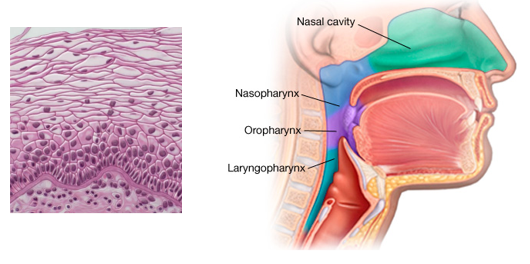
<p>Middle portion of the pharynx located posterior to the oral cavity.</p><ul><li><p>Uvula → Larynx (epiglottis)</p></li><li><p>Lined with <strong>nonkeratinized stratified squamous epithelium</strong></p><ul><li><p>Tough, but not as tough as skin</p></li></ul></li><li><p>More protective against mechanical stress as this cavity is a passageway for both air and food</p></li></ul><p></p>
29
New cards

Laryngopharynx

Bottom portion of the pharynx which extends from the hyoid bone to the esophagus.

  • Anteriorly - opens into larynx (voice box)

  • Posteriorly - opens into esophagus

  • Also a common passageway for both air and food

  • Lined with nonkeratinized stratified squamous epithelium
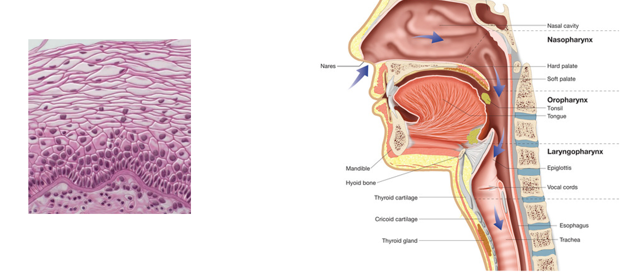
<p>Bottom portion of the pharynx which extends from the <strong>hyoid bone</strong> to the <strong>esophagus.</strong> </p><ul><li><p>Anteriorly - opens into larynx (voice box)</p></li><li><p>Posteriorly - opens into esophagus</p></li><li><p>Also a common passageway for both air and food </p></li><li><p>Lined with <strong>nonkeratinized stratified squamous epithelium </strong></p></li></ul><p></p>
30
New cards

Larynx (Voice Box)

Keeps food and liquids out of the remaining respiratory tract & houses the vocal cords

  • Superior to the vocal cords

    • Lined by stratified squamous nonkeratinized epithelium

    • Protects against mechanical stress where both food and air pass through

  • Inferior to the vocal cords

    • Lined by pseudostratified ciliated columnar epithelium

    • Cilia serve to propel mucus and debris up and out as one “clears their throat”
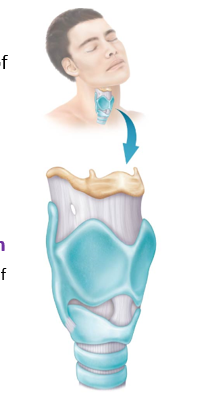
<p>Keeps food and liquids out of the remaining respiratory tract &amp; houses the vocal cords </p><ul><li><p>Superior to the vocal cords </p><ul><li><p>Lined by <strong>stratified squamous nonkeratinized epithelium</strong> </p></li><li><p>Protects against mechanical stress where both food and air pass through </p></li></ul></li><li><p>Inferior to the vocal cords</p><ul><li><p>Lined by <strong>pseudostratified ciliated columnar epithelium </strong></p></li><li><p>Cilia serve to propel mucus and debris up and out as one “clears their throat”</p></li></ul></li></ul><p></p>
31
New cards

Anatomy of the Larynx

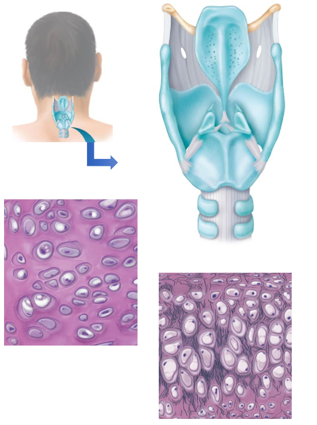
  • Contains 9 separate sections of cartilage that provide a flexible framework

    • 3 unpaired

    • 6 paired

    • Thyroid, cricoid, and most of the arytenoid cartilages are made of hyaline cartilage

    • Remaining sections are composed of elastic fibrocartilage

    • Elastic structures found within cartilaginous framework are involved in sound production

  • Supported by

    • Muscles that connect it to the surrounding neck

    • Muscles within the larynx itself

<ul><li><p>Contains 9 separate sections of cartilage that provide a flexible framework</p><ul><li><p>3 unpaired</p></li><li><p>6 paired</p></li><li><p>Thyroid, cricoid, and most of the arytenoid cartilages are made of <strong>hyaline cartilage </strong></p></li><li><p>Remaining sections are composed of <strong>elastic fibrocartilage </strong></p></li><li><p>Elastic structures found within cartilaginous framework are involved in sound production </p></li></ul></li><li><p>Supported by</p><ul><li><p>Muscles that connect it to the surrounding neck</p></li><li><p>Muscles within the larynx itself </p></li></ul></li></ul><p></p>
32
New cards

Thyroid Cartilage

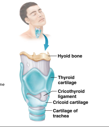
Unpaired, largest shield-like piece forming the anterior and superior walls of the larynx.

  • Connected superiorly to the hyoid bone

  • Connected inferiorly to the cricoid cartilage by a fibrous membrane

  • Contains “Adam’s Apple”

<p><strong>Unpaired</strong>, largest shield-like piece forming the anterior and superior walls of the larynx. </p><ul><li><p>Connected superiorly to the <strong>hyoid bone</strong> </p></li><li><p>Connected inferiorly to the <strong>cricoid cartilage</strong> by a fibrous membrane </p></li><li><p>Contains “Adam’s Apple”</p></li></ul><p></p>
33
New cards

“Adam’s Apple”

Anterior protrusion of the thyroid cartilage.

  • Generally more prominent in males

    • Resulting in deeper voices

  • Development is influenced by testosterone

34
New cards

Epiglottis

Unpaired, leaf-shaped flap of elastic cartilage found posteriorly to the thyroid cartilage.

  • Base attached to the posterior side of the thyroid cartilage

  • Superior free edge is not attached to surroundings

    • Free superior edge usually stands upright

    • Creates an opening called the glottis

      • Closes off when swallowing to allow food into the esophagus

      • Opens when breathing to allow air into the trachea
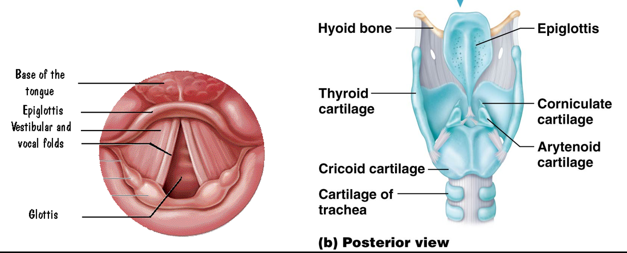
<p>Unpaired, leaf-shaped flap of elastic cartilage found posteriorly to the thyroid cartilage.</p><ul><li><p>Base attached to the posterior side of the thyroid cartilage</p></li><li><p>Superior free edge is not attached to surroundings</p><ul><li><p>Free superior edge usually stands upright</p></li><li><p>Creates an opening called the glottis</p><ul><li><p>Closes off when swallowing to allow food into the esophagus </p></li><li><p>Opens when breathing to allow air into the trachea</p></li></ul></li></ul></li></ul><p></p>
35
New cards

Swallowing Process

  • Surrounding muscles elevate the larynx

  • The glottis is closed by the epiglottis

    • Prevents food and liquids from entering the larynx
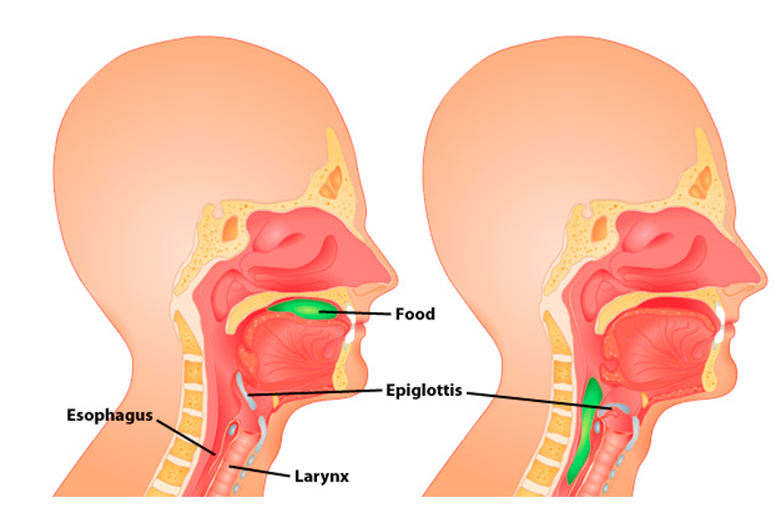
<ul><li><p>Surrounding muscles elevate the larynx</p></li><li><p>The glottis is closed by the epiglottis</p><ul><li><p>Prevents food and liquids from entering the larynx </p></li></ul></li></ul><p></p>
36
New cards

Cough Refelx

Reflex that helps to expel food and/or liquids that manage to get through the glottis and into the larynx

  • Prevents damage to the remaining respiratory tract

  • During a cough, the epiglottis quickly closes off the trachea to prevent any foreign objects or substances from getting in

    • Helps protect the lungs from infection, irritation, or injury

37
New cards

Unpaired Sections of Cartilage

  • Thyroid Cartilage

  • Epiglottis

  • Cricoid Cartilage

38
New cards

Cricoid Cartilage

Third and last of the unpaired sections of the larynx cartilage.

  • Shaped like a ring

  • Found inferior to the thyroid cartilage

    • Connected by the cricothyroid ligament
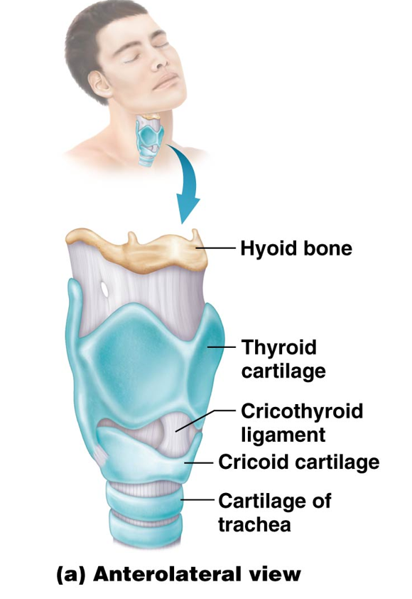
<p>Third and last of the unpaired sections of the larynx cartilage. </p><ul><li><p>Shaped like a ring</p></li><li><p>Found inferior to the thyroid cartilage </p><ul><li><p> Connected by the cricothyroid ligament</p></li></ul></li></ul><p></p>
39
New cards

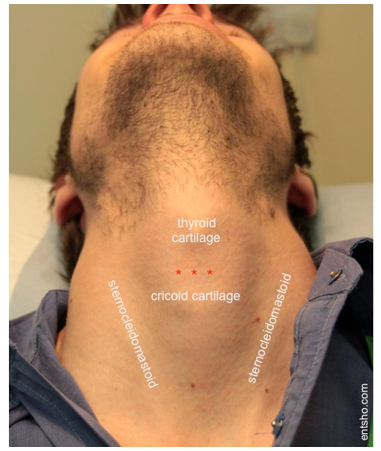
Cricothyroid Ligament

A thin membrane that connects the cricoid cartilage to the thyroid cartilage.

  • Cricothyroidotomy

    • A tube can be inserted through this ligament to provide an emergency airway
<p>A thin membrane that connects the cricoid cartilage to the thyroid cartilage. </p><ul><li><p>Cricothyroidotomy </p><ul><li><p>A tube can be inserted through this ligament to provide an emergency airway</p></li></ul></li></ul><p></p>
40
New cards

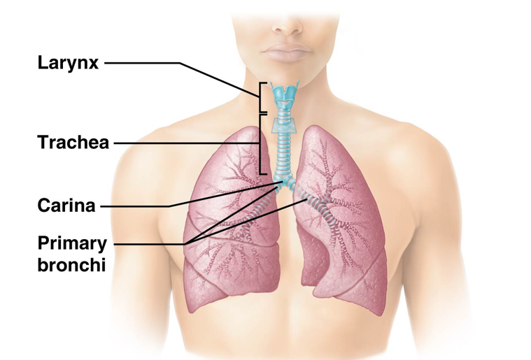
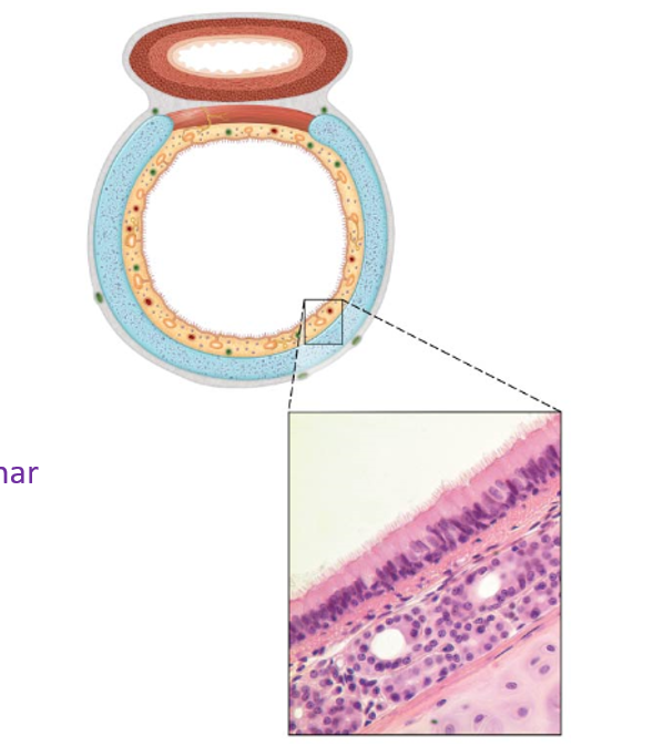
Trachea (Windpipe)

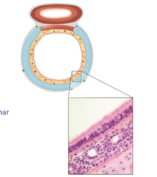
Part of the lower respiratory tract beginning in the inferior neck and extending to the mediastinum.

  • Anterior and lateral surfaces covered by hyaline cartilage rings in a C shape

  • Posterior surface covered with elastic connective tissue and smooth muscle

    • Allow esophagus to expand during swallowing

  • Supportive enough to keep the trachea open, or patent, but flexible enough to allow a change in diameter during pulmonary ventilation
<p>Part of the lower respiratory tract beginning in the inferior neck and extending to the mediastinum. </p><ul><li><p>Anterior and lateral surfaces covered by hyaline cartilage rings in a C shape</p></li><li><p>Posterior surface covered with elastic connective tissue and smooth muscle </p><ul><li><p>Allow esophagus to expand during swallowing</p></li></ul></li><li><p>Supportive enough to keep the trachea open, or patent, but flexible enough to allow a change in diameter during pulmonary ventilation </p></li></ul><p></p>
41
New cards

Carina

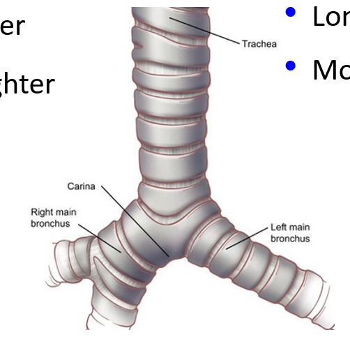
The last tracheal cartilage ring.

  • The mucosa contains sensory receptors that trigger a violent cough reflex if foreign materials contact them
<p>The last tracheal cartilage ring.</p><ul><li><p>The mucosa contains sensory receptors that trigger a <strong>violent cough reflex </strong>if foreign materials contact them</p></li></ul><p></p>
42
New cards

Histological Pattern of Trachea

  • Mucosa

    • Lined with pseudostratified ciliated columnar epithelium and goblet cells

  • Submucosa

    • Loose connective tissue & cartilage rings

  • Adventitia or Serosa

    • Dense irregular connective tissue

<ul><li><p>Mucosa</p><ul><li><p>Lined with pseudostratified ciliated columnar epithelium and goblet cells</p></li></ul></li><li><p>Submucosa</p><ul><li><p>Loose connective tissue &amp; cartilage rings </p></li></ul></li><li><p>Adventitia or Serosa</p><ul><li><p>Dense irregular connective tissue</p></li></ul></li></ul><p></p>
43
New cards

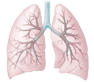
Bronchial Tree

Inside the lungs is a series of progressively smaller tubes that end in alveoli. Each bronchus branches to form this
<p>Inside the lungs is a series of progressively smaller tubes that end in alveoli. Each bronchus branches to form this </p>
44
New cards

Primary Bronchi

Air enters either the left or right primary bronchus (at the hilum). Structure due to the position of the heart in relation to the left lung

  • Right Primary Bronchus

    • Wider

    • Shorter

    • Straighter

    • More likely side for inhaling foreign objects

  • Left Primary Bronchus

    • Narrower

    • Longer

    • More Horizontal

<p>Air enters either the left or right primary bronchus (at the hilum). Structure due to the position of the heart in relation to the left lung </p><ul><li><p>Right Primary Bronchus</p><ul><li><p>Wider</p></li><li><p>Shorter</p></li><li><p>Straighter</p></li><li><p>More likely side for inhaling foreign objects</p></li></ul></li><li><p>Left Primary Bronchus</p><ul><li><p>Narrower</p></li><li><p>Longer</p></li><li><p>More Horizontal</p></li></ul></li></ul><p></p>
45
New cards

Secondary Bronchi (Lobar Bronchus)

Present inside each lung

  • Three in the right lung

  • Two in the left lung (due to the position of the heart)
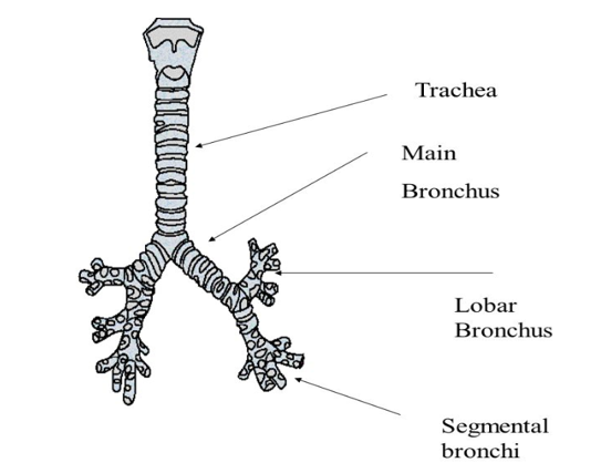
<p>Present inside each lung</p><ul><li><p>Three in the right lung </p></li><li><p>Two in the left lung (due to the position of the heart)</p></li></ul><p></p>
46
New cards

Tertiary Bronchi

  • Secondary bronchi branch into about 10 smaller tertiary bronchi per lung

  • Continue to branch into smaller and smaller branches
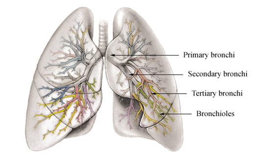
<ul><li><p>Secondary bronchi branch into about 10 smaller tertiary bronchi per lung </p></li><li><p>Continue to branch into smaller and smaller branches</p></li></ul><p></p>
47
New cards

Bronchi Histology

As airways divide and get smaller, histology changes significantly

  • Cartilage

    • C-shaped → complete rings → progressively fewer irregular plates

  • Epithelium

    • Gradually changes from pseudostratified → simple columnar cells

  • Smooth Muscle

    • Amount increases as bronchi get smaller

      • Allows control of airflow

<p>As airways divide and get smaller, histology changes significantly</p><ul><li><p>Cartilage</p><ul><li><p>C-shaped → complete rings → progressively fewer irregular plates</p></li></ul></li><li><p>Epithelium</p><ul><li><p>Gradually changes from <strong>pseudostratified → simple columnar cells</strong></p></li></ul></li><li><p>Smooth Muscle</p><ul><li><p>Amount increases as bronchi get smaller</p><ul><li><p>Allows control of airflow</p></li></ul></li></ul></li></ul><p></p>
48
New cards

Bronchioles

Smallest airways; features include:

  • Less than 1 mm in diameter

  • Lack cartilage (kind of leaky)

  • Have a thicker ring of smooth muscle

  • Simple cuboidal epithelium with few cilia & a few goblet cells

    • Cilia and goblet cells help to move air

  • Continue to branch until they become terminal bronchioles
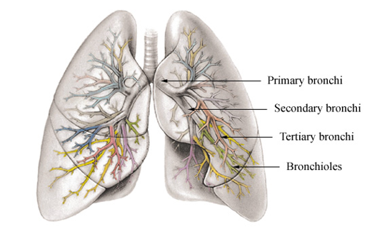
<p>Smallest airways; features include:</p><ul><li><p>Less than 1 mm in diameter</p></li><li><p>Lack cartilage (kind of leaky)</p></li><li><p>Have a thicker ring of smooth muscle</p></li><li><p><strong>Simple cuboidal epithelium</strong> with few cilia &amp; a few goblet cells</p><ul><li><p>Cilia and goblet cells help to move air</p></li></ul></li><li><p>Continue to branch until they become terminal bronchioles</p></li></ul><p></p>
49
New cards

Terminal Bronchioles

Bronchioles that branch into two or more smaller respiratory bronchioles

  • Surrounded by a thin layer of smooth muscle

  • The conducting zone of the respiratory tract ends when inspired air reaches here
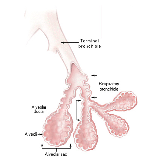
<p>Bronchioles that branch into two or more smaller respiratory bronchioles</p><ul><li><p>Surrounded by a thin layer of smooth muscle</p></li><li><p>The <strong>conducting zone</strong> of the respiratory tract ends when inspired air reaches here</p></li></ul><p></p>
50
New cards

Respiratory Bronchioles

Bronchioles that branch into two or more alveolar ducts

  • The respiratory zone begins here, with alveoli budding from the walls
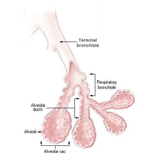
<p>Bronchioles that branch into two or more alveolar ducts</p><ul><li><p>The <strong>respiratory zone </strong>begins here, with alveoli budding from the walls</p></li></ul><p></p>
51
New cards

Alveolar Ducts

Ducts that branch off of respiratory bronchioles.

  • Have alveoli attached to their walls

  • End in alveolar sacs (grape-like clusters of alveoli)
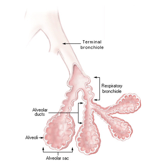
<p>Ducts that branch off of respiratory bronchioles. </p><ul><li><p>Have alveoli attached to their walls</p></li><li><p>End in <strong>alveolar sacs</strong> (grape-like clusters of alveoli)</p></li></ul><p></p>
52
New cards

Airflow to the Alveoli

Nares → Nasal Cavity → Nasopharynx → Oropharynx → Laryngopharynx → Larynx → Trachea → Primary Bronchi → Secondary Bronchi → Tertiary Bronchi → Multiple branches of Bronchi → Bronchioles → Terminal Bronchioles → Respiratory Bronchioles → Alveolar Ducts → Alveolar Sacs
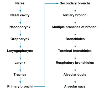
<p>Nares → Nasal Cavity → Nasopharynx → Oropharynx → Laryngopharynx → Larynx → Trachea → Primary Bronchi → Secondary Bronchi → Tertiary Bronchi → Multiple branches of Bronchi → Bronchioles → Terminal Bronchioles → Respiratory Bronchioles → Alveolar Ducts → Alveolar Sacs</p>
53
New cards

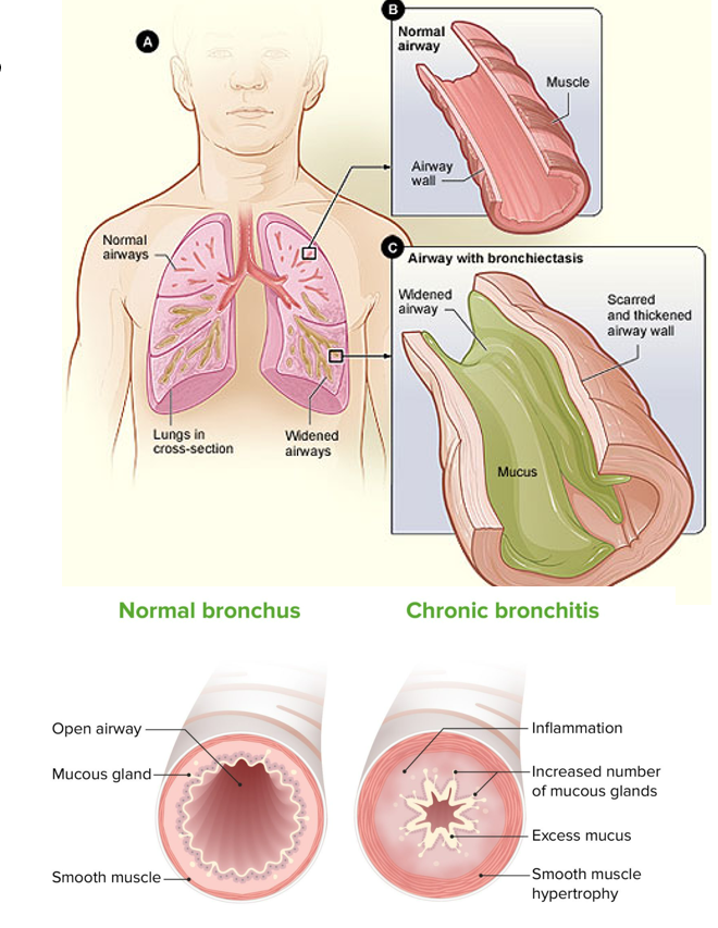
Bronchitis

Inflammation of the bronchi caused by infection or inhaled irritants. Has acute and chronic forms.
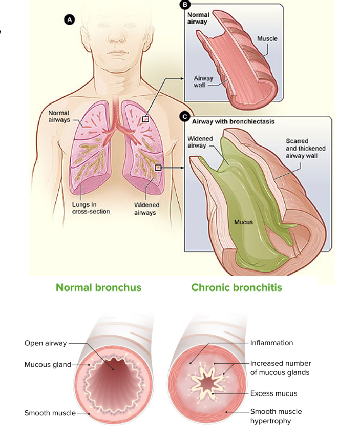
<p>Inflammation of the bronchi caused by infection or inhaled irritants. Has acute and chronic forms.</p>
54
New cards

Acute Bronchitis

  • Occurs during or after an infection

  • Coughing, sneezing, pain upon inhalation, fever

  • Most cases last 10 - 14 days

<ul><li><p>Occurs during or after an infection </p></li><li><p>Coughing, sneezing, pain upon inhalation, fever</p></li><li><p>Most cases last 10 - 14 days </p></li></ul><p></p>
55
New cards

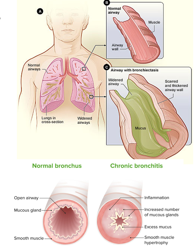
Chronic Bronchitis

  • Occurs after long-term irritant exposure

  • Large amounts of mucus and cough lasting 3 months

  • Permanent changes to bronchi occur

  • Increases the likelihood of future bacterial infections
<ul><li><p>Occurs after long-term irritant exposure</p></li><li><p>Large amounts of mucus and cough <strong>lasting 3 months</strong></p></li><li><p><strong>Permanent changes to bronchi occur</strong></p></li><li><p>Increases the likelihood of future bacterial infections</p></li></ul><p></p>
56
New cards

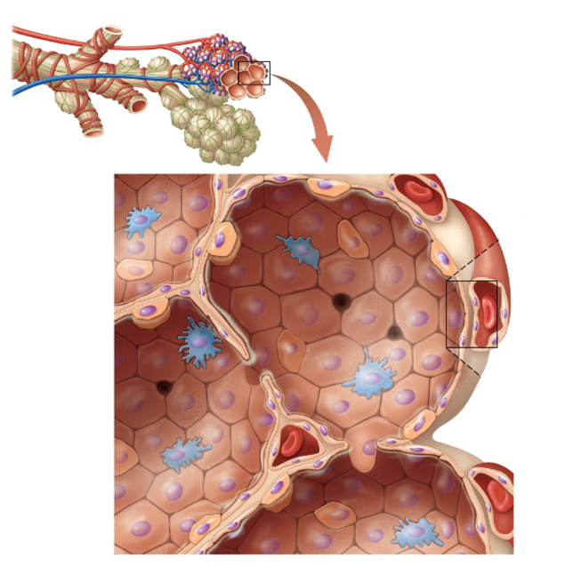
Alveoli

Round, thin-walled “bubble”-like chambers that are the final destination for inspired air within the respiratory tract.

  • Structure increases surface area → increasing gas exchange efficiency

  • Made of three different cell types:

    • Type I Alveolar Cells

    • Type II Alveolar Cells

    • Alveolar Macrophages

<p>Round, thin-walled “bubble”-like chambers that are the final destination for inspired air within the respiratory tract. </p><ul><li><p>Structure increases surface area → increasing gas exchange efficiency</p></li><li><p>Made of three different cell types:</p><ul><li><p>Type I Alveolar Cells</p></li><li><p>Type II Alveolar Cells</p></li><li><p>Alveolar Macrophages</p></li></ul></li></ul><p></p>
57
New cards

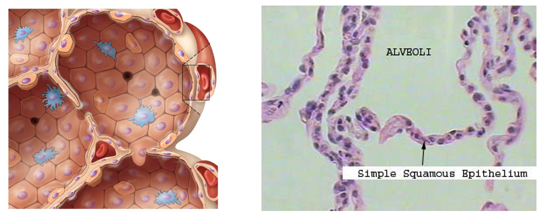
Type I Alveolar Cells

Squamous cells that account for about 90% of the cells in the alveolar wall.

  • Very Thin

  • Allow for rapid diffusion of gases across cell membranes
<p><strong>Squamous cells </strong>that account for about 90% of the cells in the alveolar wall.</p><ul><li><p>Very Thin</p></li><li><p>Allow for rapid diffusion of gases across cell membranes</p></li></ul><p></p>
58
New cards

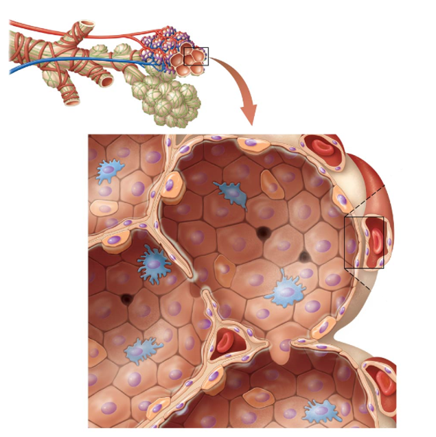
Type II Alveolar Cells

Small, cuboidal cells that account for about 10% of the cells in the alveolar wall

  • Glandular cells

  • Responsible for the synthesis of surfactant

    • Chemical that helps reduce surface tension on alveoli
<p>Small, cuboidal cells that account for about 10% of the cells in the alveolar wall </p><ul><li><p>Glandular cells</p></li><li><p>Responsible for the synthesis of surfactant </p><ul><li><p>Chemical that helps reduce surface tension on alveoli </p></li></ul></li></ul><p></p>
59
New cards

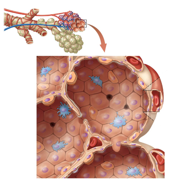
Alveolar Macrophages (Dust Cells)

Mobile phagocytes in the alveoli.

  • Derived from bone marrow cells

  • Clean up and digest debris that made its way into alveolis
<p>Mobile phagocytes in the alveoli.</p><ul><li><p>Derived from bone marrow cells</p></li><li><p>Clean up and digest debris that made its way into alveolis</p></li></ul><p></p>
60
New cards

Anatomy of the Lungs

The right and left lungs are separated by the heart and mediastinum

  • Lung’s Base

  • Superior Apex

  • Costal Surface

  • Diaphragmatic Surface

  • Mediastinal Surface

    • Hilum - triangular depression

      • Primary bronchi, blood & lymphatic vessels, and nerves enter and exit the lungs here

    • Cardiac Notch - groove in the left lung

61
New cards

Costal Surfaces

Anterior, posterior, and lateral surfaces of the lungs that come into contact with the rib cage.

62
New cards

Lobes of the Lungs

  • The right lung has three lobes

    • Superior

    • Middle

    • Inferior

    • Fissures

      • Horizontal Fissure

        • Separates the superior from the middle lobe

      • Oblique Fissure

        • Separates the middle from the inferior lobe

  • The left lung has two lobes

    • Superior

    • Inferior

    • Fissures

      • Oblique

        • Separates the superior from the inferior lobe

  • Each secondary (lobar) bronchus supplies one lobe
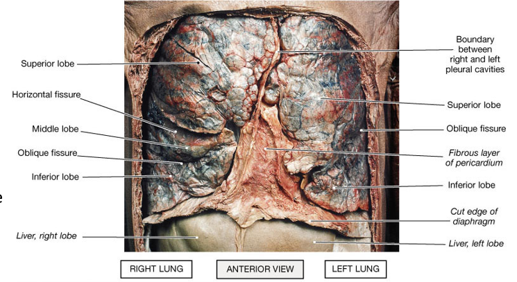
<ul><li><p>The right lung has three lobes </p><ul><li><p>Superior</p></li><li><p>Middle</p></li><li><p>Inferior </p></li><li><p>Fissures</p><ul><li><p>Horizontal Fissure </p><ul><li><p>Separates the superior from the middle lobe</p></li></ul></li><li><p>Oblique Fissure</p><ul><li><p>Separates the middle from the inferior lobe </p></li></ul></li></ul></li></ul></li><li><p>The left lung has two lobes </p><ul><li><p>Superior</p></li><li><p>Inferior </p></li><li><p>Fissures</p><ul><li><p>Oblique</p><ul><li><p>Separates the superior from the inferior lobe</p></li></ul></li></ul></li></ul></li><li><p>Each secondary (lobar) bronchus supplies one lobe </p></li></ul><p></p>
63
New cards

Smoking (Clinical Connection)

Causes respiratory changes with increased chance of:

  • Respiratory Infections

  • Cellular or Genetic Changes to the Lungs

  • Emphysema (Permanent Alveoli Damage)

  • Cancer of the Lungs, Esophagus, Stomach, and Pancreas

  • Stomach Ulcers

  • Atherosclerosis

  • Lower Birth Weight Babies in Pregnant Women

  • Poor Delivery of Oxygen and Nutrients to all Systemic Tissues

  • Bronchitis, Asthma, Ear Infections

  • All primary effects can also result from secondhand smoke
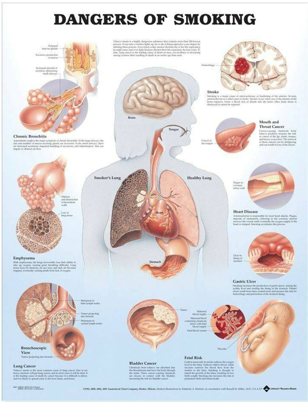
<p>Causes respiratory changes with increased chance of:</p><ul><li><p>Respiratory Infections</p></li><li><p>Cellular or Genetic Changes to the Lungs</p></li><li><p>Emphysema (Permanent Alveoli Damage)</p></li><li><p>Cancer of the Lungs, Esophagus, Stomach, and Pancreas</p></li><li><p>Stomach Ulcers</p></li><li><p>Atherosclerosis</p></li><li><p>Lower Birth Weight Babies in Pregnant Women</p></li><li><p>Poor Delivery of Oxygen and Nutrients to all Systemic Tissues</p></li><li><p>Bronchitis, Asthma, Ear Infections</p></li><li><p>All primary effects can also result from secondhand smoke </p></li></ul><p></p>
64
New cards

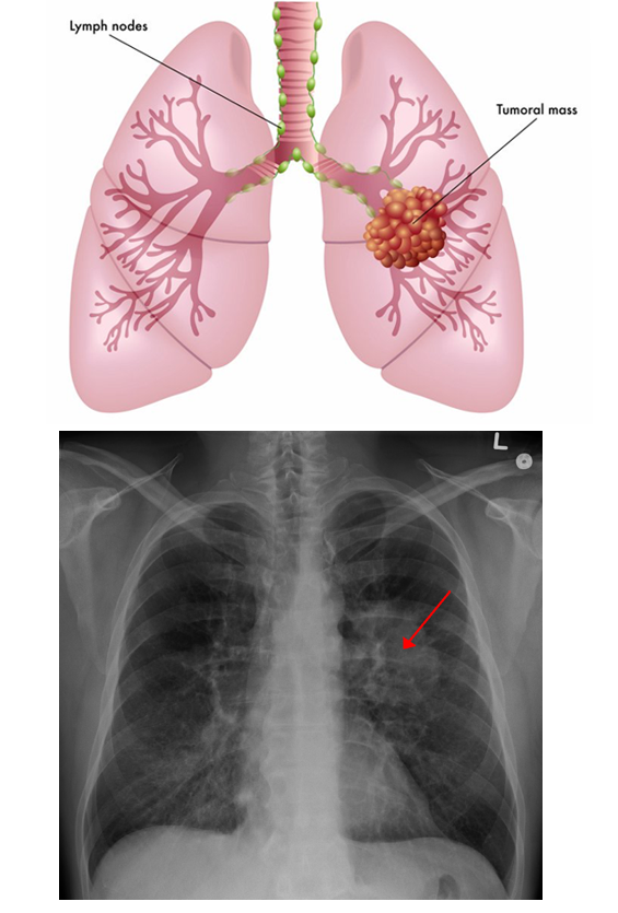
Lung Cancer (Clinical Connection)

Highly aggressive and frequently fatal malignancy originating in the respiratory epithelium.

  • Smoking causes about 85% of all lung cancer

  • Symptoms

    • Chronic Cough

    • Coughing up Blood

    • Excess Pulmonary Mucus

    • Increased Pulmonary Stress

  • Types:

    • Non-Small

    • Small Cell

<p>Highly aggressive and frequently fatal malignancy originating in the respiratory epithelium.</p><ul><li><p>Smoking causes about 85% of all lung cancer</p></li><li><p>Symptoms</p><ul><li><p>Chronic Cough</p></li><li><p>Coughing up Blood</p></li><li><p>Excess Pulmonary Mucus</p></li><li><p>Increased Pulmonary Stress</p></li></ul></li><li><p>Types: </p><ul><li><p>Non-Small</p></li><li><p>Small Cell</p></li></ul></li></ul><p></p>
65
New cards

Non-Small Cell Lung Cancer (Oat Cell)

Usually metastisizes from the lungs to the brain.

  • Cancer cells look like oats under a microscope

66
New cards

Pleural Cavity

Subdivision of the thoracic cavity located between two layers of serous membrane containing the lungs: the parietal & visceral pleura.
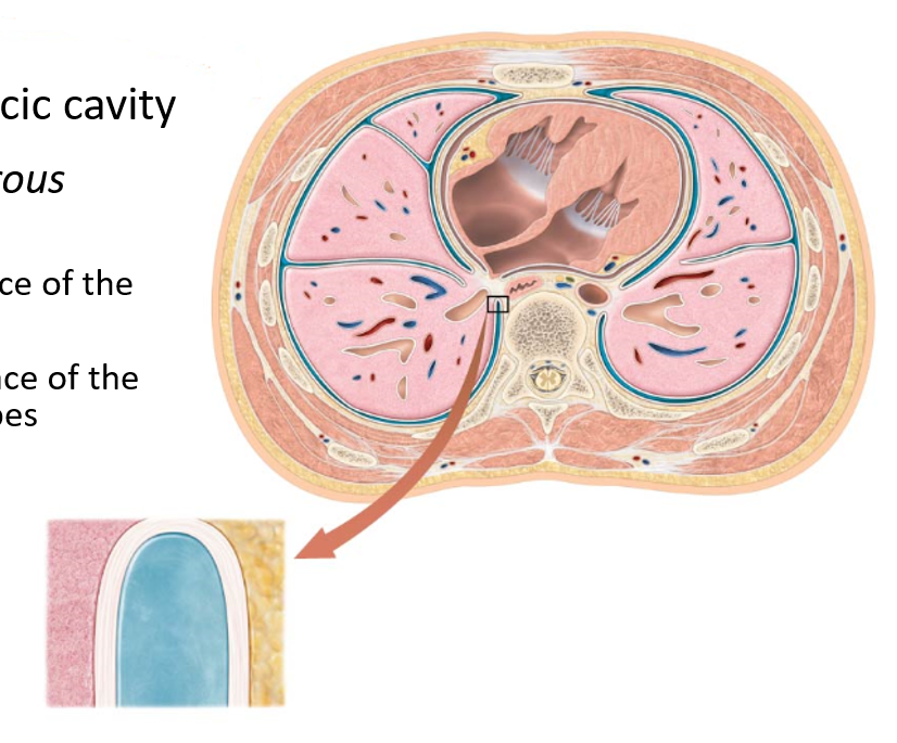
<p>Subdivision of the thoracic cavity located between two layers of serous membrane containing the lungs: the parietal &amp; visceral pleura.</p>
67
New cards

Parietal Pleura

Serous membrane lining the inner surface of the pleural cavity.
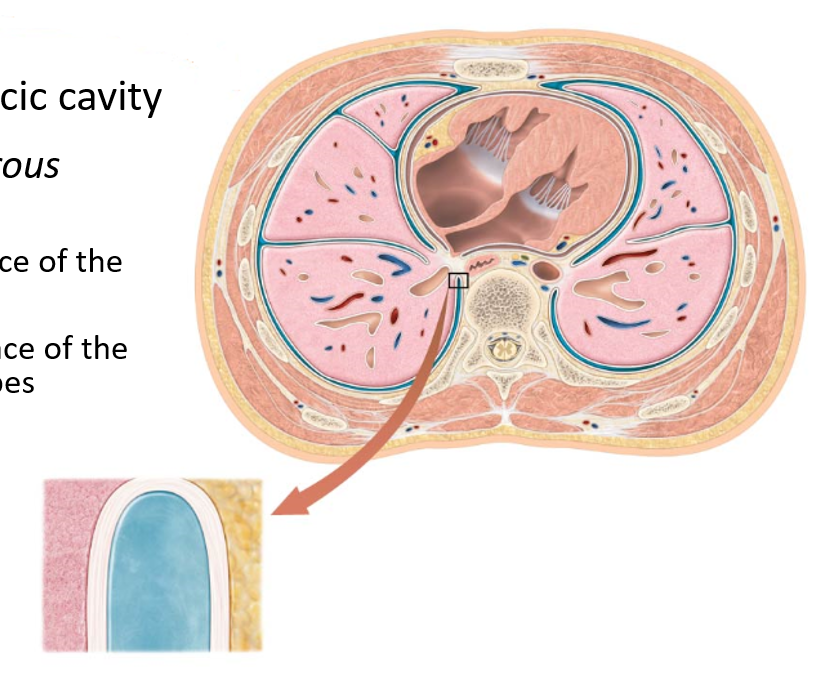
<p>Serous membrane lining the inner surface of the pleural cavity.</p>
68
New cards

Visceral Pleura

The serous membrane lining the outer surface of the lungs dives into fissures between lobes.

  • At the hilum, the parietal pleura turns over on itself to create this inner layer of membrane
<p>The serous membrane lining the outer surface of the lungs dives into fissures between lobes. </p><ul><li><p>At the hilum, the parietal pleura turns over on itself to create this inner layer of membrane</p></li></ul><p></p>
69
New cards

Pleural Fluid

A thin layer of serous fluid is secreted by the pleural membrane, lubricating the delicate surfaces of the lungs.
<p>A thin layer of serous fluid is secreted by the pleural membrane, lubricating the delicate surfaces of the lungs.</p>
70
New cards

Pleuritis

Heart failure to pneumonia can cause inflammation of the pleura.

  • Common Symptom: Pleuritic Pain

  • Treated by addressing underyling medical condition but may persist for months even after that condition has been resolved

71
New cards

Function & Composition of Regions of the Conducting and Respiratory Zones
<p></p>
72
New cards

Pressure-Volume Relationship

Provides the driving force for pulmonary ventilation.
<p>Provides the driving force for pulmonary ventilation.</p>
73
New cards

Pulmonary Ventilation (Breathing)

The first process of respiration consists of two phases

  • Inspiration or Inhalation

  • Expiration or Exhalation

  • One breath is an inhalation and an exhalation
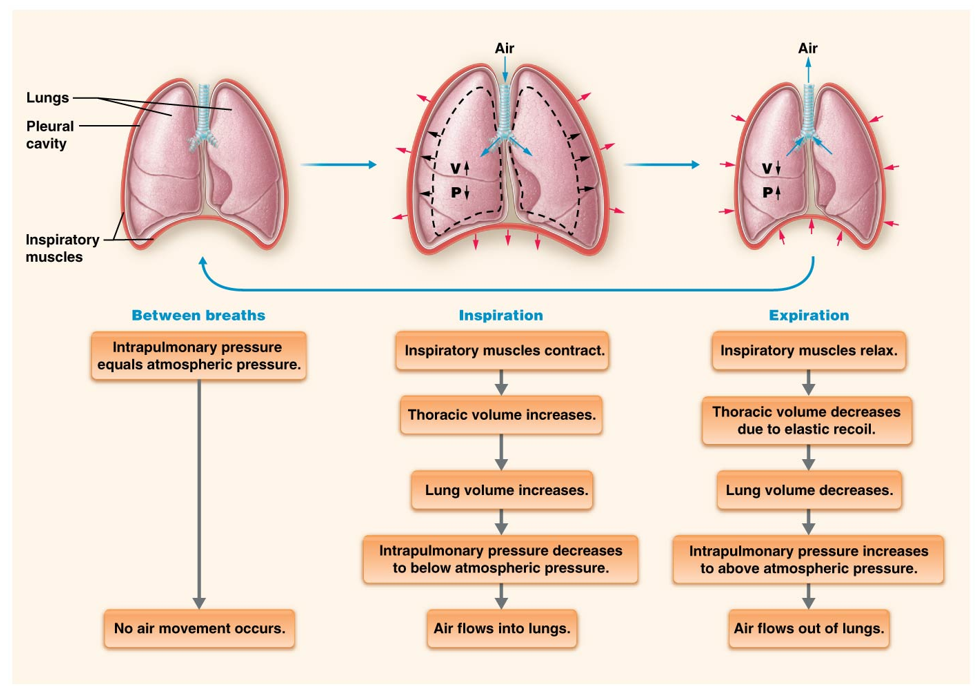
<p>The first process of respiration consists of two phases</p><ul><li><p>Inspiration or Inhalation</p></li><li><p>Expiration or Exhalation</p></li><li><p>One breath is an inhalation and an exhalation</p></li></ul><p></p>
74
New cards

Air

Mixture of gas molecules whose movement is dependent on atmospheric pressure gradients.

  • Gas molecules move from areas of high pressure to areas of low pressure

    • The body is a low-pressure system compared to the external air

      • Air is constantly being pulled in

  • Pressure gradients drive the movement of air molecules during inspiration and expiration
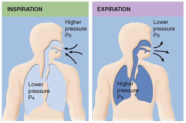
<p>Mixture of gas molecules whose movement is dependent on atmospheric pressure gradients.</p><ul><li><p>Gas molecules move from areas of high pressure to areas of low pressure</p><ul><li><p>The body is a low-pressure system compared to the external air</p><ul><li><p>Air is constantly being pulled in</p></li></ul></li></ul></li><li><p>Pressure gradients drive the movement of air molecules during inspiration and expiration</p></li></ul><p></p>
75
New cards

Boyle’s Law

A law that states that at constant temperature and number of gas molecules, the pressure and volume of a gas are inversely related such that:

  • As the volume of a container increases → the pressure the gas exerts on the container decreases

  • As the volume of a container decreases → the pressure the gas exerts on the container increases

76
New cards

Inspiratory Muscles

The lungs rely on these muscles to change their volume during ventilation.

  • Diaphragm - the main type of this muscle in humans

    • Controlled by the phrenic nerves, starts at birth

    • Sits under both lungs

  • External Intercostals

    • Controlled by the intercostal nerves

<p>The lungs rely on these muscles to change their volume during ventilation.</p><ul><li><p>Diaphragm - the main type of this muscle in humans</p><ul><li><p>Controlled by the phrenic nerves, starts at birth</p></li><li><p>Sits under both lungs </p></li></ul></li><li><p>External Intercostals</p><ul><li><p>Controlled by the intercostal nerves</p></li></ul></li></ul><p></p>
77
New cards

Inhalation

Process of taking in air in which inspiratory muscles increase lung volume indirectly by increasing the thoracic cavity size

  • Thoracic cavity expands due to contraction of external intercostals & diaphragm→ pulls on parietal pleura → pulls on visceral pleura → pulls lungs outward → increases volume of the lungs

  • Due to low pressure on the inside and higher pressure on the outside of the body
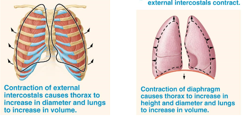
<p>Process of taking in air in which inspiratory muscles increase lung volume indirectly by increasing the thoracic cavity size</p><ul><li><p>Thoracic cavity expands due to contraction of external intercostals &amp; diaphragm→ pulls on parietal pleura → pulls on visceral pleura → pulls lungs outward → increases volume of the lungs</p></li><li><p>Due to low pressure on the inside and higher pressure on the outside of the body</p></li></ul><p></p>
78
New cards

Expiration

Normally a mostly passive process that does not utilize muscle contraction. Two things happen when inspiratory muscles relax:

  1. Diaphragm returns to its original dome shape that pushes up on lungs & external intercostals relax

  2. Elastic tissue in lungs recoils

  • Recoil & Diaphragm Relaxation Together → decrease in lung volume & rise in intrapulmonary pressure above atmospheric pressure so air flows out of the lungs

  • Due to a higher pressure inside the body than outside, inhaling air causes release of air
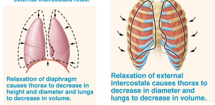
<p>Normally a mostly passive process that does not utilize muscle contraction. Two things happen when inspiratory muscles relax:</p><ol><li><p>Diaphragm returns to its original dome shape that pushes up on lungs &amp; external intercostals relax</p></li><li><p>Elastic tissue in lungs recoils</p></li></ol><ul><li><p>Recoil &amp; Diaphragm Relaxation Together → decrease in lung volume &amp; rise in intrapulmonary pressure above atmospheric pressure so air flows out of the lungs</p></li><li><p>Due to a higher pressure inside the body than outside, inhaling air causes release of air</p></li></ul><p></p>
79
New cards

Forceful Expiration

Forceful exhalation accomplished by:

  • Internal Intercoastals & abdominal muscles forcefully decrease the size of the thoracic cavity (Accessory muscles of expiration)

  • Slapping a person on the back

  • Abdominal thrusts (aka Heimlich manuever) that push up on the diaphragm

    • Used on people who are choking, in hopes that forceful expiration will dislodge obstruction and restore breathing

    • Should not be used on unconscious people

80
New cards

Skeletal Muscles of Breathing

Muscles of Quiet/Normal Breathing

  • Diaphragm

  • External Intercostals

Muscles of Forced Inspiration: Pull up and outward

  • Sternocleidomastoid

  • Scalenes

  • Serratus Posterior Superior

  • Pectoralis Minor

  • Erector Spinae

Muscles of Forced Expiration: Pull down and inward

  • Transversus Thoracis

  • Serratus Posterior Inferior

  • Internal Intercostals

  • External Oblique

  • Transversus Abdominis

<p>Muscles of Quiet/Normal Breathing</p><ul><li><p><strong>Diaphragm</strong></p></li><li><p><strong>External Intercostals</strong></p></li></ul><p>Muscles of Forced Inspiration: Pull up and outward</p><ul><li><p>Sternocleidomastoid</p></li><li><p>Scalenes</p></li><li><p>Serratus Posterior Superior</p></li><li><p>Pectoralis Minor</p></li><li><p>Erector Spinae</p></li></ul><p>Muscles of Forced Expiration: Pull down and inward</p><ul><li><p>Transversus Thoracis</p></li><li><p>Serratus Posterior Inferior</p></li><li><p>Internal Intercostals</p></li><li><p>External Oblique</p></li><li><p>Transversus Abdominis</p></li></ul><p></p>
81
New cards

Nonrespiratory Movements

Movements not intended for ventilation.

  • Include yawns, coughs, sneezes, and sighs

  • Help keep the alveoli inflated and prevent obstruction of the airways

82
New cards

Sigh

A type of nonrespiratory movement.

  • A slow and deep inspiration that is held and followed by a slow expiration.

  • Function

    • Reopens local groups of collapsed alveoli and stimulates the release of surfactant.

83
New cards

Yawn

A type of nonrespiratory movement.

  • A large sigh that takes the lung volume to its inspiratory capacity.

  • Functions

    • Opens collapsed alveoli

    • Yawning when tired occurs to minimize alveolar collapse during sleep

    • Yawning after sleep opens alveoli that have collapsed during sleep

84
New cards

Sneeze

A type of nonrespiratory movement.

  • Deep inspiration followed by a large, forceful expiration through the nose at a velocity of about 100 miles per hour.

  • Function

    • Clears foreign or irritating substances from the nasal cavity

85
New cards

Cough

A type of nonrespiratory movement.

  • Similar to a sneeze except the initial inspiration is small or absent; velocity can approach 500 miles per hour.

  • Function

    • Cleans the larynx, trachea, or lower airways

86
New cards

Factors that Affect Ventilation

Three primary physical factors influence overall effectiveness of pulmonary ventilation.

  1. Airway Resistance

  2. Alveolar Surface Tension

  3. Pulmonary Compliance

87
New cards

Airway Resistance

Defined as anything that impedes air flow through the respiratory tract

  • Factor of pulmonary ventilation effectiveness

  • Largely determined by airway diameter

  • Resistance varies during pulmonary ventilation

    • Decreases slightly during inspiration as airways are pulled open and the lungs expand

    • Increases slightly as lungs recoil and the airways narrow during expiration

  • Certain disease states may also increase airway resistance by

    • Inflammation

    • Obstruction

    • Bacterial or Viral Pathogens

<p>Defined as anything that impedes air flow through the respiratory tract</p><ul><li><p>Factor of pulmonary ventilation effectiveness</p></li><li><p>Largely determined by airway diameter</p></li><li><p>Resistance varies during pulmonary ventilation</p><ul><li><p>Decreases slightly during inspiration as airways are pulled open and the lungs expand</p></li><li><p>Increases slightly as lungs recoil and the airways narrow during expiration</p></li></ul></li><li><p>Certain disease states may also increase airway resistance by</p><ul><li><p>Inflammation</p></li><li><p>Obstruction</p></li><li><p>Bacterial or Viral Pathogens</p></li></ul></li></ul><p></p>
88
New cards

Decrease in Airway Resistance

Neurons of SNS release NE during excercise, stress, or emergency →

Triggers bronchodilation →

Increases efficiency of pulmonary ventilation

89
New cards

Diameter of Bronchioles

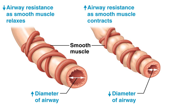
Controlled by smooth muscle contraction and relaxation

  • Relaxation (Bronchodilation) → increases the diameter of bronchioles

    • Decreases airway resistance and increases airflow

  • Contraction (Bronchoconstriction) → decreases the diameter of bronchioles

    • Increases airway resistance and decreases airflow
<p>Controlled by smooth muscle contraction and relaxation </p><ul><li><p>Relaxation (<strong>Bronchodilation</strong>) → increases the diameter of bronchioles </p><ul><li><p>Decreases airway resistance and increases airflow</p></li></ul></li><li><p>Contraction (<strong>Bronchoconstriction</strong>) → decreases the diameter of bronchioles</p><ul><li><p>Increases airway resistance and decreases airflow  </p></li></ul></li></ul><p></p>
90
New cards

Alveolar Surface Tension

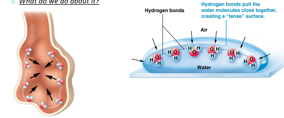
Alveoli are covered with a thin film of liquid composed mainly of water, creating a gas-water boundary.

  • Factor of pulmonary ventilation effectiveness

  • A gas-water boundary exists within each alveolus because water molecules form hydrogen bonds with each other

    • Creates surface tension

      • Makes alveoli tend to collapse (opposite walls start to cling to one another) if surface tension is not reduced

  • Managed by surfactant chemical released by type II alveolar cells
<p>Alveoli are covered with a thin film of liquid composed mainly of water, creating a gas-water boundary.</p><ul><li><p>Factor of pulmonary ventilation effectiveness</p></li><li><p>A gas-water boundary exists within each alveolus because water molecules form hydrogen bonds with each other </p><ul><li><p>Creates surface tension</p><ul><li><p>Makes alveoli tend to collapse (opposite walls start to cling to one another) if surface tension is not reduced</p></li></ul></li></ul></li><li><p>Managed by surfactant chemical released by type II alveolar cells</p></li></ul><p></p>
91
New cards

Surfactant

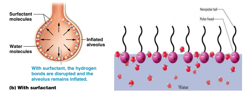
A chemical component of the liquid film coating the cells of the alveolus

  • Produced by type II alveolar cells

  • Chemical structure similar to a detergent

    • Polar and nonpolar ends

  • Disrupts water’s ability to hydrogen bond with itself

  • Reduces surface tension and allows alveolus to remain partially open even during expiration
<p>A chemical component of the liquid film coating the cells of the alveolus </p><ul><li><p>Produced by <strong>type II alveolar cells </strong></p></li><li><p>Chemical structure similar to a detergent </p><ul><li><p>Polar and nonpolar ends</p></li></ul></li><li><p>Disrupts water’s ability to hydrogen bond with itself</p></li><li><p>Reduces surface tension and allows alveolus to remain partially open even during expiration </p></li></ul><p></p>
92
New cards

Respiratory Distress Syndrome (RDS) (Clinical Connection)

  • RDS is a common breathing disorder that affects newborns

  • Most common in babies born prematurely, usually before 28 weeks of pregnancy

    • Fetal lungs start making surfactant in the 3rd trimester (26 weeks)

    • Less often, it can affect full-term newborns

  • Symptoms

    • Fast and Shallow Breathing

    • Low Oxygen Saturation

    • Grunting

  • Treatments

    • Nasal Continuous Positive Airway Pressure (nCPAP)

    • Surfactant-replacement Therapy

    • Mechanical Ventilation

93
New cards

Pulmonary Compliance

Refers to the ease with which the lungs and chest walls can stretch.

  • Factor in pulmonary ventilation effectiveness

    • Influences gas exchange effectiveness

  • Determined by three factors:

    • 1. Degree of Alveolar Surface Tension

      • Increased surface tension → resists the ability of the alveolus to inflate (decreases compliance)

    • 2. Distensibility of Elastic Tissue

      • Gives lungs the ability to stretch during inflation

      • Increased elasticity → increases compliance

    • 3. Ability of the chest wall to move or stretch during inspiration

      • Increased mobility → increases compliance

94
New cards

Spirometer

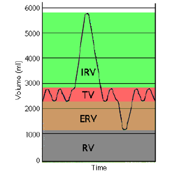
An instrument that produces a graph that records normal and forced inhalation and exhalation. Measures three volumes:

  • Tidal Volume (TV)

    • Normal Ventilation

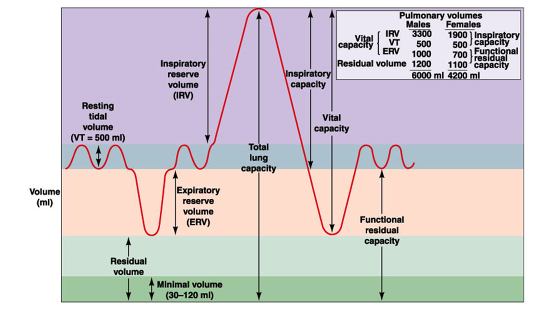
  • Inspiratory Reserve Volume (IRV)

    • Forced Inspiration

  • Expiratory Reserve Volume (ERV)

    • Forced Expiration

95
New cards

Tidal Volume (TV)

Volume of air “traded” with the atmosphere during normal quiet ventilation

  • Measured by a spirometer

  • ~500 mL of air in healthy adults

    • Only 350 mL is available for gas exchange; the remaining 150 mL is anatomical dead space
<p>Volume of air “traded” with the atmosphere during normal quiet ventilation</p><ul><li><p>Measured by a spirometer</p></li><li><p>~<strong>500 mL </strong>of air in healthy adults</p><ul><li><p><strong>Only 350 mL is available for gas exchange</strong>; the remaining 150 mL is anatomical dead space</p></li></ul></li></ul><p></p>
96
New cards

Anatomical Dead Space

Air that remains in the conducting zone (trachea, etc.) and never makes it to the respiratory zone.

  • ~150 mL of air

<p>Air that remains in the conducting zone (trachea, etc.) and never makes it to the respiratory zone.</p><ul><li><p>~<strong>150 mL of air</strong></p></li></ul><p></p>
97
New cards

Alveolar Ventilation Rate (AVR)

The volume of air that reaches the alveoli multiplied by the number of breaths per minute

  • Removes anatomical dead space from the equation

  • Averages 4.2 liters per minute

  • Health professionals prefer to think of it in this way

98
New cards

Minute Volume

Total volume of air that moves in and out of the lungs each minute.

  • The average for adults is ~ 6 liters per minute

  • MV = tidal volume (TV) * breathing/respiratory rate (RR)

<p>Total volume of air that moves in and out of the lungs each minute.</p><ul><li><p>The average for adults is ~<strong> 6 liters per minute</strong></p></li><li><p>MV = tidal volume (TV) * breathing/respiratory rate (RR)</p></li></ul><p></p>
99
New cards

Inspiratory Reserve Volume (IRV)

Volume of air that can be forcibly inspired after a normal TV inspiration.

  • Measured by a spirometer

  • Averages 2100 - 3300 mL of air depending on sexual differences, body size, and health/training
<p>Volume of air that can be forcibly inspired after a normal TV inspiration.</p><ul><li><p>Measured by a spirometer </p></li></ul><ul><li><p>Averages <strong>2100 - 3300 mL </strong>of air depending on sexual differences, body size, and health/training</p></li></ul><p></p>
100
New cards

Expiratory Reserve Volume (ERV)

Amount of air that can be forcibly expired after a normal tidal expiration

  • Measured by a spirometer

  • ERV averages 700-1200 mL of air

<p>Amount of air that can be forcibly expired after a normal tidal expiration </p><ul><li><p>Measured by a spirometer</p></li><li><p>ERV averages<strong> 700-1200 mL of air </strong></p></li></ul><p></p>